고려대학교 의과대학 내과학교실, 고려대학교의료원 안산병원 인간유전체연구소
이승현, 윤대위1, 정진용, 이경주, 김세중, 이은주, 강은해, 정기환, 이승룡, 이상엽, 김제형, 신 철, 심재정, 인광호, 강경호, 유세화
The Effects of Ethyl Pyruvate on Lipopolysaccharide-induced Acute Lung Injury
Seung Hyeun Lee, M.D., Dae Wui Yoon1, Jin Yong Jung, M.D., Kyung Joo Lee, M.D., Se Joong Kim, M.D., Eun Joo Lee, M.D., Eun Hae Kang, M.D., Ki Hwan Jung, M.D., Sung Yong Lee, M.D., Sang Yeub Lee, M.D., Je Hyeong Kim, M.D., Chol Shin, M.D., Jae Jeong Shim, M.D., Kwang Ho In, M.D., Se Hwa Yoo, M.D., Kyung Ho Kang, M.D.
Department of Internal Medicine, College of Medicine, Korea University, Seoul, Korea
1Institute of Human Genomic Study, Ansan Hospital, Korea University Medical Center, Ansan, Korea
Background: Ethyl pyruvate (EP) is a derivative of pyruvate that has recently been identified by both various in vitro and in vivo studies to have antioxidant and anti-inflammatory effects. The aim of this study was to determine the effect of EP on lipopolysaccharide (LPS)-induced acute lung injury (ALI).
Methods: 5 weeks old, male BALB/c mice were used. ALI was induced by an intratracheal instillation of LPS 0.5mg/Kg/50μL of saline. The mice were divided into the control, LPS, EP+LPS, and LPS+EP groups. In the control group, balanced salt solution was injected intraperitoneally 30 minutes before or 9 hours after the intratracheal instillation of saline. In the LPS group, a balanced salt solution was also injected intraperitoneally 30 minutes before or 9 hours after instillation the LPS. In the EP+LPS group, 40mg/Kg of EP was injected 30 minutes before LPS instillation. In the LPS+EP group, 40mg/Kg of EP was injected 9 hours after LPS instillation. The TNF-α and IL-6 concentrations in the bronchoalveolar lavage fluid (BALF), and that of NF-κB in the lung tissue were measured in the control, LPS and EP+LPS groups at 6 hours after instillation of saline or LPS, and the ALI score and myeloperoxidase (MPO) activity were measured in all four groups 24 and 48 hours after LPS instillation, respectively.
Results: The TNF-α and IL-6 concentrations were significantly lower in the EP+LPS group than in the LPS group (p<0.05). The changes in the concentration of these inflammatory cytokines were strongly correlated with that of NF-κB (p<0.01). The ALI scores were significantly lower in the EP+LPS and LPS+EP groups compared with the LPS group (p<0.05). In the EP+LPS group, the MPO activity was significantly lower than the LPS group (p=0.019).
Conclusion: EP, either administered before or after LPS instillation, has protective effects against the pathogenesis of LPS-induced ALI. EP has potential theurapeutic effects on LPS-induced ALI.
(Tuberc Respir Dis 2006; 61: 374-383)
Key words: Acute lung injury, Lipopolysaccharide, Ethyl pyruvate
Address of correspondence: Kyung Ho Kang, M.D.
Division of Pulmonary and Critical Care Medicine, Department of Internal Medicine, Korea University Guro Hospital 80, Guro-dong, Guro-gu, Seoul, 152-703, Korea
Phone: +82-2-818-6638 Fax: +82-2-865-9670 E-mail: [email protected]
Received: Jul. 19. 2006 Accepted: Sep. 18. 2006
서 론
급성폐손상 (acute lung injury, ALI)은 다양한 내 과적 혹은 외과적 손상들과 연관되어, 호흡부전, 흉부
방사선상의 양측성 미만성 폐 침윤 및 심한 저산소혈 증을 특징으로 하는 임상 증후군이다1. 병리기전에 있 어서는 폐포-모세혈관 장벽 (alveolar-capillary barrier) 의 투과성 증가와 활성화된 염증세포의 침윤, 그리고 염증 세포로부터 생성된 전염증성 매개물질 (proin- flammatory mediators), 단백분해효소 (protease) 및 활성산소종 (reactive oxygen species)이 주요한 역할 을 한다2-4. 특히 활성산소종에 의한 산화 자극은 단 백, 지질, 핵산 등 세포 구성 물질의 직접적인 손상을 유발하여 세포의 파괴 및 조직 손상에 관여한다5-7.
Pyruvate (2-oxoproprionate, CH3COCOO-)는 체내 의 대사과정 (intermediary metabolism)에서 주요한
역할을 하는 물질로, 1900년대 초부터 과산화수소에 대한 환원 작용이 있음이 알려졌다. 이후 Dobsak 등8 에 의해 pyruvate가 고반응성의 자유 라디칼과 수산 화기에 대한 제거 작용이 있음이 밝혀졌고, 이러한 특 성을 이용하여 pyruvate를 산화 손상 (oxidant injury) 과 연관된 다양한 병리 조건에서 치료 목적으로 사용 하려는 시도가 있었다9-12. 그러나 pyruvate는 용액 내 에서 자발적으로 parapyruvate로 변하는 불안정성으 로 인해 실험적 혹은 임상적 이용에 어려움이 있었다13. 최근 pyruvate 유도체인 ethyl pyruvate (EP)가 pyruvate와는 달리 Ca2+ 및 K+을 포함한 평형염액에 서 안정적으로 존재한다는 사실이 밝혀지면서 이를 이용한 다양한 연구가 진행되었다. Murine macro- phage-like RAW 264.7 세포를 이용한 in vitro 연구 및 장간막 허혈, 출혈성 쇼크, 내독소 유발 패혈증 등 의 in vivo 연구에서 EP는 염증성 시토카인 및 nuclear factor-κB (NF-κB)의 활성을 억제하고 실험 동물의 생존률을 유의하게 증가시키는 것으로 보고되
었다14-17. 폐손상과 관련된 연구로는, 괴사성 췌장염
과 동반된 간접적 급성폐손상 (indirect acute lung injury) 모델에서 EP가 폐의 투과성 증가 및 염증 반 응을 억제하여 폐손상의 정도를 감소시키는 것으로 보고된 바 있다18.
급성폐손상과 관련된 연구들에 의하면, 폐외 염증 에 의한 간접적 급성폐손상과 폐내 염증에 의한 직접 적 (direct) 급성폐손상은 병태생리, 조직소견 그리고 호흡 역학 및 치료에 대한 반응 등에 있어서 서로 다 른 것으로 보고되고 있다19,20. 따라서 본 연구는 리포 다당질로 인한 직접적 급성폐손상 모델에서 EP가 폐 손상의 기전에 미치는 영향을 연구하고자 하였다.
대상 및 방법
1. 실험 동물 및 LPS로 인한 급성폐손상 모델의 제작
실험대상 동물은 5주 령, 체중 20~25 gm, 무병균 (pathogen-free)의, 수컷 BALB/c 생쥐를 이용하였다.
LPS로 인한 급성폐손상은 생쥐를 흡입용 마취제인 enflurane으로 마취시킨 후, LPS (E.coli O55:B5,
Sigma, St. Louis, MO, USA) 0.5mg/Kg/50μL of saline을 기관 내로 투여하여 유도하였다. 이후 실험 에 적절한 폐손상의 발생 정도를 판단하기 위하여 병 리조직학적 급성폐손상 지수 (acute lung injury score, ALI score), 기관지폐포세척액 (bronchoal- veolar lavage fluid, BALF) 내의 염증성 시토카인 및 myeloperoxidase (MPO)의 활성을, LPS 투여 후 6~
72시간까지 여러 시점에서 평가 하였다.
2. 조직의 처리 및 급성폐손상 지수의 측정
폐의 적출은 pentobarbital sodium 65 mg/Kg을 복 강 내 주사하여 마취시킨 후, 기관 절개를 시행하여 삽관한 후에, 정중 흉골절개술로 흉곽을 절개하고 폐 와 심장을 함께 적출하였다. 이후 삽관을 통하여, 12-15 cmH2O의 압력으로 1% paraformaldehyde 용 액을 주입하여 폐를 팽창시킨 다음, 4% parafor- maldehyde 용액 내에서 48 시간 동안 고정한 후, 심장 및 종격동 조직을 제거한 후에, 70% alcohol in phosphate-buffered saline (PBS) 내에서 냉장 보관 하였다. 파라핀 블록은 우하엽을 파라핀에 포매하여 제작하였다.
급성폐손상 정도의 조직병리학적 고찰을 위해, 파 라핀에 포매한 폐조직을 5 μm의 두께로 잘라 Hematoxylin and Eosin (H/E) 염색을 시행하여 그 정도를 관찰하였다. 급성폐손상 지수21는 1) alveolar capillary congestion, 2) hemorrhage, 3) infiltration or aggregation of inflammatory cells in the airspace or interstitium, 4) thickness of the alveolar wall / hyaline membrane formation 등의 네 가지 지표에 대 해서, 0 = minimal (little) damage, 1+ = mild damage, 2+ = moderate damage, 3+ = severe damage, 4+ = maximal damage의 five-point scale system으로 점 수화한 후, 각 지표의 점수를 합산하여 비교하였다.
3. 기관지폐포 세척술 및 염증성 시토카인과 MPO 활 성의 측정
기관지폐포 세척술은, 마취된 상태에서 기관절개를
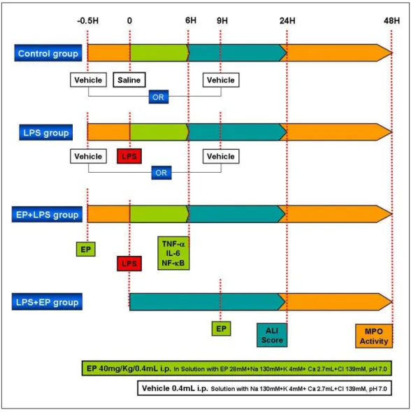
Figure 1. Study groups and protocol. The mice were divided into control, LPS, EP+LPS, and LPS+EP groups. In the control group, balanced salt solution was injected intraperitoneally 30 minutes before or 9 hours after intratracheal instillation of saline. In the LPS group, balanced salt solution was also injected intraperitoneally 30 minutes before or 9 hours after intratracheal instillation of LPS. 40mg/Kg of EP was injected 30 minutes before LPS instillation in the EP+LPS group and was injected 9 hours after LPS instillation in the LPS+EP group. The concentration of TNF-α and IL-6 in bronchoalveolar lavage fluid (BALF), and that of NF-κB in lung tissue were measured in the control, LPS and EP+LPS groups at 6 hours after intratracheal instillation of saline or LPS, and ALI score and myeloperoxidase (MPO) activity was measured in all four groups 24 and 48 hours after LPS instillation, respectively.
통하여 삽관을 한 후에, 1mL의 Ca2+, Mg2+-free phosphate-buffered saline (PBS)를 주입하여 세 차 례 반복하여 시행하였다. 모집된 BALF를 2,000×g, 4℃ 의 조건에서 10분간 원심분리한 후, 상청액 (supernatant)을 추출하여 염증성 시토카인의 농도 및 MPO의 활성을 측정하기 위하여 -70 ℃ 에 냉동보관 하였다. BALF내 염증성 시토카인의 농도는 tumor
necrosis factor-α (TNF-α) 및 interleukin-6 (IL-6) 에 대하여 Enzyme-Linked Immuno-Sorbent Assay (ELISA) (R&D Systems, MN, USA)법으로 측정하 였다. 또한 활성산소종의 주요한 원인인 활성화된 호 중구의 지표로서, MPO의 활성을 BALF내에서 측정 하였다. MPO활성은 50μL의 BALF를 microtiter plate 내에서 200μL의 O-dianisidine dihydrochloride (1.25
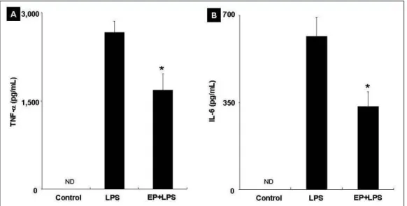
Figure 2. The concentration of tumor necrosis factor-α (TNF-α) (A) and interleukin-6 (IL-6) (B) in bronchoalveolar lavage fluid (BALF) was significantly decreased in EP+LPS group than LPS group (*p<0.05).
ND: not detected
mg/mL in PBS), 0.1% wt/vol의 bovine serum albumin (BSA) 및 H2O2 (0.05% = 0.4 mM)과 반응시 킨 후에, 450 nm에서 흡광도의 변화를 측정하여 결정 하였다22.
4. NF-κB의 측정
LPS 투여로 인한 급성폐손상의 과정에서 염증성 시토카인의 변화가 세포내 전사인자를 매개로 하는지 를 고찰하기 위하여, 폐조직에서 NF-κB의 농도를 측 정하였다. NF-κB의 농도는 적출한 양측 폐를 4℃ 에 서 manual homogenizer를 이용하여 균질 현탁액 (homogenate)을 만들고 Nuclear Extract Kit (Active Motif, Carlsbad, CA, USA)를 이용하여 핵단백을 추 출한 후, TransAMTM NF-κB p65 Transcription Factor Assay Kits (Active Motif, CA, USA) 을 이용 하여 측정하였다.
5. LPS로 인한 급성폐손상 모델의 확인
LPS로 급성폐손상을 유도한 후, BALF 내 TNF-α 및 IL-6의 농도를 6, 12, 24, 48 및 72시간의 시점에서 측정한 결과, 6시간에서 최고치에 도달한 이후에 감소
하는 추세를 보였다 (p<0.05). 또한 LPS 투여 후 6, 12, 24, 36 및 48시간의 시점에서 급성폐손상 지수로 관찰한 조직병리학적 소견은 24시간에 가장 심한 정 도를 나타낸 이후에 감소하는 경향을 보였다 (p<0.05). BALF 내 MPO 활성의 경우는 LPS 투여 후, 48시간에 가장 높은 활성을 보인 후 감소하는 추 세를 보였다 (p<0.05).
결과적으로, 본 실험의 LPS로 인한 급성폐손상 모 델에서 TNF-α 및 IL-6 등의 염증성 시토카인은 LPS 투여 후 초기인 6시간에 상승하고, 폐손상의 정도 및 MPO의 활성은, 비교적 후기인 각각 24시간 및 48시 간에 상승하는 것으로 관찰되었다.
6. 실험군의 설정
실험은 대조군 (Control group), LPS 만을 투여한 LPS군 (LPS group), LPS 투여 전 EP를 처치한 EP+LPS군 (EP+LPS group) 및 LPS 투여 후 초기 염 증성 시토카인이 최고치를 도달한 후에 EP를 처치한 LPS+EP군 (LPS+EP group)의 네 군으로 나누어 시 행하였다 (Figure 1). 대조군은 50 μL의 생리식염수를 기관 내로 투여하기 전 30분, 혹은 투여 후 9시간에 EP가 포함되지 않은 평형염액만을 복강 내 주사하였
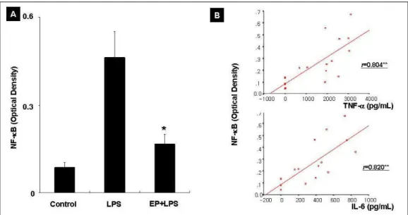
Figure 3. The concentration of nuclear factor-κB (NF-κB) in lung homogenate of the EP+LPS group was significantly lower than the LPS group and higher than the control group (*p<0.05). It was significantly correlated with tumor necrosis factor-α (TNF-α) and interleukin-6 (IL-6) (r=Spearman’s rho, **p<0.01).
다. LPS군은 5 mg/Kg의 LPS 용액 50μL를 기관 내로 투여하기 전 30분, 혹은 투여 후 9시간에 평형염액 만 을 복강 내 주사하였다. EP+LPS군은 LPS 투여 전 30 분에 40 mg/Kg의 EP를 복강 내 주사하였고, LPS+
EP군은 LPS투여 후 9시간에 EP를 복강 내 주사하였 다. BALF 내 TNF-α 및 IL-6의 농도 및 폐조직 내의 NF-κB의 농도는 대조군, LPS군 및 EP+LPS군에서 생리식염수 혹은 LPS 투여 후 6시간에 측정하였다.
급성폐손상 지수와 MPO의 활성은 네 군 모두에서, 생리식염수 혹은 LPS 투여 후, 각각 24시간 및 48시 간에 측정하였다.
7. 통 계
모든 결과는 평균±표준오차 (mean±standard error of mean, SEM)로 표시하였다. 각 군 간의 비교는 비 모수적 Kruskal-Wallis 검정과 Mann-Whitney U 검 정을 사용하였으며, 두 변수간의 상관관계는 Spear- man’s correlation coefficient를 사용하여 분석하였다.
모든 계산은 SPSS for Windows Release 10.0 (SPSS Inc. USA) 을 사용하였고, p값이 0.05이하인 경우 통 계학적 유의성이 있는 것으로 판단하였다.
결 과 1. TNF-α 및 IL-6의 농도
LPS 투여 후 6시간에 BALF에서 측정한 TNF-α의 농도는 대조군에서는 검출되지 않았고, LPS군에서는 2653.13±203.26 pg/mL, EP+LPS군에서는 1684.18±
278.76 pg/mL 로 EP+LPS군이 LPS군과 비교하여 낮 았다 (p=0.026) (Figure 2A). IL-6 역시 대조군에서 검출되지 않았고, EP+LPS군에서 333.47±59.89 pg/
mL로 LPS군의 616.39±75.67 pg/mL에 비해 낮았다 (p=0.041) (Figure 2B). 폐조직의 균질현탁액에서 측 정한 NF-κB의 농도는 대조군에서는 0.086±0.017 O.D., LPS군에서는 0.462±0.089 O.D. 및 EP+LPS군에 서는 0.167±0.032 O.D.로 세 군간에 차이를 보였다 (p=0.005 by Kruskal-Wallis test). 각 군간의 비교에 있어서는, EP+LPS군에서 LPS군과 비교하여 낮았고 (p=0.029), 대조군과 비교해서는 높았다 (p=0.038) (Figure 3A). 염증성 시토카인과 NF-κB 농도의 상관 관계를 분석한 결과, NF-κB의 농도는 TNF-α (r=0.804, p<0.01) 및 IL-6 (r=0.820, p<0.01)의 농도와 상관 관계를 보였다 (Figure 3B).
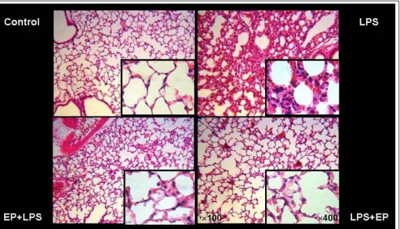
Figure 4. Histopathologic examination shows high levels of inflammatory cellular infiltration, hemorrhage, and alveolar wall thickening in the LPS group. In the EP+LPS and LPS+EP group, the degree of acute lung injury was lower and only mild inflammatory cellular infiltration was observed compared with the control group.
ALI score
Figure 5. Acute lung injury (ALI) scores were significantly different among four groups (p=0.000 by Kruskal-Wallis test). In the EP+LPS group, the score was significantly lower than the LPS group and higher than the control group (*p<0.05). And the score of the LPS+EP group was also significantly lower compared with the LPS group (**p=0.017) without significance between the control and the EP+LPS group (p>0.05).
2. 폐손상의 정도
전반적인 폐손상의 정도를 H/E 염색 하에서 관찰
한 결과, LPS군에서 폐포벽 및 폐포 내에 심한 염증 성 세포들의 침윤, 폐모세혈관 주위 및 폐포 내 출혈 과 간질의 비후 및 부종 등이 관찰되었다. 반면 EP+LPS군 및 LPS+EP군에서는 대조군과 비교하여 경도의 염증성 세포 및 간질의 비후가 관찰되기는 하 였으나, 울혈 및 부종 등 현저한 폐손상의 소견은 없 었다 (Figure 4). 이를 급성폐손상 지수로 정량화하여 비교한 결과, 대조군에서는 5.8±0.7, LPS군에서는 11.0±0.6, EP+LPS군에서는 8.0±0.6, 그리고 LPS+EP 군에서는 7.4±1.1로 네 군간에 차이를 보였다 (p=0.000 by Kruskal-Wallis test). 각 군간의 비교에 있어서는 EP+LPS군 및 LPS+EP군에서 LPS군과 비 교하여 낮았고 (p<0.05), 대조군과 비교하여서는 EP+LPS군에서 높았으나 (p=0.030), LPS+EP군과는 차이가 없었다 (p=0.329). EP+LPS군과 LPS+EP군 간 에 차이는 관찰되지 않았다 (p=0.841) (Figure 5).
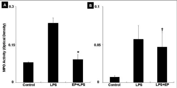
3. MPO 활성
LPS투여 48시간 후에 측정한 BALF내 MPO의 활 성은 대조군, LPS군 및 EP+LPS군의 비교에 있어서
Figure 6. The myeloperoxidase (MPO) activity in bronchoalveolar lavage fluid (BALF) was significantly lower in the EP+LPS group compared with the LPS group (*p=0.019) (A). However, in the LPS+EP group, the activity showed only decreasing trends without significant difference (†p=0.931) (B)
는, 각각 0.079±0.003 O.D., 0.235±0.02 O.D., 0.091±
0.019 O.D.로 세 군간에 차이가 있었고 (p=0.016 by Kruskal-Wallis test), 각 군간의 비교에 있어서는 EP+LPS군에서 LPS군과 비교하여 낮았고 (p=0.019), 대조군과 차이는 없었다 (p=0.394) (Figure 6A). 그러 나 대조군, LPS군 및 LPS+EP군간의 비교에 있어서 는, 각각 0.007±0.002 O.D., 0.057±0.018 O.D., 0.047±0.019 O.D.로 세 군간에 통계적으로 차이가 없 었으며 (p=0.053 by Kruskal-Wallis test), LPS+EP군 에서 LPS군과 비교하여 감소하는 경향을 보였으나 통계적으로 유의하지는 않았다 (p=0.931) (Figure 6B).
고 찰
본 연구의 결과에 의하면, LPS로 인한 급성폐손상 모델에서 EP의 전처치는 NF-κB를 매개로 하여 TNF-α 및 IL-6의 염증성 시토카인과 급성폐손상지 수, MPO의 활성을 유의하게 감소시켰다. 또한, 초기 염증성 시토카인이 상승한 이후에, EP를 후처치한 경 우에도 급성폐손상 지수를 유의하게 감소시켜, EP가 LPS로 인한 급성폐손상에 대해서 예방 및 치료 효과 가 있는 것으로 관찰되었다.
Pyruvate는 체내의 해당 경로 (glycolytic pathway)
의 마지막 단계에서 생성되는 물질로, 항산화 및 자유 라디칼에 대한 제거 기능이 있는 것으로 알려져 있다
23. 1991년 Salahudeen 등9 이 처음으로 활성산소종에 의한 급성 신부전 모델에서 sodium pyruvate가 신기 능 보전에 효과가 있음을 보고한 이후에, 심근10, 장11, 간12 등의 허혈/재관류 모델에서 pyruvate가 장기 손 상과 기능 부전 예방에 효과가 있음이 밝혀졌다. 그러 나 이러한 효과에도 불구하고 pyruvate는 용액 내에 서 aldol-like condensation을 거쳐 parapyruvate로 변 하는 불안정성으로 인해 임상적 이용에는 제약이 있 었다13.
2001년 Sims 등14은 Ca2+ 및 K+을 포함한 balanced salt solution에 pyruvate의 유도체인 EP를 혼합하여 만든 Ringer’s ethyl pyruvate solution (REPS)이 보 다 안정적이고 효과적으로, 장간막의 허혈/재관류 모 델에서 장점막의 손상 억제 효과가 있음을 처음으로 보고하였다. 이후 Yang 등17은 생쥐를 이용한 출혈성 쇼크 모델에서 EP 사용군이 대조군에 비해 유의하게 생존률이 증가하고, 염증성 시토카인의 생성 및 염증 반응에서 중요한 전사 인자인 NF-κB 활성이 감소함 을 보고하였다. 비슷한 시기에 Ulloa 등16은 LPS로 유 발한 내독소혈증 모델에서 LPS 투여 전 뿐만 아니라 투여 수시간 후 EP를 투여하였을 때도 생존률의 향상
을 보여 지연 치료 (delayed treatment)도 효과가 있 음을 확인하였다. 폐손상과 관련해서 Yang 등18은 급 성 괴사성 췌장염을 이용한 간접적 급성폐손상 모델 에서 EP가 폐포 투과성 및 염증 반응을 억제함을 보 고한 바 있다.
급성폐손상은 폐 실질의 직접적인 손상에 의해 발 생하는 일차성 또는 직접적 급성폐손상 (primary or direct ALI)과 전신적인 염증 반응에 의해 이차적으로 발생하는 이차성 또는 간접적 급성폐손상 (secondary or indirect ALI)으로 구분된다1. 직접적 그리고 간접 적 급성폐손상은 병태생리, 조직소견 그리고 호흡 역 학 및 치료에 대한 반응에 있어서 서로 다르다는 것이
밝혀졌고20,24,25 특히 폐내 염증 반응의 정도는 직접적
급성폐손상 시 더욱 심한 것으로 알려져 있다26. 따라서 항산화 및 항염증 작용을 가지는 것으로 입 증된 EP가 LPS로 인한 직접적 급성폐손상 모델에서 염증 반응 및 조직 손상을 억제하고 이 과정에서 염증 성 시토카인의 생성에 NF-κB가 관여할 것이라는 가 정 하에 본 연구를 진행하였다. 결과적으로 EP는 염 증 반응의 초기에 생성되어 작용을 나타내는 TNF-α 및 IL-6 와 같은 시토카인의 생성을 억제하였고 이는 세포 내 전사 인자인 NF-κB를 매개로 이루어지는 것 으로 관찰되었으며, 급성폐손상 지수 및 MPO 활성으 로 대표되는 후기 염증 반응에도 효과가 있음이 확인 되었다. 또한 EP를 전처치한 경우뿐만 아니라 후처치 하였을 때에도 염증 반응과 조직 손상이 감소되어 지 연 치료 효과도 있음을 확인하였다.
EP의 항산화 및 항염증 효과의 기전은 명확하지 않 지만, NF-κB를 매개로 하는 것으로 보고되고 있다.
활성산소종은 염증 반응에 있어서 세포 내 그리고 세 포 외 구조물에 직접적인 손상을 일으킬 뿐만 아니라 세포 내의 염증성 신호 전달 경로, 특히 NF-κB와 관 련된 세포 내 신호 전달계의 활성화에 관여한다27-29. 이러한 효과는 활성산소종이 NF-κB 활성화에 중요 한 IκB의 인산화 과정에 관여함으로써 나타나는 것으 로 추측된다28-30. 최근 murine macrophage-like RAW 264.7 cell을 이용한 in vitro 연구31에서는 EP 가 NF-κB의 p65 subunit의 oligonucleotide sequence 를 변형시킴으로써 DNA 결합을 억제시킴이 밝혀졌
다. 이는 EP가 활성산소종을 매개로 한 간접적인 방 법이 아닌 직접적인 방법으로 NF-κB의 활성을 억제 함을 시사한다. 본 연구에서도 EP의 전처치가 염증성 시토카인 및 NF-κB의 증가를 억제하고, 염증성 시토 카인의 증가 및 감소가 NF-κB와 유의한 상관관계가 있어, EP의 작용 기전에 있어 NF-κB의 활성이 직․
간접적으로 밀접한 관련이 있는 것으로 보인다.
그간 다양한 치료적 접근과 중환자 관리에 있어 괄 목할 만한 발전이 있었음에도 불구하고, 최근까지도 급성폐손상에 의한 사망률은 여전히 높은 실정이다.
현재까지 급성폐손상이나 패혈증에 대한 치료적 시도 는 대부분이 염증성 시토카인의 상승을 억제하거나 체내에서 제거하려는 노력이 대부분이었다32. 그러나 염증성 시토카인은 대부분 염증 반응 초기 수시간 내 에 상승하여 이후 체내에서의 농도가 떨어지고 임상 적으로 급성폐손상이나 패혈증은 염증 반응이 상당히 진행된 이후 치료적 개입이 시작되므로 치료의 효과 를 기대하기 힘들었다. 따라서 이미 초기 염증성 시토 카인이 상승한 이후에 치료하였을 경우에도 효과가 있었고16, 본 연구의 급성폐손상 모델에서도 역시 동 일한 결과를 보인 EP는, 향후 패혈증 및 다양한 원인 으로 인한 급성폐손상 환자의 치료 및 예후의 개선에 있어서 주요한 치료적 잠재력이 있을 것으로 판단된 다.
요 약
연구배경: 급성폐손상에서 활성산소종에 의한 산 화 손상은 주요한 역할을 한다. Ethyl pyruvate (EP) 는 체내에서 생성되는 pyruvate의 유도체로 항산화 및 항염증 효과가 있음이 알려졌다. 저자들은 리포다 당질에 의한 급성폐손상 모델에서 EP가 염증반응에 미치는 영향을 연구하고자 하였다.
방 법: 5주 령의 BALB/c 생쥐를 이용하여 리포다 당질을 기관 내로 투여하여 급성폐손상을 유도하였 다. 대조군, LPS군, EP+LPS군, LPS+EP군으로 나누 어 기관지폐포세척액에서 TNF-α, IL-6 및 myelo- peroxidase (MPO)의 활성을, 폐조직에서 급성폐손상 지수와 NF-κB의 농도를 측정하였다.
결 과: EP+LPS군에서 TNF-α 및 IL-6의 농도는 LPS군과 비교하여 감소하였고 (p<0.05) 이들 염증성 시토카인의 농도의 변화는 NF-κB의 농도의 변화와 상관 관계를 보였다 (p<0.01). 급성폐손상 지수는 EP+LPS군 및 LPS+EP군에서 LPS군과 비교하여 낮 았고 (p<0.05) MPO활성은 EP+LPS군에서 LPS군에 비해 낮았다 (p<0.05).
결 론: EP는 LPS로 인한 급성폐손상에 있어서 예 방 및 치료 효과가 있는 것으로 판단된다.
참 고 문 헌
1. Bernard GR, Artigas A, Brigham KL, Carlet J, Falke K, Hudson L, et al. The American-European Con- sensus Conference on ARDS: definitions, mechanisms, relevant outcomes, and clinical trial coordination. Am J Respir Crit Care Med 1994;149:818-24.
2. Sibille Y, Reynolds HY. Macrophages and polymor- phonuclear neutrophils in lung defense and injury.
Am Rev Respir Dis 1990;141:471-501.
3. Ware LB, Matthay MA. The acute respiratory distress syndrome. N Engl J Med 2000;342:1334-49.
4. Pugin J, Verghese G, Widmer MC, Matthay MA. The alveolar space is the site of intense inflammatory and profibrotic reactions in the early phase of acute respiratory distress syndrome. Crit Care Med 1999;27:304-12.
5. Janero DR, Hreniuk D, Sharif HM. Hydroper- oxide-induced oxidative stress impairs heart muscle cell carbohydrate metabolism. Am J Physiol 1994;266:
C179-88.
6. Mohr S, Stamler JS, Brune B. Posttranslational modification of glyceraldehyde-3-phosphate dehydro- genase by S-nitrosylation and subsequent NADH attachment. J Biol Chem 1996;271:4209-14.
7. Mahadev K, Zilbering A, Zhu L, Goldstein BJ.
Insulin-stimulated hydrogen peroxide reversibly inhi- bits protein-tyrosine phosphatase 1b in vivo and enhances the early insulin action cascade. J Biol Chem 2001;276:21938-42.
8. Dobsak P, Courderot-Masuyer C, Zeller M, Vergely C, Laubriet A, Assem M, et al. Antioxidative properties of pyruvate and protection of the ischemic rat heart during cardioplegia. J Cardiovasc Pharmacol 1999;34:
651-9.
9. Salahudeen AK, Clark EC, Nath KA. Hydrogen peroxide-induced renal injury: a protective role for pyruvate in vitro and in vivo. J Clin Invest 1991;
88:1886-93.
10. Bunger R, Mallet RT, Hartman DA. Pyruvate- enhanced phosphorylation potential and inotropism in normoxic and postischemic isolated working heart:
near-complete prevention of reperfusion contractile failure. Eur J Biochem 1989;180:221-33.
11. Cicalese L, Lee K, Schraut W, Watkins S, Borle A, Stanko R. Pyruvate prevents ischemia-reperfusion mucosal injury of rat small intestine. Am J Surg 1996;171:97-100.
12. Sileri P, Schena S, Morini S, Rastellini C, Pham S, Benedetti E, et al. Pyruvate inhibits hepatic ischemia- reperfusion injury in rats. Transplantation 2001;72:
27-30.
13. Vonkorff RW. Pyruvate-C14, purity and stability. Anal Biochem 1964;8:171-8.
14. Sims CA, Wattanasirichaigoon S, Menconi MJ, Ajami AM, Fink MP. Ringer's ethyl pyruvate solution ameliorates ischemia/reperfusion-induced intestinal mucosal injury in rats. Crit Care Med 2001;29:1513-8.
15. Tawadrous ZS, Delude RL, Fink MP. Resuscitation from hemorrhagic shock with Ringer's ethyl pyruvate solution improves survival and ameliorates intestinal mucosal hyperpermeability in rats. Shock 2002;17:
473-7.
16. Ulloa L, Ochani M, Yang H, Tanovic M, Halperin D, Yang R, et al. Ethyl pyruvate prevents lethality in mice with established lethal sepsis and systemic inflammation. Proc Natl Acad Sci U S A 2002;99:
12351-6.
17. Yang R, Gallo DJ, Baust JJ, Uchiyama T, Watkins SK, Delude RL, et al. Ethyl pyruvate modulates inflammatory gene expression in mice subjected to hemorrhagic shock. Am J Physiol Gastrointest Liver Physiol 2002;283:G212-21.
18. Yang R, Uchiyama T, Alber SM, Han X, Watkins SK, Delude RL, et al. Ethyl pyruvate ameliorates distant organ injury in a murine model of acute necrotizing pancreatitis. Crit Care Med 2004;32:1453-9.
19. Pelosi P, D'Onofrio D, Chiumello D, Paolo S, Chiara G, Capelozzi VL, et al. Pulmonary and extrapul- monary acute respiratory distress syndrome are different. Eur Respir J Suppl 2003;42:48s-56s.
20. Rocco PR, Zin WA. Pulmonary and extrapulmonary acute respiratory distress syndrome: are they diffe- rent? Curr Opin Crit Care 2005;11:10-7.
21. Imanaka H, Shimaoka M, Matsuura N, Nishimura M, Ohta N, Kiyono H. Ventilator-induced lung injury is associated with neutrophil infiltration, macrophage activation, and TGF-beta 1 mRNA upregulation in rat lungs. Anesth Analg 2001;92:428-36.
22. Hirano S. Migratory responses of PMN after
intraperitoneal and intratracheal administration of lipopolysaccharide. Am J Physiol 1996;270:L836-45.
23. Melzer E, Schmidt HL. Carbon isotope effects on the decarboxylation of carboxylic acids: comparison of the lactate oxidase reaction and the degradation of pyruvate by H2O2. Biochem J 1988;252:913-5.
24. Pelosi P, Caironi P, Gattinoni L. Pulmonary and extrapulmonary forms of acute respiratory distress syndrome. Semin Respir Crit Care Med 2001;22:
259-68.
25. Terashima T, Matsubara H, Nakamura M, Sakamaki F, Waki Y, Soejima K, et al. Local pseudomonas ins- tillation induces contralateral lung injury and plasma cytokines. Am J Respir Crit Care Med 1996;153:
1600-5.
26. Menezes SL, Bozza PT, Neto HC, Laranjeira AP, Negri EM, Capelozzi VL, et al. Pulmonary and extrapulmonary acute lung injury: inflammatory and ultrastructural analyses. J Appl Physiol 2005;98:
1777-83.
27. Verhasselt V, Vanden Berghe W, Vanderheyde N, Willems F, Haegeman G, Goldman M. N-acetyl- L-cysteine inhibits primary human T cell responses at the dendritic cell level: association with NF-kappaB
inhibition. J Immunol 1999;162:2569-74.
28. Schoonbroodt S, Ferreira V, Best-Belpomme M, Boelaert JR, Legrand-Poels S, Korner M, et al.
Crucial role of the amino-terminal tyrosine residue 42 and the carboxyl-terminal PEST domain of I kappa B alpha in NF-kappa B activation by an oxidative stress. J Immunol 2000;164:4292-300.
29. Livolsi A, Busuttil V, Imbert V, Abraham RT, Peyron JF. Tyrosine phosphorylation-dependent activation of NF-kappa B: requirement for p56 LCK and ZAP-70 protein tyrosine kinases. Eur J Biochem 2001;268:
1508-15.
30. Rahman I, Mulier B, Gilmour PS, Watchorn T, Donaldson K, Jeffery PK, et al. Oxidant-mediated lung epithelial cell tolerance: the role of intracellular glutathione and nuclear factor-kappaB. Biochem Pharmacol 2001;62:787-94.
31. Han Y, Englert JA, Yang R, Delude RL, Fink MP.
Ethyl pyruvate inhibits nuclear factor-kappaB- dependent signaling by directly targeting p65. J Pharmacol Exp Ther 2005;312:1097-105.
32. Riedemann NC, Guo RF, Ward PA. Novel strategies for the treatment of sepsis. Nat Med 2003;9:517-24.