Tuberc Respir Dis 2010;69:337-347
CopyrightⒸ2010. The Korean Academy of Tuberculosis and Respiratory Diseases. All rights reserved.
리포다당질로 유도된 급성 폐손상 후 섬유화증식에서 Transglutaminase-2의 역할
고려대학교 안산병원 호흡기내과
김제형The Role of Transglutaminase-2 in Fibroproliferation after Lipopolysaccharide-induced Acute Lung Injury
Je Hyeong Kim, M.D., Ph.D.
Division of Pulmonary, Sleep and Critical Care Medicine, Department of Internal Medicine, Korea University Ansan Hospital, Ansan, Korea
Background: Transglutaminase-2 (TG-2) has been reported to play an important role in the process of fibrosis.
However, TG-2 studies on fibroproliferation of acute lung injury (ALI) are absent. The purpose of this study was to investigate the role of TG-2 in the fibroproliferation of lipopolysaccharide (LPS)-induced ALI.
Methods: The male C57BL/6 mice of 5 weeks age were divided into 3 groups; control group (n=30) in which 50 μL of saline was given intratracheally (IT), LPS group (n=30) in which LPS 0.5 mg/kg/50 μL of saline was given IT, and LPS+Cyst group treated with intraperitoneal 200 mg/kg of cystamine, competitive inhibitor of TG-2, after induction of ALI by LPS. TG-2 activity and nuclear factor (NF)-κB were measured in lung tissue homogenate.
Tumor necrosis factor (TNF)-α, interleukin (IL)-1β, IL-6, myeloperoxidase (MPO), and transforming growth factor (TGF)-β1 were measured using bronchoalveolar lavage fluids. Histopathologic ALI score and Mallory’s phospho- tunistic acid hematoxylin (PTAH) for collagen and fibronectin deposition were performed.
Results: The TG-2 activities in the LPS group were significantly higher than the control and LPS+Cyst groups (p<0.05). The TNF-α and IL-1β concentrations and NF-κB activity were lower in the LPS+Cyst group than the LPS group (p<0.05). The LPS+Cyst group showed lower MPO, ALI score, TGF-β1 concentration, and Mallory’s PTAH stain than the LPS group, but the differences were not significant (p>0.05).
Conclusion: Inhibition of TG-2 activity in the LPS-induced ALI prevented early inflammatory parameters, but had limited effects on late ALI and fibroproliferative parameters.
Key Words: Acute Lung Injury; Lipopolysaccharides; Inflammation; Fibrosis
Address for correspondence: Je Hyeong Kim M.D., Ph.D.
Division of Pulmonary, Sleep and Critical Care Medicine, Department of Internal Medicine, Korea University Ansan Hospital, Gojan 1-dong, Danwon-gu, Ansan 425-707, Korea Phone: 82-31-412-5950, Fax: 82-31-413-5950
E-mail: [email protected] Received: Oct. 5, 2010
Accepted: Oct. 5, 2010
서 론
지난 수십 년간 급성 폐손상(acute lung injury, ALI) 및 급성호흡곤란증후군(acute respiratory distress syndrome,
ARDS)의 병리기전에 대한 연구와 치료적 접근 및 중환자 관리에 있어서 괄목할만한 발전이 있었음에도 불구하고, ARDS 환자들의 사망률은 41% 정도로 매우 높다1. 현재까 지 알려진 바에 따르면, 전통적인 기계환기 양식으로 인한 폐손상(ventilator-induced lung injury, VILI)이 환자들의 사망에 있어서 주요한 역할을 하는 것으로 보고되고 있다.
따라서 VILI를 최소화하기 위해, 저일호흡량환기법(low tidal volume ventilation)이 모색되었고, 대규모 임상연구 에서 환자들의 사망률을 유의하게 감소시켰다2. 하지만, 폐의 심한 비균질성으로 인하여 그 효과가 제한적이어서, ARDS의 병리기전에 대한 추가적인 연구와, 이에 근거한
다각적인 치료방법들을 모색하는 것이 필요하다.
ALI 및 ARDS의 급성기 후, 일부 환자들은 후유증 없이 증식기(proliferative phase)를 거쳐 치유되기도 하지만, 다른 생존자들에서는 폐섬유화(fibrotic phase)3,4 및 이로 인한 폐기능 장애5,6를 갖는 경우가 흔하다. 섬유화는 지속 적이고 심한 염증이 치유되지 않음으로써 발생하는, 조직 손상에 대한 stereotypic response로, 최근 연구들에 의하 면, collagen turnover가 ARDS의 비교적 초기에 증가하는 것으로 보고되고 있다7,8. Transglutaminase (Tgase, TG) 는 특이 펩티드에 결합된 glutamine residue의 γ-carbox- amine group과 다양한 amine들 사이의 결합을 촉진하는 효소로9, 이 중 type 2 Tgase (TG-2)는 일차적으로는 손상 의 예방과 방어 및 복구를 촉진하는 데 있어서 주요한 역 할을 하는 것으로 여겨졌으나10, 최근의 연구들에 의하면 비정상적으로 과도하게 유발될 경우에는 neurodegener- ative diseases, atherosclerosis, inflammatory diseases 및 autoimmune diseases 등과 같은 질병의 발생에 주요한 역할을 하는 것으로 보고되고 있다11. 또한 collagen, fi- bronectin, laminia, nidogen 및 proteoglycan 등의 matrix protein들과 비가역적으로 결합하거나12, 세포 외 기질의 proteolytic enzyme에 대한 저항성을 증가시켜, 심한 세포 외기질의 침착을 유도13하여 조직의 섬유화에 관여하는 것으로 제시되고 있다. 실제로 실험적인 신장14 및 간15 섬 유화에 있어서 TG-2가 주요한 역할을 하고, 억제제를 이 용하여, 그 활성을 억제하였을 경우, 섬유화의 정도가 감 소하는 것으로 보고된 바 있다16,17. TG-2는 collagen syn- thesis 및 epithelial-mesenchymal transition의 증가에 있 어서 주요한 매개체인 transforming growth factor (TGF)- β1와 밀접한 연관관계가 있어, 비활성화 상태의 TGF-β1 를 활성화시키는 데 있어서 주요한 역할을 하고18, TGF-β 1은 TG-2의 주요한 유도인자인 것으로 보고되고 있어19,20, TGF-β1와 TG-2가 조직 섬유화과정에서 상호 긴밀한 기 능을 하는 것으로 판단된다. 하지만, 이와 같은 TG-2의 기능 및 역할에도 불구하고 폐질환에 있어서 TG-2에 대한 연구는 매우 드물고, 특히, ALI 후 폐섬유화의 병리기전에 서 TG-2의 역할을 고찰한 연구는 없다.
본 연구는 리포다당질(lipopolysaccharide, LPS)로 유도 된 급성 폐손상에서 폐조직 내 TG-2의 활성을 급성 폐손 상 및 섬유화 표지자 등의 활성 및 발현과 비교하고, TG-2 억제제의 효과를 고찰함으로써, 급성 폐손상의 급성 염증 및 폐섬유화 과정에서 TG-2의 역할을 연구하고자 하였다.
대상 및 방법
1. 리포다당질로 인한 급성 폐손상의 유도 및 폐의 적출
5주령의 수컷 C57BL/6 mice를 pentobarbital sodium 65 mg/kg로 마취시키고, midcervical incision을 통해 기 관을 노출 시킨 후, 26 G microsyringe를 이용하여 LPS (
E. coli
0127:B8; Sigma, St. Louis, MO, USA) in 50 μL of saline을 기관 내로 주사하여 유도하였다. 폐의 적출은 마취가 되어있는 상태에서, 기관절개를 통해 angiocath를 고정한 후, 복강을 절개하여 대동맥 및 하대정맥을 절단하 여 충분히 실혈시킨 후에, 정중 흉골절개술로 흉곽을 절개 하고, 폐와 심장을 함께 적출한 후, 기관에 고정된 angio- cath를 통해서 15 cmH2O의 압력으로 4% paraformalde- hyde 용액을 주입하여 폐를 팽창시킨 다음, 4% paraf- ormaldehyde 용액 내에서 48시간 동안 고정하였다. 이후 심장 및 종격동 조직을 제거한 후에, 좌측폐를 파라핀에 포매하였다.2. 급성 폐손상 지수
21및 Mallory's phosphotunstic acid hematoxylin 염색
22급성 폐손상 정도의 조직학적 고찰을 위해, 파라핀에 포매된 폐조직을 5 μm의 두께로 잘라 Hematoxylin and Eosin (H&E) 염색을 시행하여, 1) alveolar capillary con- gestion, 2) hemorrhage, 3) infiltration or aggregation of inflammatory cells in the airspace or interstitium, 4) thickness of the alveolar wall/hyaline membrane for- mation 등의 네 가지 지표에 대해서, 0, minimal (little) damage; 1+, mild damage; 2+, moderated damage;
3+, severe damage; 4+, maximal damage의 five-point scale system으로 점수화하여 비교하였다. 섬유화반응으 로 인한 collagen 및 fibronectin 침착은, fibronectin에 대 한 단클론항체(monoclonal antibody)를 이용한 면역조직 화학염색(immunohistochemical stain)인 Mallory's phos- photunstic acid hematoxylin (PTAH) 염색을 시행한 후, 이 미지분석시스템(Image-ProⓇ Plus; MediaCybenetics Inc., Bethesda, MD, USA)을 이용하여 분석하였다.
3. 기관지 폐포 세척술 및 기관지 폐포 세척액 분석
기관지 폐포 세척술(bronchoalveolar lavage, BAL)은 1 mL의 PBS (pH 7.4)를 3차례 기도 내로 주입하여, 회수된 기관지폐포세척액을 4oC에서, 10분간 5,000 rpm으로 원Figure 1. (A) The time course of transglutaminase (TG)-2 activity. Thirty six hours after intratracheal instillation of lip- opolysaccharide (LPS, 0.5 mg/kg in 50 μL of saline), TG-2 activity showed significantly high activity compared with the control, 3, 6, 12, and 24 hours (*p<0.05). At 48 hours and 72 hours, TG-2 activities decreased, but not significant compared with the activity at 36 hours (†p>0.05). (B) The response of TG-2 activity for LPS doses. Instillation of LPS 1.0 mg/kg in 50 μL of saline showed an increased trend of TG-2 activity, but not significant compared with the activ- ity of LPS 0.5 mg/kg (‡p=0.248). (C) The dose response of TG-2 activity for cystamine. At the cystamine dose of 200 mg/kg, TG-2 activity decreased significantly compared with the LPS group (§p=0.009), which was not different with the control group (∥p=0.806).
심분리한 후, 상층액은 분석을 위해 -70oC에 보관하였 다. BAL fulid (BALF) 내의 tumor necrosis factor (TNF)- α, interleukin (IL)-1β, IL-6 및 TGF-β1 농도의 측정은 enzyme- linked immunosorbent assay (ELISA; R & D Systems, Minneapolis, MN, USA)법으로 시행하였고, 활 성산소종의 주요한 원인인 활성화된 호중구의 지표로서, myeloperoxidase (MPO)의 활성을, 50 μL의 BALF를 mi- crotiter plate 내에서 200 μL의 O-dianisidine dihydro- chloride (1.25 mg/mL in PBS), 0.1% wt/vol의 bovine serum albumin (BSA) 및 H2O2 (0.05%=0.4 mM)과 반응 시킨 후에, 450 nm에서 흡광도의 변화를 측정하여 결정하 였다.
4. Nuclear factor (NF)-κB activity의 측정
Tissue homogenate에서 NF-κB의 활성의 측정은 정중
흉골절개술로 흉곽을 절개하고, 폐와 심장을 함께 적출한 후, 폐조직만을 절제하여, lung tissue homogenate에서 nu- clear protein extraction kit (Active Motif, Carlsbad, CA, USA)를 이용하여, nuclear protein을 추출하여, NF-κB의 DNA binding capacity를 이용한 TransAMTM NF-κB Kit (Active Motif)으로 NF-κB의 활성을 측정하였다.
5. TG-2 활성의 측정 및 TG-2 억제제
TG-2의 활성은 lung tissue homogenate에서 fluore- scence based protein arrays를 이용하여 측정하였다.
Fibrinogen과 incubation한 amine modified glass slide를 제작한 후, lung tissue homogenate를 CaCl2와 thrombin 이 포함된 reaction buffer와 혼합하고, reaction mixture를 array well에 투여하였다. TG-2의 촉매반응에 의해서 fi- brinogen내로 incorporation된 pentylamine을 Cy3-conju-
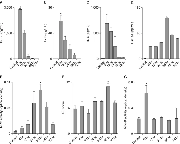
Figure 2. The time course of acute lung injury (ALI) parameters. The concentrations of tumor necrosis factor (TNF)-α (A), interleukin (IL)-1β (B), and IL-6 (C) in bronchoalveolar lavage fluid (BALF) and nuclear factor (NF)-κB activity (G) in lung tissue homogenates showed peaks at 6 hours after lipopolysaccharide (LPS) administration. Transforming growth factor (TGF)-β1 (D) and myeloperoxidase (MPO) activity (E) were highest at 36 hours, and ALI score (F) showed peak at 48 hours (*p<0.05, compared with the control and other time points).
gated streptavidin으로 probe한 후, 543 nm에서 fluo- rescence scanner (GenePix Personal 4100A; Molecular Devices, Sunnyvale, CA, USA)로 스캔한 후, 그 활성도를 상대형광단위(relative fluorescence units, RFU)/mg of protein로 구하였다. TG-2 억제제는 TG activity에 대한 competitive inhibitor23인 cystamine (Sigma)을 복강 내로 투여하였다.
6. 실험 모델의 형성
LPS 0.5 mg/kg in 50 μL of saline을 기관 내로 주사한 후, TG-2의 활성을 3, 6, 12, 24, 36, 48 및 72시간 시점에 서 측정한 결과, 36시간에 최고 활성을 보였고(p<0.05,
compared with the control, 3, 6, 12, and 24 hours time points), 48시간 및 72시간에는 감소하였으나, 36시간 활 성과 유의한 차이는 없었다(p>0.05) (Figure 1A). LPS에 대한 용량반응을 관찰하기 위하여, LPS 1.0 mg/kg in 50 μL of saline을 기관 내로 투여한 후, 36시간에 TG-2의 활성을 측정한 결과 LPS 0.5 mg/kg in 50 μL of saline의 경우와 비교하여, 경도의 증가는 있었으나 통계적으로 유 의한 차이는 보이지 않아(Figure 1B), LPS 0.5 mg/kg in 50 μL of saline를 급성 폐손상의 유도용량으로 결정하였 다. LPS 투여 후, cystamine 10, 50, 100 및 200 mg/kg을 복강 내로 투여한 후, 36시간 후에 측정한 TG-2의 활성은, 200 mg/kg을 투여한 경우에, LPS 단독 투여군과 비교하
Figure 3. Overall study design. Control: control group; LPS: lipopolysaccharide group; LPS+Cyst: LPS+cystamine group; BAL: bronchoalveolar lavage; TNF-α: tumor necrosis factor-α; IL-1β: interleukin-1β; IL-6, interleunkin-6; BALF:
bronchoalveolar lavage fluid; NF-κB: nuclear factor-κB; TGF-β1: transforming growth factor-β1; Mallory's PTAH:
Mallory's phosphotunstic acid hematoxylin; ALI: acute lung injury.
여 유의하게 감소하였으며(p=0.009), 대조군과 차이가 없 어(p=0.806), cystamine 200 mg/kg을 치료용량으로 결정 하였다(Figure 1C). 각종 급성 폐손상의 지표들을 LPS 0.5 mg/kg in 50 μL of saline을 기관 내로 주사한 후, 6, 12, 24, 36, 48 및 72시간 시점에서 고찰하였다. TNF-α, IL-1 β, IL-6 및 NF-κB activity는 6시간에서 최고 농도 및 활 성을 보였다. MPO활성과 TGF-β1의 농도는 36시간에 제 일 높았고, 급성 폐손상 지수(ALI score)는 48시간에 최고 를 나타냈다(p<0.05, compared with the control and other time points) (Figure 2).
7. 실험 디자인
급성 폐손상 지표, TG-2 활성 및 cystamine 용량 반응 에 대한 실험결과들을 근거로 다음과 같은 디자인으로 실 험을 진행하였다. 정상대조군(n=30), LPS 0.5 mg/kg in 50 μL of saline을 기관 내로 주사하여 급성 폐손상을 유 도한 LPS군(n=30) 및 LPS를 기관 내로 투여한 후 200 mg/kg의 cystamine을 복강 내로 투여한 LPS+Cyst군 (n=30)으로 나누어 실험하였다. 각 군은 다시 BALF에서 TNF-α, IL-1β 및 IL-6 측정을 위한 6시간 BAL set (n=6/group), MPO 활성 및 TGF-β1 측정을 위한 36시간 BAL set (n= 6/group), NF-κB activity 측정을 위한 6시간 tissue homogenate set (n=6/group), Mallory's PTAH 염
색을 위한 36시간 tissue set (n=6/group) 및 급성 폐손상 지수 측정을 위한 48시간 tissue set (n=6/group)들로 나 누었다(Figure 3).
8. 통계
모든 결과는 평균±표준편차로 표시하였다. 각 군 간의 비교는 비모수적 Kruskal-Wallis 검정과 Mann-Whitney U 검정을 사용하였으며, 모든 계산은 SPSS for Windows Release version 12.0 (SPSS Inc., Chicago, IL, USA)을 사 용하였고, p값이 0.05 이하인 경우 통계학적 유의성이 있 는 것으로 판단하였다.
결 과
1. TG-2 활성
정상대조군, LPS군 및 LPS+Cyst군에서 TG-2의 활성 은, 각각 5408.8±235.63 RFU/mg of protein, 7185.79±
371.13 RFU/mg of protein 및 5829.2±496.08 RFU/mg of protein으로 세 군 간에 유의한 차이가 있었다(p=0.011 by Kruskal-Wallis test). LPS군과 비교하여 LPS+Cyst군 에서 TG-2의 활성이 유의하게 감소하였으며(p=0.009), 대조군과 LPS+Cyst군 간에는 유의한 차이가 없었다 (p=0.806) (Figure 4A).
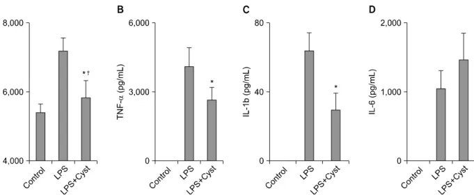
Figure 4. Transglutaminase (TG)-2 activity (A), tumor necrosis factor (TNF)-α (B), and interleukin (IL)-1β (C) were sig- nificantly decreased in the lipopolysaccharide (LPS)+Cyst group compared with the LPS group (*p<0.05). IL-6 (D) concentration was higher than the LPS group, but not significant (†p>0.05).
Figure 5. Nuclear factor (NF)-κB activity (A) was significantly decrease in the lipopolysaccharide (LPS)+Cyst group compared with the LPS group (*p=0.028). Myeloperoxidase (MPO) activity (B) and transforming growth factor (TGF)-β1 concentration (C) in the LPS+Cyst group was lower than the LPS group, but not significant (†p>0.05).
2. 염증성 시토카인
LPS 투여 후 6시간에 BALF에서 측정한 TNF-α, IL-1β 및 IL-6의 농도는 정상대조군에서는 측정되지 않았고, TNF-α의 농도는 LPS군 및 LPS+Cyst군에서 각각 4,105.1±811.91 pg/mL, 2,650.2±530.57 pg/mL로 LPS
+Cyst군에서 유의하게 낮았고(p=0.14) (Figure 4B), IL-1 β의 농도 역시 64.2±10.17 pg/mL, 29.7±9.67 pg/mL로 LPS+Cyst군에서 유의하게 낮았다(p=0.14) (Figure 4C).
하지만 IL-6의 농도는 LPS군 및 LPS+Cyst군에서 각각 1,050.2±261.99 pg/mL, 1,468.5±381.28 pg/mL로 LPS
+Cyst군에서 오히려 높은 경향을 보이기는 했지만, 통계 적으로 유의하지는 않았다(p=0.083) (Figure 4D).
3. NF-κB 활성, MPO 활성 및 ALI 지수
Lung tissue homogenate에서 LPS 투여 후 6시간에 측 정한 NF-κB의 활성은 정상대조군, LPS군 및 LPS+Cyst 군에서, 각각 0.0289±0.0053 optical density (O.D.), 0.2728±0.0629 O.D. 및 0.1757±0.0477 O.D로 세 군 간 에 유의한 차이가 있었고(p=0.001 by Kruskal-Wallis test), LPS군과 비교하여 LPS+Cyst군에서 NF-κB의 활성 이 유의하게 감소하였다(p=0.028) (Figure 5A). LPS 투여
Figure 6. Histopathologic examination shows significantly higher levels of intra-alveolar exudates, inflammatory infiltration, hemorrhage, and interstitial edema in the lipopolysaccharide (LPS) group (B), compared with the control group (A). The LPS+Cyst group (C) showed similar findings with the LPS group. Acute lung injury (ALI) score (D) was not different between the LPS and LPS+Cyst groups (*p=0.171) (H&E stain, ×100).
후 36시간에 BALF에서 측정한 MPO의 활성은 세 군에서, 각각 0.0324±0.0151 O.D., 0.1246±0.0461 O.D. 및 0.1094±0.0414 O.D로 유의한 차이가 있었지만(p=0.004 by Kruskal-Wallis test), LPS군과 비교하여 LPS+Cyst군 에서 MPO의 활성이 감소하는 경향을 보이기는 했지만 통 계적 유의성은 없었다(p=0.754) (Figure 5B). 전반적인 폐 손상의 정도를 H&E 염색 하에서 고찰한 결과, 정상대조 군(Figure 6A)과 비교하여 LPS군(Figure 6B)에서 폐포 전 반에 다량의 삼출액 및 초자막의 형성과 폐포벽 및 폐포 내에 많은 염증성 세포들의 침윤, 폐 모세혈관 주위 및 폐포 내 출혈과 간질의 비후 및 부종 등이 관찰되었다.
LPS+Cyst군에서(Figure 6C) 현미경으로 관찰한 폐손상 의 정도는 LPS군과 비교하여 뚜렷한 차이가 없었으며, 이 를 ALI 지수로 정량화하여 비교한 결과, 정상대조군, LPS 군 및 LPS+Cyst군에서, 각각 4.83±0.7, 11.8±2.4 및
9.8± 1.3으로 세 군 간에 유의한 차이가 있었으나(p=0.03 by Kruskal-Wallis test), LPS군과 LPS+Cyst군 사이에 유 의한 차이는 없었다(p=0.171) (Figure 6D).
4. TGF-β1 및 Mallory's PTAH 염색
LPS 투여 후 36시간에 BALF에서 측정한 TGF-β1의 농 도는 정상대조군, LPS군 및 LPS+Cyst군에서, 각각 58.2±7.92 pg/mL, 144.3±50.32 pg/mL 및 118.2±24.29 pg/mL로 유의한 차이가 있었다(p=0.004 by Kruskal- Wallis test). 하지만 LPS군과 비교하여 LPS+Cyst군에서 TGF-β1의 활성이 감소하는 경향을 보이기는 했지만 통 계적 유의성은 없었다(p=0.347) (Figure 5C). Mallory's PTAH 염색(Figure 7)을 시행하여 collagen 및 fibronectin 침착을 현미경으로 고찰한 결과, 갈색으로 염색된 양성염 색의 정도가 LPS군(Figure 7B) 및 LPS+Cyst군(Figure
Figure 7. Mallory's phosphotunstic acid hematoxylin (PTAH) stain (×400). The percent area of positivity (brown color
▲) was significantly larger in the LPS group (B) than the control group (A). Between the LPS and LPS+Cyst (C) groups, the area was similar (D, *p=0.423).
7C)에서 정상대조군(Figure 7A)과 비교하여 증가하였으 며, 이미지분석시스템을 이용하여 정량화한 결과, 세 군에 서 각각 1.35±0.53% (of stained area), 5.17±0.52% 및 5.0±0.56%로 유의한 차이가 있었으나(p=0.003 by Krus- kal-Wallis test), LPS군과 비교하여 LPS+Cyst군에서 염색 의 정도가 감소하는 경향을 보이기는 했지만 통계적 유의 성은 없었다(p=0.423) (Figure 7D).
고 찰
리포다당질로 유도된 급성 폐손상에서 폐조직 내 TG-2 의 활성을 고찰한 결과, 정상대조군과 비교하여, TG-2의 활성이 유의하게 증가하였으며, TG-2 억제제에 의해서 TG-2의 활성이 현저히 감소하였다. 급성 폐손상의 초기 염증성 지표인 BALF 내 TNF-α 및 IL-1β, 그리고 염증성
시토카인의 전사 인자인 NF-κB의 활성이 TG-2 억제제에 의해 유의하게 감소되었으나, 36시간의 BALF 내 MPO 활 성 및 48시간의 병리조직학적 소견은 감소하는 경향만을 보였다. 또한 섬유화와 관련된 지표인 TGF-β1 및 Mal- lory's PTAH 염색의 경우에 있어서도 현저한 감소는 관찰 되지 않았다.
일반적으로 ARDS의 병리단계는 급성삼출기(early exu- dative phase), 증식기(proliferative phase) 및 섬유화기 (fibrotic phase)로 구분되는데, 섬유화기에는 collagen deposition을 동반한 진행성의 폐섬유화 및 미세혈관의 폐쇄가 발생한다24. 이는 지속적이고 심한 염증이 치유되 지 않음으로써 발생하는 반응으로, 과거에는 ALI 혹은 ARDS 발생 약 1주일 후부터 시작되는 것으로 여겨졌으나, 최근의 연구들에 의하면, collagen turnover가 ARDS의 비 교적 초기에 증가하는 것으로 보고되었다7,8,25. 과도한 섬
유화증식 반응(fibroproliferative response)은 폐의 고유기 능인 가스교환을 감소시키고, 임상적으로는 심한 저산소 증, 폐유순도의 감소 및 폐혈관 저항의 증가를 초래하며26, ARDS 환자들의 사망률과 밀접한 연관성을 보인다27. 하지 만, 임상적 중요성에도 불구하고, ALI 및 ARDS와 연관된 대부분의 연구는 급성 삼출기의 염증반응에 집중되어 있 었고, 섬유화증식 및 이로 인한 폐섬유화에 대한 연구는 상대적으로 매우 부족한 상황이다. 결과적으로, 최근까지 도 폐포의 회복과 진행성 섬유화를 결정하는 정확한 기전 은 불확실한 상태로, 폐섬유화 증식을 조절하는 기전을 밝히는 것은 ALI 및 ARDS의 합병증으로서 폐섬유화가 발 생하는 환자들에 대한 새로운 치료법을 모색하는 데 있어 서 매우 중요하다. 일반적으로 급성 염증반응 후의 섬유 화증식은 interstitial fibroblast가 폐포 내 혈전으로 이동하 면서 시작된다. 이후 interstitial fibroblast는 myofibro- blast로 분화되며, 이 과정에서 cytoskeletal organization 의 증가와 새로운 matrix receptor 발현이 증가된다.
Myofibroblast는 증가된 세포표면의 receptor들을 매개로 하여 interstitium으로부터 airspace 내의 provisional ma- trix로 이동함으로써, 결과적으로 가스교환 표면의 efface- ment를 초래하며, 섬유화반응은 미세혈관에서도 진행하 여 폐고혈압증이 발생하기도 한다28. 이 과정에서 TGF-β 1는 세포외기질(extracellular matrix) 분자의 유전자 발현 에 영향을 미치고, fibroblast의 증식을 유발하며, 구조유 지세포(structural cell) 내에서의 자가분비자극(autocrine stimulation) 등의 기전을 통해서, 결체조직의 침착을 동반 한 폐조직의 섬유화증식 및 개형(remodeling)에 있어서 주요한 역할을 한다28.
TG는 특이 펩티드에 결합된 glutamine residue의 γ- carboxamine group과 다양한 amine들 사이의 결합을 촉 진하는 효소로9, 포유류에서는 총 8종류의 TG가 보고되고 있다29. 이 중 TG-2는 내피세포 및 평활근세포에서는 정상 적으로 발현되나, 이외의 세포들에서는 서로 다른 신호전 달을 통해서 그 발현이 유도되어, 일차적으로는 손상의 예방과 방어 및 복구를 촉진하는 데 있어서 주요한 역할을 하는 것으로 여겨졌으나10, 최근의 연구들에 의하면 비정 상적으로 과도하게 유발될 경우에는 각종 질환의 발생에 주요한 역할을 하는 것으로 보고되고 있다11. 특히, TG-2 는 LPS30, oxidative stress31, UV32, calcium ionophores33, retinoic acid34, TNF-α 및 IL-6 등의 염증성 시토카인35,36, 바이러스 감염37 등의 자극들로 인하여 활성화되는 것으 로 알려져, LPS로 유도되는 급성 폐손상 및 이로 인한 염
증반응에 있어서 주요한 역할을 할 것으로 판단된다. 본 연구의 결과에 있어서도 LPS의 기관 내 투여로 유도된 급 성 폐손상에서 TG-2의 활성이, TNF-α 및 IL-1β 등의 염 증성 시토카인의 증가와 동반하여 현저하게 증가되었으 며, TG-2 억제제로 인해 감소되는 소견을 보였다. 또한 이와 같은 변화가 조직 내 NF-κB의 증가 및 감소와 일치 하는 소견을 보였다. 잘 알려진 바대로, ARDS의 병리기전 과 연관된 다양한 시토카인의 발현에 있어서 NF-κB는 필 수적인 전사인자 중의 하나이다. 최근 TG-2의 활성도의 증가가 I-κB의 polymerization을 통해 세포질 내의 유리 I-κBα를 감소시킴으로써, 결과적으로 활성화된 NF-κB 의 핵 내로의 이동을 촉진하고38, TG-2 promoter가 NF-κ B binding motif를 가지고 있음으로써, 직접적으로 NF-κ B의 활성을 유도함으로써 염증반응을 유발하거나 악화시 키는 것으로 보고된 바 있다15.
각종 섬유화성 질환에서 TGF-β1는 콜라겐 합성 및 ep- ithelial-mesenchymal transition의 증가에 있어서 주요한 매개체로서, 기본적인 기능의 특성상 TG-2와 밀접한 연관 관계가 보고되었다. 일부 연구에 따르면 TG-2가 비활성화 상태의 TGF-β1를 활성화시키는 데 있어서 주요한 기여 인자인 것으로 보고하고 있는가 하면18, 다른 연구들에 있 어서는 TGF-β1가 TG-2의 주요한 유도인자인 것으로 보 고하고 있어서19, TGF-β1와 TG-2가 조직 섬유화 과정에 서 상호 긴밀한 기능을 하는 것으로 판단된다. 하지만, 이 와 같은 TG-2의 기능 및 역할에도 불구하고 폐질환에 있 어서 TG-2에 대한 연구는 매우 드물어서, Griffin 등39이 파라콰트(paraquat)로 유도된 폐섬유화 모델에서 plasma transglutaminase (Factor XIIIa, different subtype of TG with TG-2)가 폐섬유화에 기여함을 보고한 바 있고, Suh 등40이 복강 내 LPS 투여로 유도된 급성 폐손상 모델에서 anti-TG activity peptide가 몇몇 염증성 지표들을 감소시 키는 것으로 보고한 것 이외에는, 폐질환, 특히, ALI 후 폐섬유화의 병리기전에서 TG-2의 역할을 고찰한 연구는 거의 없는 상황이다. 본 연구에서는 LPS로 유도된 급성 폐손상 모델에서 초기 섬유화성 기전에 있어서, TG-2의 역할을 급성 염증성 지표 및 섬유화성 지표인 TGF-β1과 Mallory's PTAH 염색과의 변화와 비교하여 고찰한 결과, 일부 초기 염증성 지표와는 동반되어 증가하거나 감소하 는 것이 관찰되었으나, TGF-β1과 Mallory's PTAH 염색 과의 관계에 있어서는, TG-2가 감소함에도 불구하고, 현 저하게 감소되는 소견이 없어, TG-2가 LPS로 인한 급성 폐손상 후 초기 섬유화 기전에 있어서, 주요한 역할은 없
을 수 있는 것으로 관찰되었다. 이와 같은 결과의 원인으 로 추정할 수 있는 것은 LPS 0.5 mg/kg in 50 μL of saline 를 기관 내 투여로 급성 폐손상을 유도하였을 경우, TG-2 의 활성이 정상대조군과 비교하여, 통계적으로 유의하게 증가하기는 하였지만, 그 정도가 약 145% 정도로, 병리기 전에 관련된 다른 지표들의 변화와 비교하여 현저히 낮아 서, 이를 억제함에도 불구하고, 병리현상에 대한 치료효과 는 없었던 것으로 판단된다. 이와 같은 TG-2 활성의 변화 는 LPS 1.0 mg/kg 투여하였을 경우에도 비슷하여, 향후 TG- 2가 급성 폐손상의 염증성 병리기전 및 섬유화성 병리 기전에 미치는 영향을 적절하게 연구하기 위해서는 TG-2 의 활성이 좀 더 현저하게 증가하는 모델을 개발하고, 이를 이용하여 고찰하는 것이 필요할 것으로 판단된다.
참 고 문 헌
1. Rubenfeld GD, Caldwell E, Peabody E, Weaver J, Mar- tin DP, Neff M, et al. Incidence and outcomes of acute lung injury. N Engl J Med 2005;353:1685-93.
2. Ware LB, Matthay MA. The acute respiratory distress syndrome. N Engl J Med 2000;342:1334-49.
3. Desai SR, Wells AU, Rubens MB, Evans TW, Hansell DM. Acute respiratory distress syndrome: CT abnormal- ities at long-term follow-up. Radiology 1999;210:29-35.
4. McHugh LG, Milberg JA, Whitcomb ME, Schoene RB, Maunder RJ, Hudson LD. Recovery of function in survi- vors of the acute respiratory distress syndrome. Am J Respir Crit Care Med 1994;150:90-4.
5. Davidson TA, Caldwell ES, Curtis JR, Hudson LD, Steinberg KP. Reduced quality of life in survivors of acute respiratory distress syndrome compared with crit- ically ill control patients. JAMA 1999;281:354-60.
6. Herridge MS, Cheung AM, Tansey CM, Matte-Martyn A, Diaz-Granados N, Al-Saidi F, et al. One-year outcomes in survivors of the acute respiratory distress syndrome.
N Engl J Med 2003;348:683-93.
7. Armstrong L, Thickett DR, Mansell JP, Ionescu M, Hoyle E, Billinghurst RC, et al. Changes in collagen turnover in early acute respiratory distress syndrome.
Am J Respir Crit Care Med 1999;160:1910-5.
8. Marshall RP, Bellingan G, Webb S, Puddicombe A, Goldsack N, McAnulty RJ, et al. Fibroproliferation oc- curs early in the acute respiratory distress syndrome and impacts on outcome. Am J Respir Crit Care Med 2000;162:1783-8.
9. Griffin M, Casadio R, Bergamini CM. Transglutamin-
ases: nature's biological glues. Biochem J 2002;368:377- 96.
10. Folk JE, Finlayson JS. The epsilon-(gamma-glutamyl)ly- sine crosslink and the catalytic role of transglutami- nases. Adv Protein Chem 1977;31:1-133.
11. Kim SY, Jeitner TM, Steinert PM. Transglutaminases in disease. Neurochem Int 2002;40:85-103.
12. Aeschlimann D, Thomazy V. Protein crosslinking in as- sembly and remodelling of extracellular matrices: the role of transglutaminases. Connect Tissue Res 2000;41:
1-27.
13. Johnson TS, Skill NJ, El Nahas AM, Oldroyd SD, Thomas GL, Douthwaite JA, et al. Transglutaminase transcription and antigen translocation in experimental renal scarring. J Am Soc Nephrol 1999;10:2146-57.
14. Shweke N, Boulos N, Jouanneau C, Vandermeersch S, Melino G, Dussaule JC, et al. Tissue transglutaminase contributes to interstitial renal fibrosis by favoring accu- mulation of fibrillar collagen through TGF-beta activa- tion and cell infiltration. Am J Pathol 2008;173:631-42.
15. Mirza A, Liu SL, Frizell E, Zhu J, Maddukuri S, Martinez J, et al. A role for tissue transglutaminase in hepatic injury and fibrogenesis, and its regulation by NF-kap- paB. Am J Physiol 1997;272:G281-8.
16. Johnson TS, Fisher M, Haylor JL, Hau Z, Skill NJ, Jones R, et al. Transglutaminase inhibition reduces fibrosis and preserves function in experimental chronic kidney disease. J Am Soc Nephrol 2007;18:3078-88.
17. Qiu JF, Zhang ZQ, Chen W, Wu ZY. Cystamine amelio- rates liver fibrosis induced by carbon tetrachloride via inhibition of tissue transglutaminase. World J Gastroen- terol 2007;13:4328-32.
18. Annes JP, Munger JS, Rifkin DB. Making sense of latent TGFbeta activation. J Cell Sci 2003;116:217-24.
19. Shin DM, Jeon JH, Kim CW, Cho SY, Lee HJ, Jang GY, et al. TGFbeta mediates activation of transglutaminase 2 in response to oxidative stress that leads to protein aggregation. FASEB J 2008;22:2498-507.
20. Verderio EA, Johnson T, Griffin M. Tissue transgluta- minase in normal and abnormal wound healing: review article. Amino Acids 2004;26:387-404.
21. Kim JH, Suk MH, Yoon DW, Kim HY, Jung KH, Kang EH, et al. Inflammatory and transcriptional roles of poly (ADP-ribose) polymerase in ventilator-induced lung injury. Crit Care 2008;12:R108.
22. Rojas M, Woods CR, Mora AL, Xu J, Brigham KL.
Endotoxin-induced lung injury in mice: structural, func- tional, and biochemical responses. Am J Physiol Lung Cell Mol Physiol 2005;288:L333-41.
23. Jeitner TM, Delikatny EJ, Ahlqvist J, Capper H, Cooper AJ. Mechanism for the inhibition of transglutaminase 2 by cystamine. Biochem Pharmacol 2005;69:961-70.
24. Tomashefski JF Jr. Pulmonary pathology of acute respi- ratory distress syndrome. Clin Chest Med 2000;21:435- 66.
25. Chesnutt AN, Matthay MA, Tibayan FA, Clark JG. Early detection of type III procollagen peptide in acute lung injury. Pathogenetic and prognostic significance. Am J Respir Crit Care Med 1997;156:840-5.
26. Steinberg KP, Hudson LD. Acute lung injury and acute respiratory distress syndrome: the clinical syndrome.
Clin Chest Med 2000;21:401-17.
27. Martin C, Papazian L, Payan MJ, Saux P, Gouin F.
Pulmonary fibrosis correlates with outcome in adult res- piratory distress syndrome: a study in mechanically ventilated patients. Chest 1995;107:196-200.
28. Dhainaut JF, Charpentier J, Chiche JD. Transforming growth factor-beta: a mediator of cell regulation in acute respiratory distress syndrome. Crit Care Med 2003;31 Suppl 4:S258-64.
29. Grenard P, Bates MK, Aeschlimann D. Evolution of transglutaminase genes: identification of a transgluta- minase gene cluster on human chromosome 15q15.
Structure of the gene encoding transglutaminase X and a novel gene family member, transglutaminase Z. J Biol Chem 2001;276:33066-78.
30. Park KC, Chung KC, Kim YS, Lee J, Joh TH, Kim SY.
Transglutaminase 2 induces nitric oxide synthesis in BV-2 microglia. Biochem Biophys Res Commun 2004;
323:1055-62.
31. Lee ZW, Kwon SM, Kim SW, Yi SJ, Kim YM, Ha KS.
Activation of in situ tissue transglutaminase by intra- cellular reactive oxygen species. Biochem Biophys Res Commun 2003;305:633-40.
32. Shin DM, Jeon JH, Kim CW, Cho SY, Kwon JC, Lee HJ, et al. Cell type-specific activation of intracellular
transglutaminase 2 by oxidative stress or ultraviolet irra- diation: implications of transglutaminase 2 in age-re- lated cataractogenesis. J Biol Chem 2004;279:15032-9.
33. Lorand L, Weissmann LB, Epel DL, Bruner-Lorand J.
Role of the intrinsic transglutaminase in the Ca2+-medi- ated crosslinking of erythrocyte proteins. Proc Natl Acad Sci U S A 1976;73:4479-81.
34. Scott KF, Meyskens FL Jr, Russell DH. Retinoids in- crease transglutaminase activity and inhibit ornithine decarboxylase activity in Chinese hamster ovary cells and in melanoma cells stimulated to differentiate. Proc Natl Acad Sci U S A 1982;79:4093-7.
35. Esposito C, Paparo F, Caputo I, Porta R, Salvati VM, Mazzarella G, et al. Expression and enzymatic activity of small intestinal tissue transglutaminase in celiac disease. Am J Gastroenterol 2003;98:1813-20.
36. Kuncio GS, Tsyganskaya M, Zhu J, Liu SL, Nagy L, Thomazy V, et al. TNF-alpha modulates expression of the tissue transglutaminase gene in liver cells. Am J Physiol 1998;274:G240-5.
37. Roberts ES, Zandonatti MA, Watry DD, Madden LJ, Henriksen SJ, Taffe MA, et al. Induction of pathogenic sets of genes in macrophages and neurons in Neuro- AIDS. Am J Pathol 2003;162:2041-57.
38. Lee J, Kim YS, Choi DH, Bang MS, Han TR, Joh TH, et al. Transglutaminase 2 induces nuclear factor-kap- paB activation via a novel pathway in BV-2 microglia.
J Biol Chem 2004;279:53725-35.
39. Griffin M, Smith LL, Wynne J. Changes in trans- glutaminase activity in an experimental model of pul- monary fibrosis induced by paraquat. Br J Exp Pathol 1979;60:653-61.
40. Suh GY, Ham HS, Lee SH, Choi JC, Koh WJ, Kim SY, et al. A Peptide with anti-transglutaminase activity de- creases lipopolysaccharide-induced lung inflammation in mice. Exp Lung Res 2006;32:43-53.