․교신저자: Jin-ju Kim Hoegi-dong, Dongdaemun-gu, Seoul, Republic of Korea Department of Oriental Physiology, College of Pharmacy, Kyung-Hee University TEL: 82-2-961-9437 FAX: 82-2-968-0560 E-mail: [email protected], [email protected]
Effects of Opuntia Ficus - indica on Ovalbumin-induced Asthma Model
Je-hyeon Ra1, Feng-yan Shen2, Seong-gi Jung2, Jin-ju Kim1
1Dept. of Oriental Physiology, College of Pharmacy, Kyung-Hee University
2Division of Allergy and Respiratory System, Dept. of Oriental Internal Medicine, College of Oriental Medicine, Kyung-Hee University
ABSTRACT
Objective :
This study was undertaken to investigate effects of Opuntia ficus-indica(OP)extract on an asthma murine model.
Methods :
The total number of cells and eosinophils in BALF and the infiltration of inflammatory cells into lung tissues were determined by hematoxylin and eosin staining.
Results :
The number of OVA-induced total cells and eosinophils, a phenomenon of asthma, were decreased by treatment of the animals with OP extract (200 ㎎/㎏) (respectively, p < 0.001 and p<0.01). Furthermore, we showed that OP extract reduced the increased immune cell infiltration induced by ovalbumin (p < 0.01). The levels of interleukin (IL)-4 in BAL fluid were measured by enzyme-linked immunosorbent assay, because the eosinophilia is associated with a T helper (Th) 2 response including IL-4. The level of OVA-induced IL-4 was decreased by OP extract in BAL fluid (p < 0.05). We investigated whether OP extract reduced nitrite (NO) production on lipopolysaccharide (LPS)-stimulated RAW 264.7 sells, because asthma is an inflammatory disease. The level of LPS-induced NO production was decreased by OP extract (50, 100 and 200 mg/ml) on RAW 264.7 cells (respectively, p < 0.05, p<0.01andp<0.05).
Conclusions :
Our results indicate that OP extract has an inhibitory effect on lung eosinophilia of asthma and suggest that OP extract is a therapeutic candidate in the treatment of inflammatory disease, including asthma.
Key words : Asthma, Opuntiaficus-indica, Eosinophil, Immune cell infiltration
Ⅰ. Introduction
Asthma is the most common chronic respiratory
disease in industrialized society and has emerged
as a public health problem in the world. Although
numbers of asthma patients receive conventional
medical treatment, effects of therapy are frequently
poor
1. In addition, frequently poor effect of
conventional medical treatment simulated interest
in complementary and alternative medicine (CAM)
being used for the treatment of asthma
2,3; herbal medicine is popular in asthma patients
4.
Opuntia ficus-indica (OP) is a member of the Cactaceae family and is an important forage crop for livestock in many arid and semi-arid regions.
It is widely distributed in the world, including Mexico
5. Baekryunchihyo-tang including OP, widely used for asthma in Kyunghee oriental hospital, has anti-inflammatory effects in HMC-1
6. However, many studies are needed to understand effects of OP. Thus, we investigated the effects of OP on an ovalbumin (OVA)-induced asthma model.
Allergic asthma is an inflammatory disease of the airways, associated with enhanced T helper (Th)2 lymphocytes response to allergens, leading to eosinophilic infiltration, airway hyper-reponsiveness, reversible airway obstruction
7. It is reported that interleukin-4 (IL-4), an Th2 cytokine, was increased in asthma
8, and that this played important roles in the phenomenon of asthma
9. In addition, nitrite (NO) were significantly higher in the asthma group than that in the control group
10. Thus, we investigated whether OP reduced NO production on LPS-stimulated RAW 264.7 cells.
In the present study, we show that OP extract inhibited the infiltration of total cells and eosinophils, and decreased the expression of IL-4 in bronchoalveolar lavage fluid (BALF) in an ovalbumin (OVA)-induced asthma model. Furthermore, OP extract reduced LPS-induced NO production on RAW 264.7 macrophages.
Thus, OP extract is a therapeutic candidate for the treatment of allergic airway disease.
Ⅱ. Materials and methods
1. Preparation of the OP extract
OP was collected from July to August in Jeju-Do
(Korea) was used in this study. The OP extract were prepared as follows. OP was dried in the shade and then grinded. The OP powder was weighed precisely to 20 g and was boiled in 200 ㎖ distilled water (DW) for 2 h. The aqueous phase was concentrated with a rotary vacuum evaporator and then was freeze-dried. The freeze-dried OP extract were weighed precisely to 100 ㎎ and were dissolved in 10 ㎖ DW using a stirrer, overnight, at room temperature. The dissolved solution was centrifuged for 10 min at 3000 rpm. The supernatant was sterilized by passing through a 0.22 ㎛ syringe filter and used in the experiments.
2. Induction of asthma in Balb/C mice and drug administration
Six-weeks-old female Balb/C mice were purchased from Orient Bio Inc. (Korea). The animals were housed under specific pathogen free conditions (about 22 ± 1°C). The mice were divided into four groups (n=5), and airway inflammation was induced by OVA (GradeV;Sigma-Aldrich,MO,USA) in three groups using the method described by Fig.
1. Briefly, each mouse was immunized by intraperitoneal (i.p.) injection of 100㎍ of chicken OVA with 20
㎎ aluminum hydroxide (Sigma-Aldrich, MO, USA) on days 0 and 6. The OVA-challenged mice were exposed with 5 ㎎/㎖ OVA in phosphate buffer saline (PBS) via intra-nasalatthreetimesper week from days 20 to 62. Two groups of asthma- induced mice were pre-treated with PBS or with 200 ㎎/㎏ of OP extract at OVA-challenge time between days 48 and 62 via orogastric gavage, respectively. The control group was sensitized with only aluminum hydroxide and challenged with PBS without treatment via orogastric gavage.
Mice were sacrificed on day 65.
3. Collection of BALF
BALF was collected by the lung lavage with 0.9 ㎖ of PBS via the trachea. After three lavages, approximately 0.7 ㎖ of BALF was recovered and centrifuged for 10 min at 400 g and 4°C. The supernatant was stored at –70°C for the measurement of cytokines. The cells in the BALF were resuspended in 100 ㎕ of PBS for total cell and differential counts. After the measurement of total cell number using a neubauer hemocytometer (Hausser Scientific, PA, USA), the cells suspended in PBS were applied to a slide by cytospinning (3min at 400 rpm) for hematoxylin and eosin staining purposes.
The percentage of eosinophils was determined, and the absolute leukocyte count was calculated by multiplying eosinophils percentage by the total cell count.
4. Histological analysis
Lung tissues were detached from the mice and fixed with 4% formaldehyde for overnight at 4°C.
The fixed tissues were embedded in paraffin and cut into 4 ㎛ sections with a microtome (Leica, Nussloch, Germany). The sections were placed on glass slides, deparaffinized, and stained with hematoxylin and eosin (Sigma-Aldrich, MO, USA) in order to examine the cells that had infiltrated into the peribronchial connective tissues. A peribronchial cell count based on a five-point scoring system was performed as previously described, to estimate the severity of leukocyte infiltration
11. The scoring system was: 0, no cells; 1, a few cells; 2, a ring of cells 1 cell layer deep; 3, a ring of cells 2–4 cell layers deep; and 4, a ring of cells more than 4 cell layers deep. The leukocyte scoring was examined in three independent fields of lung section from each mouse.
5. Cell culture and viability assay
RAW 264.7 cells obtained from the American Type Culture Collection (VA, USA) were cultured in 96-well plates (1 × 10
4cells per well) with DMEM (Hyclone, UT, USA) supplemented with 10% heat-inactivated fetal bovine serum (FBS) (Hyclone, UT, USA), 100U/㎖ penicillin, 100 ㎍/
㎖ streptomycin for overnight. Cells were pretreated with indicated concentrations of OP extract for 1 h, followed by stimulation with or without LPS (1
㎍/㎖). After 24 h incubation, the medium was removed, the cells were washed, and 0.5 ㎎/㎖ of 3-(4,5-dimethylthiazol-2-yl)-2,5-diphenyltetrazoliu m bromide (MTT) (Sigma-Aldrich, MO, USA) was added to each well and incubated for an additional 4 h to form formazan crystals
12. Formazan crystals were dissolved in DMSO and quantified at 570 ㎚ using an ELx800 reader (Bio-Tek Instruments, VT, USA). The formation of formazan in the control group was taken as 100% viability.
6. NO quantification
RAW 264.7 cells were adjusted to 4 × 10
5cells/
㎖ in 24 well plate (Corning incorporated, MA, USA) with DMEM (Hyclone, UT, USA) supplemented with 10% heat-inactivated fetal bovine serum (FBS) (Hyclone, UT, USA), 100 U/㎖ penicillin, 100 ㎍/㎖ streptomycin for overnight. The medium was changed into DMEM without FBS and incubated for 4h. The cells were pretreated with various concentrations of OP extract for 1 h, followed by stimulation with LPS (1 ㎍/㎖) for 24 h. The medium was taken.
7. Measurement of IL-4 and NO
The concentrations of IL-4 and NO were measured
by a sandwich ELISA using Mouse CytoSet Kit
(Invitrogen, CA, USA) and Griess reagent system (Promega, WI, USA) respectively. All assays were performed in duplicate. The concentration of each protein was calculated from the standard curve.
8. Statistical analysis
Statistical analysis of the data was carried out using the Prism 4 software (GraphicPad Software Inc., CA, USA). Data are presented as mean±S.E.M, and statistical analysis was performed by one-way ANOVA for multiple comparisons. Results with p <0.05 were considered statistically significant.
Ⅲ. Results
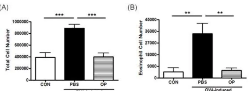
1. The infiltration of eosinophils into the airways To investigate the effect of OP extract in an OVA-induced asthma model, we observed the number of total cells and eosinophils in the BALF. There were a few total cells and eosinophils in the control group, but OVA significantly infiltrated the numbers of total cells and eosinophils in the asthma group (respectively, p < 0.001 and p < 0.01). In contrast, these groups treated with OP extract (200 ㎎/㎏) via orogastric gavage markedly inhibited OVA-induced infiltration of total cells and eosinophils, similar to control group (respectively, p < 0.001 and p < 0.01) (Fig. 1). These results reveal that OP extract exerted anti-inflammatory effects in anasthma model.
Fig. 1. Effect of OP Extract on the Recruitment of Inflammatory Cells in BALF Obtained from OVA-induced Asthma Model.
CON, sensitized and treated with PBS without drug administration; PBS, OVA-induced asthma model treated with PBS; OP, OVA-induced asthma model treated with 200 mg/kg of OP. The number of total cell (A) and eosinophil (B) in the fluid was counted after hematoxylin and eosin stain. The results were expressed as the means ± S.E.M.. **
p
<0.01and***p
<0.001.2. The pathologic progress in lung tissues
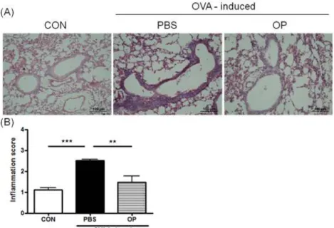
To examine the inhibitory effects of OP extract on the histological features of the OVA-induced asthma model, we stained lung tissues using hematoxylin-eosin staining solution. Although the lung tissues from the OVA-treated group revealed
a narrowing of bronchi by infiltration of leukocytes,
OP extract markdly reduced the degree of inflammatory
cell infiltration (Fig. 2A). A peribronchial cell count
based on a five-point scoring system was performed
as described in material and methods. In the asthma
group, the inflammation score was significantly increased
at 2.52 in comparison with control group at 1.12 ( p < 0.001). In contrast, the OP extract group was significantly reduced at 1.48 ( p < 0.01) (Fig. 2B).
These data indicate that OP extract reduced the pathological degree in the OVA-induced asthma model.
Fig. 2. Effect of OP Extract on Airway Inflammation Caused by Cell Infiltration in Lung Tissue.
To confirm the pathological changes in lung tissues, we performed hematoxylin and eosin staining (A) and cored the inflammatory cells in filtration in the lung tissues (B) as described in method. CON, sensitized and treated with PBS without drug administration; PBS, OVA-induced asthma model treated with PBS; OP, OVA-induced asthma model treated with 200 mg/kg of OP. Magnification × 200.
3. The expression of IL-4 in BALF
Allergic asthma is associated with an enhanced Th2 lymphocytes response to allergens. IL-4, as represented Th2 cytokine, plays important roles in the phenomenon of asthma. Because the OP extract reduced the pathological degree in lung tissues of the OVA-induced asthma model, we investigated whether the OP extract decreased the expression of IL-4 in BALF. The results showed that the OVA increased the level of IL-4 in BALF ( p < 0.01) and OP extract significantly decreased the OVA- increased protein level of IL-4 in BALF by 39%
( p < 0.01) (Fig. 3).
4. Viability of RAW 264.7 cells
Because, the asthma is an inflammatory disease,
coupled with significantly increasing nitrite (NO)
in the asthma group than that in the control group,
we measured NO production with or without various
concentration of OP extract on LPS-stimulated RAW
264.7 cells. First of all, to investigate whether OP
extract affect cell viability on RAW 264.7 cells,
the cell was incubated with indicated-concentrations
of OP extract. At the concentrations tested, OP
extract did not affect cell viability as measured by MTT assay-method(Fig. 4).
Fig. 3. Effect of OP Extract on the Expression of IL-4 in BALF.
BALF was collected after the cells in the fluid were liminated. The protein expression level IL-4 in BALF was analyzed by ELISA. The data are expressed as the means ± S.E.M..
*
p
<0.05 and **p
<0.01. CON, sensitized and treated with PBS without drug administration;PBS, OVA-induced asthma model treated with PBS; OP, OVA-induced asthma model treated with 200 mg/kg of OP.
Fig. 4. Effect of OP Extract on Cell Viability.
Cell viability was evaluated by MTT assay 24 h after OP treatment without (A) or with (B) lipopolysaccharide (LPS) in RAW 264.7 macropharge.
The data are expressed as the means ± S.E.M..
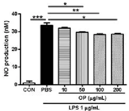
5. NO production on RAW 264.7 cells
To investigate the effect of OP extract on NO production, nitrite, a stable end-product of NO, in the culture media was assayed. As shown in Fig.
5, LPS increased a 32-fold in NO production compared with the control ( p < 0.001). The increase was inhibited by various concentrations (50, 100, 200 ㎍/㎖) of OP extract in a dose-dependent manner, except for 10 ㎍/㎖ (respectively, p <
0.05, p <0.01 and p <0.05).
Fig. 5. Effect of OP Extract on Nitrite (NO) Production in LPS-stimulated RAW 264.7 Macrophages.
NO production was measured by the Griess reaction assay. The data are expressed as the means ± S.E.M..
*
p
<0.05,**p
<0.01and***p
<0.001.Ⅳ. Discussion and conclusion
Asthma is one of chronic diseases, and was
characterized by reversible airway obstruction and
airway inflammation
13. Airway inflammation is an
important factor in the pathology of asthma,
coupled with current target of treatment for the
suppression of asthma
14. Since there is an increasing number of asthmatics who have received medical treatment but have not shown effective improvement, an interest for CAM as a potential asthma therapy was increasing
15-17. In this regard, there is an increasing need for new therapeutics from CAM to be used as adjuncts. The medicine from the crude drug has two benefits. One is fewer side effects and the other is more effective with or without current medications. Although the value of Chinese herbal medicines in therapies is uncertain, a number of studies have positively reported on the benefits of herbal medicine in the asthma model
18-20.
There were numbers of study on OP and OP was reported until a recent day. However, there was a few of study on the anti-inflammatory effect of OP
21,22. In addition, many studies have reported that OP is effective for treatment of several diseases.
Especially, OP exhibit the antioxidant
23,24and anti-inflammatory effects
25,26. However, no study was reported on asthma. In the present study, we investigated whether OP extract is effective in the OVA-induced asthma murine model and the effect is anti-inflammatory, particularly focusing on the possibility of OP extract as a therapeutic agent.
The migration of inflammatory cells into the lung is an important step in pathogenesis of asthma.
In particular, the recruitment and activation of eosinophils into the airways of asthma patients is a contributing causative agent to the histopathology and lung dysfunction
27. Thus, the number of total cells and eosinophils in our OVA-induced asthma model was investigated. In the asthma group, the total cells and eosinophils were induced by OVA, compared to the control group; however the total cells and eosinophils were decreased by the OP
extract (Fig. 1A and B). Furthermore, the OP extract significantly decreased the infiltration of inflammatory cells into lung tissue; this corresponded with the results of Fig.1 (Fig. 2).
These results indicated that PN extract has an anti-inflammatory effect for the development of asthma.
Th2 lymphocytes produce IL4 and therefore play an important role in allergic disease such as asthma
28. In particular, the regulation of IL-4 expression is very interesting as target for treatment of asthma. IL-4 acts as a growth factor for Th2 cells, and plays a role in the induction of immunoglobulin class switching to immunoglobulin E, which plays a critical role in mediating allergic responses. We therefore measured the IL-4 expression in BALF of asthma model. OP extract reduced the induced IL-4 in BALF by OVA (Fig.
3). These results suggest that OP extract may regulate the response of Th2 lymphocytes. Because, the significantly increasing nitrite (NO) in the asthma patient and the possible potential of nitric oxide (NO) in the processes of atopic inflammation was reported by a number of studies, NO is a target for treatment for asthma
29,30. We measured NO production with or without various concentration of OP extract on LPS-stimulated RAW 264.7 cells.
The level of LPS-induced NO production was decreased by OP extract (50, 100 and 200 ㎍/㎖) on RAW 264.7 cells (Fig. 5). This result suggests that OP extract reduced NO production, a key molecule released during chronic inflammatory events. In another study, OP extract has anti-inflammatory effect on the production of NO, prostaglandin E
2(PGE
2) and reactive oxygen species (ROS) in
human chondrocytes, corresponding to ourstudy
30.
These results, are shown in Fig 5, suggest that OP
extract has protective effect on asthma by reducing a key molecule released during inflammatory events, such as NO, PGE
2and ROS.
In conclusion, we discovered that OP extract reduced the infiltration of inflammatory cells into lung tissues, the expression of IL-4 in BALF from OVA-induced asthma model. Furthermore, the level of LPS-induced NO production was decreased by OP extract on RAW 264.7 cells. Taken together,
OP extract exerts an anti-inflammatory effect during asthma and could be considered as a therapeutic candidate for the treatment of allergic airway disease.
Acknowledgement
Grant from Seoul R&BD Program Republic of
Korea.
Ovalbumin으로 유도된 천식 마우스 모델에서 백련초의 효과
라제현1, 심봉암2, 정승기2, 김진주1
1경희대학교 약학대학 한방생리학 교실, 2경희대학교 한의과대학 폐계내과학 교실
ABSTRACT
목 적 :
이 연구는 백련초의 천식마우스 모델에 대한 효과를 조사하기 위해 수행하였다.
방 법 :
폐 세척액의 전체 세포와 호산구 수 그리고 폐조직으로의 면역세포의 침윤은 헤마톡실린과 에오신 염색으로 관찰
하였다. Interleukin (IL)-4의 발현량은 Enzyme-linked immunosorbent assay을 이용하여 측정하였으며, NO는 Griess reagent을 이용하였다.
결 과 :
천식 병리현상의 하나인 Ovalbumin (OVA)에 의한 전체 세포와 호산구 수의 증가는 백련초 추출물 (200 ㎎/㎏) 을 처리한 군에서 감소하였다 (respectively,p<0.001andp<0.01). 또한, OVA에 의한 증가된 면역세포의 침투는 백련초 추출물에 의해 감소하였다 (p < 0.01). 호산구의 침윤증상은 interleukin (IL)-4 그리고 5를 포함한 T helper (Th) 2 반응과 관련있기 때 문에 폐세척액에서 IL-4의 발현량을 조사하였다. OVA에 증가한 IL-4는 백련초 추출물에 의해 감소하였다 (p < 0.05). 천식이 염증성 질병이기 때문에, lipopolysaccharide (LPS)로 자극한 RAW 264.7 세포에서 nitrite (NO) 생성을 살펴보았다. LPS에 의 해 증가한 NO 생성은 백련초 추출물 (50, 100 and 200 mg/ml)에서 감소하였다 (respectively. p < 0.05, p<0.01andp<0.05).
결 론 :