Induced Acute Lung Injury
Jae Yeol Kim, M.D., Jae Chul Choi, M.D., Young Woo Lee, M.D., Jae Woo Jung, M.D., Jong Wook Shin, M.D., In Won Park, M.D., Byoung Whui Choi, M.D.
Department of Internal Medicine, Chung-Ang University College of Medicine
출혈성 및 내독소 투여로 유발된 급성폐손상에서 heparin의 항염증효과
중앙대학교 의과대학 내과학교실
김재열, 최재철, 이영우, 정재우, 신종욱, 박인원, 최병휘
배 경 : 급성 폐손상은 폐내, 외의 원인질환들에 의해 폐포-모세혈관의 투과성이 증가하며, 폐부종에 의해 급성 저산소성 호흡곤란이 유발되는 증후군이다. 헤파린은 항응고작용 외에 자체적으로 항염증효과를 가지고 있으나, 염증성질환에 헤 파린을 투여하면 출혈성 합병증이 발생하기 때문에 실제로 임상에서 이용하는데 제약이 있다. 하지만 헤파린에서 2-O와 3-O sulfate를 제거하면, 항응고 효과가 제거되고 항염증효과는 지니고 있는 비항응고성 헤파린 (nonanticoagulant heparin)으로 변화한다. 본 연구에서는 흰쥐에게 내독소 (LPS)를 투여하거나, 출혈성 쇼크를 일으켜서 유발된 급성폐손상 에서 비항응고성 헤파린의 치료효과를 살펴보았다.
방 법 : 각 군당 5 마리 이상의 흰쥐 (Balb/c mouse)를 이용하였다. 미정맥 (tail vein)을 통해 생리식염수 또는 비항응고성 헤파린 (50 mg/kg)을 투여한 직후에 내독소를 복강으로 투여하거나 (1 mg/kg), 심장천자를 통해 총 혈액의 1/3 정도로 제거 하여 출혈성 쇼크를 유도하여 급성폐손상을 유발하였다. 내독소 투여 또는 출혈성 쇼크 유발 1 시간 후에 흰쥐를 희생시키 고 폐를 적출하였고, 폐의 염증성 변화는 사이토카인 (TNF-α, MIP-2, IL-1β)을 측정하여 살펴보았고, 폐손상의 정도는 myeloperoxidase (MPO) assay와 wet-to-dry weight ratio를 측정하여 알아보았다.
결 과 : 내독소를 투여한 흰쥐의 폐에서 대조군의 폐에 비해 사이토카인의 발현이 증가하고 (TNF-α; 196.1±10.8 vs 83.7±18.4 pg/ml, MIP-2; 3,000±725 vs 187±26 pg/ml, IL-1β; 6,500±1167 vs 266±25 pg/ml, p<0.05, respectively), 폐의 MPO 활성이 증가하였다 (27.9±6.2 vs 10.5±2.3 U/g of lung protein, p<0.05). 출혈성 쇼크를 일으킨 흰쥐의 폐에서 대조군 의 폐에 비해 사이토카인의 발현은 증가되지 않았으나, MPO 발현은 증가되었다 (16.5±3.2 vs 10.5±2.3 U/g of lung protein, p<0.05). 내독소 투여 또는 출혈성 쇼크에 의해 급성폐손상이 유발된 흰쥐에서 생리적 식염수를 투여하거나 비항 응고성 헤파린을 투여한 군 사이에 사이토카인의 발현이나 MPO 활성에 의미있는 차이는 관찰되지 않았다.
결 론 : 이상의 결과로 비항응고성 헤파린은 내독소를 투여하거나 출혈성 쇼크를 일으키고 한 시간 뒤에 측정한 흰쥐의 급성폐손상에서 의미있는 치료효과를 보이지 않았다. (Tuberc Respir Dis 2006; 60: 49-56)
Key words : 급성폐손상, 비항응고성 헤파린, TNF-α, MIP-2, IL-1β, MPO assay
Acknowledgement : This research was supported by the Chung-Ang University Research Grants in 2005.
Address for correspondence : Jae Yeol Kim, M.D., Internal Medicine, Chung Ang University Hospital, 224-1 HeukSeok Dong, DongJak Gu, Seoul, Korea, 156-755
Phone : 82-2-6299-1396 Fax : 82-2-825-7571 E-mail : [email protected]
Received : Nov. 28. 2005 Accepted : Jan. 4. 2006
Introduction
Acute lung injury (ALI), which is called as acute respiratory distress syndrome (ARDS) in its most severe clinical manifestation, affects around 150,000 patients per year in the U.S, with recent mortality
rates being over 30 percent
1,2. At present, there is
no effective treatment for ALI except the low tidal
volume ventilation, which is rather a way of redu-
cing artificially induced ventilation-associated lung
injury (VLI) than an active, specific way of treat-
ment. ALI is characterized by neutrophil accumula-
tion in the lungs, interstitial edema, disruption of
epithelial integrity, and leakage of protein into the
alveolar space
3-6. Infection, associated with endoto-
xemia, and blood loss are frequent predisposing fa-
ctors to the development of ALI. In experimental
settings, endotoxemia or hemorrhage produces
ALI
7. Neutrophils, which play a central role in the
ALI, produce proinflammatory mediators, including
cytokines such as tumor necrosis factor α (TNF-α) and macrophage inflammatory peptide-2 (MIP- 2) and demonstrate increased activation of transcri- ptional regulatory factors including cyclic adenosine monophosphate (cAMP) response element binding protein (CREB) and nuclear factor kappa B (NF-κ B)
8-10.
Heparin presents a dazzling array of properties.
In lung disease, it is used as an anticoagulant for thromboembolism, but its polyanionic nature con- fers a wide variety of other actions not related to anticoagulation
11. Heparin is a potent antiinflamma- tory agent that inhibits neutrophil-derived elasta- se
12, complement activation
13, tumor necrosis fac- tor-induced lung edema
14, L- and P-selectins
15, le- ukocyte rolling
16and neutrophil-induced injury of pulmonary alveolar epithelium
17. However, therape- utic potential of heparin as an antiinflammatory treatment in lung injury is limited by its inherent anticoagulant activity. Attachment of heparin to antithrombin III and some other proteins is criti- cally dependent on binding energies conferred by specific saccharide sequences or charged side groups, and anticoagulant activity can be removed from heparin by partial chemical desulfation
18. However, removal of sulfates may have variable effects on other heparin-related activities, which appear to be related to simple charge neutralization of cationic proteins by the anionic polysaccha- ride
11,14. This change often results in the loss of important pharmacological activities. Lyophilization of porcine mucosal heparin under extreme alkaline conditions (pH>13) produces a nonanticoagulant heparin (NCH) remarkable for the selective loss of only 2-O and 3-O sulfates, leaving 6-O and N- sulfates intact. Selectively O-desulfated heparin re- tains potent activity as an inhibitor of neutrpohil protease, elastase and cathepsin G
19. It also inhibi- ted translocation of the NF-κB from the cytoplasm
to the nucleus in human endothelial cells and atte- nuated myocardial reperfusion injury
20. It is plausi- ble that NCH could show therapeutic effects on in- flammatory diseases of lung. However, as far as authors have searched, there has been no report which evaluated the therapeutic effects of NCH on animal models of the acute lung injury.
In the present study, we evaluated the therapeu- tic effects of selectively O-desulfated heparin on the mouse model of acute lung injury developed by endotoxemia or hemorrhage.
Materials and Methods
Mice. Male BALB/c mice, 8-12 week of age, were purchased from Harlan Sprague Dawley (In- dianapolis, IN). The mice were kept on a 12:
12-hour, light-dark cycle with free access to food and water. At least five mice were used for each experimental group. All experiments were conduc- ted in accordance with institutional review board- approved protocols.
Materials. Nonanticoagulant heparin (2-O,3-O desulfated porcine intestinal heparin, NCH) was kindly donated by Dr. Thomas Kennedy (Universi- ty of North Carolina, Chapel Hill, NC, USA). Es- cherichia coli 0111:B4 endotoxin (LPS) was pur- chased from Sigma (St. Louis, MO). Isofluorane was obtained from Abbott Laboratories (Chicago, IL, USA). Bicinchoninic acid (BCA) protein assay reagent was purchased from Pierce (Rockford, IL).
All other reagents were purchased from Sigma un- less otherwise noted in the text.
Model for Hemorrhage. Either PBS (200 μl) or
NCH (6.25 mg/ml, 200 μl, 50 mg/kg) was injected
into the tail vein. Just after the injection of either
solution, hemorrhage model was induced into the
mouse. In brief, mice were anesthetized with in-
haled isofluorane. Cardiac puncture was used to
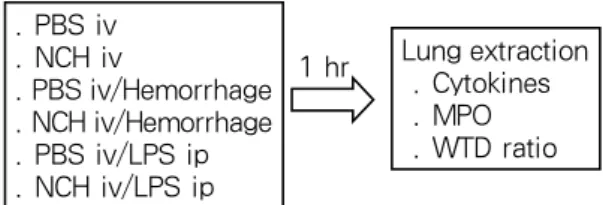
. PBS iv . NCH iv
. PBS iv/Hemorrhage . NCH iv/Hemorrhage . PBS iv/LPS ip . NCH iv/LPS ip
Lung extraction . Cytokines . MPO . WTD ratio 1 hr
Figure 1. Design of the experiment. Mice were di- vided into 6 groups and each group was composed of 5 mice. Nonanticoagulant heparin (NCH, 6.25 mg/ml, 200 μl, 50 mg/kg) or PBS (200 μl) was injected into the tail vein. Immediately after the injection of NCH or PBS, hemorrhage or LPS-injection was followed. One hour after the induction of hemorrhage or LPS-injec- tion, mice were anesthetized and lungs were removed for the evaluation of cytokines, MPO assay and wet-to-dry ratio.
remove 30% of the calculated total blood volume (0.27 ml/10g body wt) over 60 seconds into a heparinized syringe. One hour after the induction of hemorrhage, mice were again anesthetized with isofluorane, and the previously removed blood was infused into the retro-orbital venous plexus. The sham procedure involved cardiac puncture under isofluorane anesthesia, without blood removal, followed by the second episode of anesthesia and retroorbital puncture 1 hour later. One hour after the induction of hemorrhage, Mice were anestheti- zed with inhaled isofluorane and chest was opened and flushed by infusing 10 ml of PBS into beating right ventricle. After then, lungs were removed and stored at -70 ℃ before being used for cytokines and MPO assay (Figure 1).
Model of endotoxemia. Either PBS (200 μl) or NCH (6.25 mg/ml, 200 μl, 50 mg/kg) was injected into the tail vein. Just after the injection of either solution, endotoxemia-induced lung injury was ma- de by injecting LPS (125 μg/ml, 200 μl, 1 mg/kg) into the peritoneum. One hour after the injection of LPS, mice were anesthetized with inhaled isofluo- rane and chest was opened and flushed by infusing 10 ml of PBS into beating right ventricle. After then, lungs were removed and stored at -70 ℃ before being used for cytokines and MPO assay
(Figure 1).
Preparation of lung homogenate for ELISA.
Lung tissues were homogenized in ice cold lysis buffer (50 mM HEPES, 150 mM NaCl, 10% glyce- rol, 1% Triton X-100, 1.5 mM MgCl
2, 1 mM EGTA, 1 mM sodium vanadate, 10 mM sodium pyrophos- phate, 10 mM NaF, 300 µM p-nitrophenyl phospha- te, 1 mM PMSF, 10 µg/ml leupeptin, 10 µg/ml ap- rotinin, pH 7.3) containing 1 mM of protease inhi- bitor cocktail (Sigma, St Louis, MO). Homogenates were centrifuged at 14,000 g for 15 minutes and supernatants were collected. The protein concent- ration of each sample was assayed using the micro BCA protein assay kit standardized to BSA, accor- ding to manufacturer’s protocol (Pierce, Rockford, IL).
Cytokine ELISA. Immunoreactive TNF-α, MIP- 2 and IL-1β were quantitated using commercially available ELISA kits (R&D Systems, Minneapolis, MN), according to manufacturer's instructions.
Myeloperoxidase assay: MPO activity was as- sayed as described as follows. Lung tissue was homogenized in 1.0 ml of 50 mM potassium phos- phate buffer (pH 6.0) containing 10 mM N-ethy- lmaleimide for 30 seconds on ice. The homogenate was centrifuged at 12,000 g for 30 minutes at 4 ℃.
The proteinous pellet was homogenized once more in ice-cold buffer, and the homogenate was centri- fuged once more at 12,000 g for 30 minutes at 4 ℃.
The pellet was resuspended and sonicated on ice
for 90 seconds in 10 times vol of hexadecyltrime-
thylammonium bromide buffer (0.5% hexadecyltri-
methylammonium in 50 mM potassium phosphate,
pH 6.0). Samples were incubated in a water bath
(56 ℃) for 2 hours and then centrifuged at 12,000
g for 10 minutes. The supernatant was collected for
assay of MPO activity as determined by measuring
the H
2O
2-dependent oxidation of o-DA (3,3'-dime-
thoxybenzidine dihydrochloride) at 460 nm.
0 50 100 150 200 250
PBS NCH PBS/Hmr NCH/Hmr PBS/LPS NCH/LPS
*
Figure 2. NCH treatment did not reduce TNF-α expressions in the lung after LPS injection. Mice were injected with either PBS (200 μl) or NCH (50 mg/kg).
Hemorrhage or LPS-injection (1 mg/kg, ip) was followed immediately. One hour after the induction of hemorrhage of LPS-injection, mice were anestheti- zed and the expression of TNF-α was evaluated by ELISA method. *; <0.05. PBS; phosphate buffered sa- line, NCH; nonanticoagulant heparin, PBS/ Hmr; PBS injection followed by hemorrhage, NCH/ Hmr; NCH injection followed by hemorrhage, PBS/ LPS; PBS injection followed by LPS injection, NCH/ LPS; NCH injection followed by LPS injection
0 500 1000 1500 2000 2500 3000 3500 4000 4500 5000
PBS NCH PBS/Hmr NCH/Hmr PBS/LPS NCH/LPS
*
Figure 3. NCH treatment did not reduce MIP-2 expressions in the lung after LPS injection. Mice were injected with either PBS (200 μl) or NCH (50 mg/kg).
Hemorrhage or LPS-injection (1 mg/kg, ip) was fol- lowed immediately. One hour after the induction of hemorrhage of LPS-injection, mice were anestheti- zed and the expression of MIP-2 was evaluated by ELISA method. *; <0.05
0 1000 2000 3000 4000 5000 6000 7000 8000 9000
PBS NCH PBS/Hmr NCH/Hmr PBS/LPS NCH/LPS
*
Figure 4. NCH treatment did not reduce IL-1β ex- pressions in the lung after LPS injection. Mice were injected with either PBS (200 μl) or NCH (50 mg/kg). Hemorrhage or LPS-injection (1 mg/kg, ip) was followed immediately. One hour after the in- duction of hemorrhage of LPS-injection, mice were anesthetized and the expression of IL-1β was eva- luated by ELISA method. *; <0.05
Wet-to-Dry Lung Weight Ratios. Lungs were excised, rinsed briefly in PBS, blotted, then wei- ghed to obtain the "wet" weight. Lungs were dried in an oven at 80℃ for 7 days to obtain the "dry"
weight.
Statistical Analysis. Data are expressed as means
± SEM. ANOVA was performed with SPSS Win-
dows 9.0 statistical analysis software, and a diffe- rence was accepted as significant if the p value was less than 0.05, as verified by Duncan and Tukey post hoc test.
Results
The expressions of lung cytokines were increa- sed in endotoxemia model
The expression of TNF-α in PBS injected mice (control group) was 83.7±18.4 pg/ml/g of lung pro- tein and that of LPS injected mice was 191.6±10.8 pg/ml/g of lung protein (p<0.05 compared to con- trol group or hemorrhage group). In the while, lung TNF-α level in mice with hemorrhage was 91.1±6.3 pg/ml/g of lung protein (p>0.05 compared to con- trol group) (Figure 2). The lung MIP-2 expressions in the control, hemorrhage, and endotoxemic mice were 187±26, 200±22, and 3000±725 pg/ml/g of lung protein, respectively. The level of lung MIP-2 in the endotoxemic mice was significantly higher than those of control group or hemorrhage group (p<
0.05) (Figure 3). The level of IL-1β in endotoxemic
mice (6,500±1167 pg/ml/g of lung protein) was
significantly elevated compared to those of control
0 5 1 0 1 5 2 0 2 5 3 0 3 5 4 0
P B S N C H P B S / H m r N C H / H m r P B S / L P S N C H / L P S
*
Figure 5. NCH treatment did not reduce neutrophil recruitment in the lung after LPS injection. Mice were injected with either PBS (200 μl) or NCH (50 mg/kg). Hemorrhage or LPS-injection (1 mg/kg, ip) was followed immediately. One hour after the in- duction of hemorrhage of LPS-injection, mice were anesthetized and lung neutrophil recruitment was evaluated by MPO assay. *; <0.05
4.3 4.4 4.5 4.6 4.7 4.8 4.9
PBS NCH PBS/Hmr NCH/Hmr PBS/LPS NCH/LPS
Figure 6. Either hemorrhage nor LPS-injection did not increase lung leakage at one hour after insults.
Mice were injected with either PBS (200 μl) or NCH (50 mg/kg). Hemorrhage or LPS-injection (1 mg/
kg, ip) was followed immediately. One hour after the induction of hemorrhage of LPS-injection, mice were anesthetized and lung leakage was evaluated by wet-to-dry ratio.
group (266±25 pg/ml/g of lung protein) or hemo- rrhage group (224±24 pg/ml/g of lung protein) (p<
0.05) (Figure 4).
NCH treatment had no effects on the expre- ssions of lung cytokines
There was no difference in the expressions of lung cytokines between mice injected by PBS or NCH in control states. In detail, lung TNF-α, MIP- 2, and IL-1β levels of PBS- vs NCH-injected mice were 83.7±18.4 vs 57.8±10.4, 187±26 vs 168±33, and 266±25 vs 204±28 pg/ml/g of lung protein, respecti- vely (Figure 2, 3, and 4). Both in hemorrhage and endotoxemic models, NCH injection didn't affect the expressions of three cytokines. In hemorrhage model, lung TNF-α levels in PBS-injected or NCH-injected mice were 91.1±6.3 and 87.5±5.5 pg/ml/g of lung protein (p>0.05) (Figure 2). Those of MIP-2 levels were 200±22 and 231±41 pg/ml/g of lung protein (p>0.05) (Figure 3). Lung IL-1β levels were 224±24 and 492±44 pg/ml/g of lung protein (p>0.05) (Figure 4). In endotoxemic model, lung TNF-α levels in PBS-injected or NCH-injec- ted mice were 192±11 and 199±6 pg/ml/g of lung protein (p>0.05) (Figure 2). Those of MIP-2 levels
were 3000±725 and 3791±779 pg/ml/g of lung pro- tein (p>0.05) (Figure 3). Lung IL-1β levels were 6500±1167 and 7080±447 pg/ml/g of lung protein (p>0.05) (Figure 4).
NCH treatment did not reduce the recruitment of neutrophils in the lung
The MPO activities in PBS- or NCH-injected control mice were 10.5±2.3 and 5.9±0.7 U/g of lung (p>0.05). Those of PBS- or NCH-injected mice in hemorrhage model were 16.5±3.2 and 21.2±3.0 U/g of lung, which were higher than those of control group but no significant difference between them (p>0.05). In endotoxemic model, MPO activities were increased both in PBS-injected (27.9±6.2 U/g of lung, p<0.05 compared to control group) and NCH-injected mice (32.1±3.6 U/g of lung, p<0.05 compared to control group). However, there was no significant difference in MPO activity between PBS-injected and NCH-injected mice in endotoxe- mic model (p>0.05) (Figure 5).
Either hemorrhage or endotoxemic model didn't increase in the leakage of lung
Lung leakage was evaluated by wet-to-dry ratio.
The wet-to-dry ratios of PBS- or NCH-injected mice in control mice were 4.66±0.06 and 4.68±0.05 (p>0.05). Those of PBS- or NCH-injected mice in hemorrhage mice were 4.73±0.05 and 4.63±0.05 (p>
0.05). In endotoxemic mice, they were 4.72±0.03 and 4.77±0.04 (p>0.05) (Figure 6).
Discussion
Sepsis, which is one of the most important cau- ses of acute lung injury, is the leading cause of death in critically ill patients world-widely. In the United States, sepsis develops in 750,000 people an- nually, and more than 210,000 of them die
21. Despite more than 20 years of extensive research, sepsis and systemic inflammatory response syndrome (SIRS) remain the chief causes of death in inten- sive care units, with mortality rates between 30 to 70%. Further while, according to a recent report, the incidence is rising at rates between 1.5% and 8% annually
22. In the while, hemorrhage is often associated with severe acute lung injury
23. The is- chemia and reperfusion that occur with hemorrhage result in production of reactive oxygen species (ROS) that are thought to cause some of the lung injury that follows hemorrhage
24. A common con- sequence of sepsis or hemorrhage associated with systemic inflammation is multiple organ dysfunc- tion syndrome (MODS)
25. Whereas the clinical ma- nifestations of this syndrome are inconsistent, alte- rations in pulmonary functions are almost always observed
26,27. For example, systemic administration of LPS and/or hemorrhagic shock induced an inc- rease in the expression of inflammatory cytokines (TNF-α, IL-6) in serum and in the lung as early as 1 hour after insults
7,28,29. Components of the pulmo- nary dysfunction associated with MODS include widening of the alveolar-arterial PO
2gradient, decreased lung compliance and pulmonary edema.
Most clinical trials targeting blockade of specific inflammatory mediators have not been successful except activated protein C (APC), which has recen- tly been approved for treatment of severe sepsis. In the same sense, transfusion is the sole therapeutic option for the victims of major trauma which doe- sn't prevent the development of acute lung injury after severe bleeding. In the context of these dire situations in the management of sepsis- and hemo- rrhage-associated acute lung injury, we tried to evaluate the therapeutic potential of NCH on acute lung injury. Previously in in vitro study, NCH not only decreased the neutrophil-induced injury of pulmonary alveolar epithelium
17, but also prevented ischemia-reperfusion injury of the lung
30,31. With these actions, heparin might pose an ideal possible treatment for acute lung injury. With these in mind, we evaluated the therapeutic effects of NCH on the two kinds of acute lung injury model (hemorrhage and LPS injection). Both model induced the in- crease of MPO activity in the lung and in the LPS injection model, the expression of lung cytokines (TNF-α, MIP-2, IL-1β) increased. These results imply that hemorrhage and endotoxemia could re- sult in the development of acute lung injury. Unfor- tunately, however, NCH failed to reduce the expre- ssion of lung cytokines (Figure 2, 3, 4) and neutro- phil recruitment (Figure 5). By the way, both mo- dels for acute lung injury, endotoxemia and hemo- rrhage, didn't even increase the leakage of the lung (Figure 6), which was evaluated with wet-to-dry ratio. The validity of this method for the evaluation of lung leakage needs to be tested in the future.
The failure of NCH to reduce the inflammation of
lung might be related it's inefficacy in the in viro
model of acute lung injury. However, because we
tried a fixed dose of NCH (50 mg/kg) at fixed time
point (one hour after the induction of acute lung
injury), it is still a too hasty conclusion to give off
the hope for NCH as a potential agent in the treatment of acute lung injury.
In conclusion, NCH failed to reduce acute lung injury associated with endotoxemia and hemorrha- ge. However, considering the fact that the history of therapeutic intervention for sepsis has been re- ferred to as ‘the graveyard for pharmaceutical com- panies’, this result is not quite surprising. Still the research to find the exact time point of injection and effective dose of NCH needs to be continued.
Reference
1. Repine JE. Scientific perspectives on adult respirato- ry distress syndrome. Lancet 1992;339:466-9.
2. Kollef MH, Schuster DP. The acute respiratory dist- ress syndrome. N Engl J Med 1995;332:27-37.
3. Ware LB, Matthay MA. The acute respiratory dist- ress syndrome. N Engl J Med 2000;342:1334-49.
4. Chollet-Martin S, Jourdain B, Gibert C, Elbim C, Chastre J, Gougerot-Pocidalo MA. Interactions bet- ween neutrophils and cytokines in blood and alveolar spaces during ARDS. Am J Respir Crit Care Med 1996;154:594-601.
5. Goodman RB, Strieter RM, Martin DP, Steinberg KP, Milberg JA, Maunder RJ, et al. Inflammatory cyto- kines in patients with persistence of the acute respi- ratory distress syndrome. Am J Respir Crit Care Med 1996;154:602-11.
6. Suter PM, Suter S, Girardin E, Roux-Lombard P, Grau GE, Dayer JM. High bronchoalveolar levels of tumor necrosis factor and its inhibitors, interleukin-1, interferon, and elastase, in patients with adult respi- ratory distress syndrome after trauma, shock, or sep- sis. Am Rev Respir Dis 1992;145:1016-22.
7. Abraham E, Carmody A, Shenkar R, Arcaroli J. Neut- rophils as early immunologic effectors in hemorrhage- or endotoxemia-induced acute lung injury. Am J Phy- siol Lung Cell Mol Physiol 2000;279:L1137-45.
8. Parsey MV, Tuder RM, Abraham E. Neutrophils are major contributors to intraparenchymal lung IL-1 be- ta expression after hemorrhage and endotoxemia. J Immunol 1998;160:1007-13.
9. Shenkar R, Abraham E. Mechanisms of lung neutro- phil activation after hemorrhage or endotoxemia: ro- les of reactive oxygen intermediates, NF-kappa B, and cyclic AMP response element binding protein. J
Immunol 1999;163:954-62.
10. Xing Z, Jordana M, Kirpalani H, Driscoll KE, Schall TJ, Gauldie J. Cytokine expression by neutrophils and macrophages in vivo: endotoxin induces tumor necrosis factor-alpha, macrophage inflammatory pro- tein-2, interleukin-1 beta, and interleukin-6 but not RANTES or transforming growth factor-beta 1 mRNA expression in acute lung inflammation. Am J Respir Cell Mol Biol 1994;10:148-53.
11. Jaques LB. Heparins: anionic polyelectrolyte drugs.
Pharmacol Rev 1979;31:99-166.
12. Rao NV, Kennedy TP, Rao G, Ky N, Hoidal JR. Sul- fated polysaccharides prevent human leukocyte elas- tase-induced acute lung injury and emphysema in ha- msters. Am Rev Respir Dis 1990;142:407-12.
13. Weiler JM, Edens RE, Linhardt RJ, Kapelanski DP.
Heparin and modified heparin inhibit complement ac- tivation in vivo. J Immunol 1992;148:3210-5.
14. Hocking DC, Ferro TJ, Johnson A. Dextran sulfate in- hibits PMN-dependent hydrostatic pulmonary edema induced by tumor necrosis factor. J Appl Physiol 1991;70:1121-8.
15. Skinner MP, Lucas CM, Burns GF, Chesterman CN, Berndt MC. GMP-140 binding to neutrophils is inhi- bited by sulfated glycans. J Biol Chem 1991;266:
5371-4.
16. Ley K, Cerrito M, Arfors KE. Sulfated polysacchari- des inhibit leukocyte rolling in rabbit mesentery ve- nules. Am J Physiol 1991;260:H1667-73.
17. Simon RH, DeHart PD, Todd RF 3rd. Neutrophil-in- duced injury of rat pulmonary alveolar epithelial cells. J Clin Invest 1986;78:1375-86.
18. Inoue Y, Nagasawa K. Selective N-desulfation of he- parin with dimethyl sulfoxide containing water or me- thanol. Carbohydr Res 1976;46:87-95.
19. Fryer A, Huang YC, Rao G, Jacoby D, Mancilla E, Whorton R, et al. Selective O-desulfation produces no- nanticoagulant heparin that retains pharmacological activity in the lung. J Pharmacol Exp Ther 1997;
282:208-19.
20. Thourani VH, Brar SS, Kennedy TP, Thornton LR, Watts JA, Ronson RS, et al. Nonanticoagulant hepa- rin inhibits NF-kappaB activation and attenuates my- ocardial reperfusion injury. Am J Physiol Heart Circ Physiol 2000;278:H2084-93.
21. Angus DC, Linde-Zwirble WT, Lidicker J, Clermont G, Carcillo J, Pinsky MR. Epidemiology of severe se- psis in the United States: analysis of incidence, out- come, and associated costs of care. Crit Care Med 2001;29:1303-10.
22. Martin GS, Mannino DM, Eaton S, Moss M. The epi- demiology of sepsis in the United States from 1979
through 2000. N Engl J Med 2003;348:1546-54.
23. Rainer TH, Lam PK, Wong EM, Cocks RA. Deriva- tion of a prediction rule for post-traumatic acute lung injury. Resuscitation 1999;42:187-96.
24. McCord JM. Oxygen-derived free radicals in postis- chemic tissue injury. N Engl J Med 1985;312:159-63.
25. Deitch EA. Multiple organ failure: pathophysiology and potential future therapy. Ann Surg 1992;216:
117-34.
26. Rangel-Frausto MS, Pittet D, Costigan M, Hwang T, Davis CS, Wenzel RP. The natural history of the sy- stemic inflammatory response syndrome (SIRS): a prospective study. JAMA 1995;273:117-23.
27. Sands KE, Bates DW, Lanken PN, Graman PS, Hi- bberd PL, Kahn KL, et al. Epidemiology of sepsis syndrome in 8 academic medical centers. JAMA 1997;
278:234-40.
28. Ayala A, Perrin MM, Meldrum DR, Ertel W, Chaudry
IH. Hemorrhage induces an increase in serum TNF which is not associated with elevated level of endo- toxin. Cytokine 1990;2:170-4.
29. Hierholzer C, Kalff JC, Omert L, Tsukada K, Loeffert JE, Watkins SC, et al. Interleukin-6 production in he- morrhagic shock is accompanied by neutrophil recru- itment and lung injury. Am J Physiol 1998;275:
L611-21.
30. Friedrichs GS, Kilgore KS, Manley PJ, Gralinski MR, Lucchesi BR. Effects of heparin and N-acetyl heparin on ischemia/reperfusion-induced alterations in myo- cardial function in the rabbit isolated heart. Circ Res 1994;75:701-10.
31. Black SC, Gralinski MR, Friedrichs GS, Kilgore KS, Driscoll EM, Lucchesi BR. Cardioprotective effects of heparin or N-acetylheparin in an in vivo model of my- ocardial ischaemic and reperfusion injury. Cardiovasc Res 1995;29:629-36.