• 검색 결과가 없습니다.

EphA2 Receptor Signaling Mediates Inflammatory Responses in Lipopolysaccharide-Induced Lung Injury

N/A
N/A
Protected

Academic year: 2021

Share "EphA2 Receptor Signaling Mediates Inflammatory Responses in Lipopolysaccharide-Induced Lung Injury"

Copied!
9
0
0

로드 중.... (전체 텍스트 보기)

전체 글

(1)

EphA2 Receptor Signaling Mediates Inflammatory Responses in

Lipopolysaccharide-Induced Lung Injury

Ji Young Hong, M.D., Mi Hwa Shin, Kyung Soo Chung, M.D., Eun Young Kim, M.D., Ji Ye Jung, M.D., Young Ae Kang, M.D., Ph.D., Young Sam Kim, M.D., Ph.D., Se Kyu Kim, M.D., Ph.D., Joon Chang, M.D., Ph.D. and Moo Suk Park, M.D., Ph.D.

Division of Pulmonology, Department of Internal Medicine, Yonsei University College of Medicine, Seoul, Korea

Background: Eph receptors and ephrin ligands have several functions including angiogenesis, cell migration, axon guidance, fluid homeostasis, oncogenesis, inflammation and injury repair. The EphA2 receptor potentially mediates the regulation of vascular permeability and inflammation in response to lung injury.

Methods: Mice were divided into 3 experimental groups to study the role of EphA2 signaling in the lipopolysaccharide (LPS) -induced lung injury model i.e., IgG+phosphate-buffered saline (PBS) group (IgG instillation before PBS exposure), IgG+LPS group (IgG instillation before LPS exposure) and EphA2 monoclonal antibody (mAb)+LPS group (EphA2 mAb pretreatment before LPS exposure).

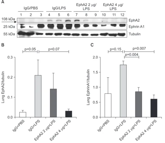
Results: EphA2 and ephrinA1 were upregulated in LPS-induced lung injury. The lung injury score of the EphA2 mAb+LPS group was lower than that of the IgG+LPS group (4.30±2.93 vs. 11.45±1.20, respectively; p=0.004). Cell counts (EphA2 mAb+LPS: 11.33×10

4

±8.84×10

4

vs. IgG+LPS: 208.0×10

4

±122.6×10

4

; p=0.018) and total protein concentrations (EphA2 mAb+LPS: 0.52±0.41 mg/mL vs. IgG+LPS: 1.38±1.08 mg/mL; p=0.192) were decreased in EphA2 mAb+LPS group, as compared to the IgG+LPS group. In addition, EphA2 antagonism reduced the expression of phospho-p85, phosphoinositide 3-kinase 110γ, phospho-Akt, nuclear factor κB, and proinflammatory cytokines.

Conclusion: This results of the study indicated a role for EphA2-ephrinA1 signaling in the pathogenesis of LPS-induced lung injury. Furthermore, EphA2 antagonism inhibits the phosphoinositide 3-kinase–Akt pathway and attenuates inflammation.

Keywords: Lipopolysaccharides; Lung Injury; EphA2 Protein

Introduction

The Eph tyrosine kinase receptor and ephrin ligand are cell surface-bound and are involved in cell-to-cell communica- tion

1,2

. The influence of Eph ephrin activation differs depend- ing on the cell type and environment. Eph- ephrin signaling contributes to several functions including vasculogenesis, angiogenesis, cell migration, axon guidance, fluid homeostasis and repair after injury

1-3

. A great deal of recent research has fo- cused on the complex role of Eph and ephrin in malignancy

4,5

. According to several studies, Eph receptors and ephrin ligands affect multiple oncogenic signaling pathways such as MAPK/

ERK, phosphoinositide 3-kinase (PI3K), E-cadherin and integ- rin/FAK/paxillin

4,6-8

. In addition to bidirectional signaling, Eph Copyright © 2015

The Korean Academy of Tuberculosis and Respiratory Diseases.

All rights reserved.

Address for correspondence: Moo Suk Park, M.D., Ph.D.

Division of Pulmonology, Department of Internal Medicine, Yonsei University College of Medicine, 50 Yonsei-ro, Seodaemun-gu, Seoul 120- 752, Korea

Phone: 82-2-2228-1955, Fax: 82-2-393-6884 E-mail: [email protected]

Received: Dec. 16, 2014 Revised: Mar. 30, 2015 Accepted: Apr. 27, 2015

cc It is identical to the Creative Commons Attribution Non-Commercial License (http://creativecommons.org/licenses/by-nc/4.0/).

(2)

receptors and ephrin ligands function independently of each other or in conjunction with other cell surface communica- tion systems

1

.

Eph-Ephrin proteins may play a role in inflammation through phenotypic changes in the vascular endothelium, which allow movement of inflammatory cells into the injured tissue

3

. There are studies on the role of EphA2-ephrinA1 sig- naling in lung injury

9-11

. These studies have shown that both EphA2 and ephrinA1 increase in bleomycin-induced lung injury and lung injury due to viral infection and hypoxia

9,10

, contrary to counter-directed changes in expressional regula- tion of Eph and ephrin during lipopolysaccharide (LPS) fever in rats

11

. In the bleomycin-induced lung injury model, EphA2 knockout mice showed reduced permeability and less inflam- matory response than wild type mice

9

. Similarly, in lung injury due to viral infection and hypoxia, EphA2 antagonism with EphA2/Fc and anti-EphA2 antibody reduced vascular leakage and albumin extravasation

10

.

In this study, we investigated the expression of Eph-ephrin proteins and the effect of EphA2 monoclonal antibody (mAb) pretreatment in an LPS induced lung injury model. Also, we studied the association between EphA2 signaling and other known signal pathways of LPS-induced lung injury.

Materials and Methods

1. Experimental animals

Experiments were conducted in accordance with proto- cols approved by the institutional animal care committee of the Medical College of Yonsei University. Wild type male C57BL/6J mice, 8–10 weeks of age and weighing 20–24 g were purchased from Orient Bio (Seongnam, Korea). All animals were supplied with food and water and were subjected to a similar day and night light cycle.

2. Study design

Animals were studied in three experimental groups: the IgG+phosphate-buffered saline (PBS) group, the IgG+LPS group and the EphA2 mAb+LPS group. The EphA2 mAb+LPS group was divided into two subgroups according to the dose of EphA2 mAb (2 μg and 4 μg). Under inhalation anesthesia using isoflurane, the mice inhaled 4 μg of mouse IgG (Cat#

ab37355; Abcam, Cambridege, MA, USA) or monoclonal rat EphA2 antibody (Cat# MAB639; R&D Systems, Minneapolis, MN, USA) in 50 μL PBS 1 hour prior to instillation experi- ments

10

. After pretreatment, mice underwent intranasal instil- lation of 50 μL PBS or Escherichia coli LPS (E.coli 0127: B8;

Sigma, St. Louis, MO, USA) 40 μg/g in 50 μL PBS. The intrana- sal administration was performed by gradually releasing the solution into the nostril with the help of microsyringe (Cat#

7637-01; Hamilton Company, Reno, NV, USA)

12

. The rate of release was adjusted to allow the mouse to inhale the solution completely without trying to form bubbles.

All mice were sacrificed 48 hours after treatment and lung tissue was harvested for analysis. For bronchoalveolar lavage fluid (BALF) studies, lungs were lavaged via the trachea with two 1-mL aliquots of saline. After the BALF was centrifuged, the cell pellets were used for differential cell counts and the supernatants were used for quantitative analysis of protein by the Bradford protein assay. While left lung tissue was used for histological studies, right lung tissue was prepared for Western blotting and quantitative cytokine enzyme-linked immuno- sorbent assay (ELISA).

3. Histopathology

Left lungs were inflated and fixed with 4% paraformalde- hyde before being embedded in paraffin and sectioned. Semi- quantitative grading of lung injury on hematoxylin and eosin–

stained sections was performed as previously described

13

. 4. Western blotting and cytokine ELISA

The right lung homogenate lysates were centrifuged at 13,000 ×g for 30 minutes at 4

o

C. Equal amounts of protein were separated by sodium dodecyl sulfate polyacrylamide gel electrophoresis and transferred to a nitrocellulose membrane before immunoblotting with primary antibodies, as indicated.

Membranes were incubated with anti-rabbit or anti-mouse IgG horseradish peroxidase conjugated antibodies and de- veloped using a Super-Signal West Pico chemiluminescence detection kit (Pierce, Rockford, IL, USA). The band images were quantified using the Alpha Ease FC software version 4.1.0 (Alpha Innotech, San Leandro, CA, USA).

The antibodies used in this study included rabbit EphA2

(1:1,000, Thermo Fisher Scientific, Bremen, Germany), goat

ephrinA1 (1:1,000, Thermo Fisher Scientific), rabbit phos-

phate–Akt (1:1,000, Cell Signaling Technologies, Beverly, MA,

USA), rabbit Akt (1:1,000, Cell Signaling Technologies), rabbit

phosphate-p85 (1:500, Cell Signaling Technologies), rabbit

p85 (1:1,000, Cell Signaling Technologies), rabbit PI3 kinase

110γ (1:1,000, Cell Signaling Technologies), rabbit α-tubulin

(1:1,000, Cell Signaling Technologies), rabbit phosphate–p65

nuclear factor κB (NF-κB; 1:500, Thermo Fisher Scientific),

rabbit p65 NF-κB (1:500, Thermo Fisher Scientific), mouse

phosphate-IκBα (1:500, Cell Signaling Technologies) and

mouse IκBα (1:500, Cell Signaling Technologies). Measure-

ment of keratinocyte chemoattractant (KC/CXCL1), macro-

phage inflammatory protein 2 (MIP-2), interleukin (IL)-1β, IL-

6, and tumor necrosis factor α (TNF-α) were performed on

whole-lung homogenates using quantified ELISA kits (MIL-

LIPLEX MAG Mouse Cytokine/Chemokine kit from Millipore,

Billerica, MA, USA).

(3)

5. Statistical analysis

Statistical analysis was performed using Prism version 5.0 (Graphpad Software, Durham, NC, USA). Two group com- parisons were performed with an unpaired student t test. Data are expressed as mean±SD for each group. Differences were considered significant at p<0.05.

Results

1. EphA2 and ephrinA1 are upregulated in LPS induced lung injury

As shown in Figure 1, Western blotting studies demonstrat- ed a significant increase in EphA2 and ephrinA1 protein ex- pression after LPS challenge. Both EphA2 and ephrinA1 pro- tein expression were reduced when EphA2 mAb was instilled as pretreatment. Also, the degree of decline was higher in the 4 μg EphA2 mAb subgroup than the 2 μg mAb subgroup in both EphA2 and ephrinA1 (Figure 1).

2. EphA2 antagonism attenuates LPS induced lung injury In the IgG+PBS group, neutrophil recruitment, alveolar wall

thickening and hemorrhage were not observed, contrary to the results of the IgG+LPS group (Figure 2A), where hyaline membrane formation, neutrophils in the alveolar and intersti- tium space and thickening of alveolar septa were remarkably increased (Figure 2B). The EphA2 mAb+LPS groups demon- strated reduced neutrophil infiltration and alveolar thickening compared with the IgG+LPS group (Figure 2C, D).

Figure 3 shows the lung injury scores, bronchoalveolar la- vage cell counts and protein concentrations of the four groups.

The lung injury score of the EphA2 mAb 4 μg+LPS group was lower than that of the IgG+LPS group (4.30±2.93 vs.11.45±1.20, respectively; p=0.004). LPS caused a significant increase in cell counts and concentration of total protein in the lung lavage fluid. LPS-induced lung injury was attenuated by pretreat- ment with EphA2 mAb. Cell counts were significantly less (EphA2 mAb 4 μg+LPS: 11.33×10

4

±8.84×10

4

vs. IgG+LPS:

208.0×10

4

±122.6×10

4

; p=0.018) and concentrations of total protein were lower (EphA2 mAb 4 μg+LPS: 0.52±0.41 mg/mL vs. IgG+LPS: 1.38±1.08 mg/mL; p=0.192) in EphA2 mAb+LPS group, compared with IgG+LPS group.

3. EphA2 regulates expression of the PI3K-Akt pathway To elucidate whether the LPS-induced injury related signal pathway is regulated by EphA2-ephrinA1 signaling, the PI3K-

Figure 1. The expression of EphA2 and

ephrinA1 in lung lysates after lipopoly-

saccharide (LPS) exposure, as shown

by Western blots (A) and densitometry

(B, C). IgG pretreatment increased the

expression of EphA2 and ephrinA1 after

LPS exposure; whereas, EphA2 mono-

clonal antibody pretreatment reduced

the expression of EphA2 and ephrinA1

after LPS exposure. Values are presented

as mean±SD. PBS: phosphate buffered

saline.

(4)

Akt pathway was assessed by Western blot analysis in the IgG+PBS, IgG+LPS, and EphA2 mAb+LPS groups (Figures 4, 5B).

Compared to the IgG+PBS group, the IgG+LPS group showed significantly increased phospho-p85 and PI3K 110γ and phospho-Akt. After EphA2 mAb pretreatment, the expres- sion of PI3K 110γ and phospho-Akt by LPS challenge were reduced, which indicated the inhibitory effect of EphA2 mAb (PI3K 110γ, p=0.06; p-Akt, p=0.002). The regulatory subunit PI3K phospho-p85 was not influenced by EphA2 mAb pre- treatment.

4. EphA2 antagonism decreased NF-κB expression and cytokine production

To determine whether EphA2 mAb has a protective func- tion in inflammatory response, we assessed the expression of p65 NF-κB, IκB protein and cytokine levels (IL-1β, IL-6, KC, MIP-2, and TNF-α) by Western blot analysis and ELISA.

EphA2 antagonism led to a decrease of p65 NF-κB and cyto- kine production in the EphA2 mAb+LPS group compared to the IgG+LPS group (Figures 5B, 5C, 6).

5. EphA2-ephrinA1 signaling in LPS-induced lung injury EphrinA1-dependent EphA2 activation may be an unrec- Figure 2. The effect of EphA2 monoclonal antibody pretreatment in lipopolysaccharide (LPS)-induced lung injury. Mice were intranasally treated with IgG or EphA2 antibody. One hour later, they were intranasally treated with phosphate buffered saline (PBS) or 40 μg LPS in PBS.

The lungs were removed after 24 hr and stained with hematoxylin and eosin (H&E). (A) IgG+PBS group, (B) IgG+LPS group, (C) EphA2 Ab 2

μg+LPS group, and (D) EphA2 Ab 4 μg+LPS group, n=4 per group. The IgG+LPS group (B) showed obvious neutrophil infiltration and alveo-

lar septal infiltration; however, the EphA2 Ab+LPS groups (C, D) showed attenuated lung injury (H&E stain, ×400).

(5)

Figure 3. The effect of EphA2 monoclonal antibody pretreatment on semiquantitative lung injury scores (A), total bronchoalveolar fluid (BALF) cell count (B) and BALF protein concentration (C). In the lipopolysaccharide (LPS)-induced lung injury model, EphA2 antagonism with EphA2 monoclonal antibody led to improved lung injury scores, decreased BALF cell counts and decreased BALF proteins (p<0.05, ex- cept BALF protein), as compared to IgG pretreatment. Values are presented as mean±SD (n=4 per group).

Figure 4. The expression of phos-

phoinositide 3-kinase (PI3K) protein

(pp85 and 110γ) in lung lysates after lipo-

polysaccharide (LPS) exposure, as shown

by Western blots (A) and densitometry

(B, C). Phosphorylated 110γ (catalytic

subunit of PI3K) levels were increased in

mice with LPS exposure (IgG+LPS), as

compared with controls (IgG+phosphate

buffered saline [PBS]). Activation of

P110γ was reduced in response to EphA2

monoclonal antibody pretreatment

(EphA2+LPS) (C). However, the expres-

sion of p85 (regulatory subunit of PI3K)

protein was not consistently influenced

by EphA2 monoclonal antibody (B). Val-

ues are presented as mean±SD.

(6)

ognized contributor to LPS-induced lung injury. Antagoniz- ing EphA2 not only resulted in diminished expression of the PI3Kγ isoform, but it also led to decreased NF-κB activation and expression of proinflammatory cytokines through the PI3K-Akt pathway. Crosstalk of EphA2 signaling with the PI3K- Akt pathway may contribute to the neutrophil-driven inflam- matory process in LPS-induced lung injury (Figure 7).

Discussion

Although acute lung injury accounts for significant morbid- ity and mortality in critically ill patients, there are no specific effective therapies

14

. Thus, it is important to find a therapeutic target and verify the effectiveness of related treatment in acute lung injury.

This study demonstrates that the expression of the EphA2 receptor and ephrinA1 ligand is upregulated in an LPS-in- duced lung injury model. Blockage of EphA2 activation leads to reduction of protein leakage, recruitment of inflammatory cells and cytokine production. Furthermore, the EphA2 recep- tor is involved in the PI3K/Akt signal pathway in LPS-induced NF-κB activation.

Eph receptors and ephrin ligands represent the largest family of receptor tyrosine kinases that regulate important processes during embryonic neuronal development, angio- genesis and oncogenesis

1,2

. Emerging evidence suggests that Eph/ephrin plays a key role in inflammation

3

and previous studies have established the role of EphA2 in postnatal lung injury

9-11,15

.

Contrary to our results that both EphA2 and ephrinA1

increased after LPS exposure, Ivanov et al.

11

demonstrated

Figure 5. The expression of Akt, p65 nuclear factor κB (NF-κB), and IκB in lung lysates after lipopolysaccharide (LPS) exposure, as shown

by representative Western blots (A) and densitometry (B–D). The amount of phosphorylated Akt was increased in mice with LPS exposure

(IgG+LPS), as compared with controls (IgG+PBS). LPS-induced activation of Akt was reduced in response to EphA2 monoclonal antibody

pretreatment (EphA2+LPS) (B). The expression of phosphorylated NF-κB protein was similar to that of Akt (C). The expression of phosphor-

ylated IκB after LPS exposure was increased in mice with EphA2 monoclonal antibody pretreatment (D). Values are presented as mean±SD.

(7)

opposing changes in expressional regulation of the EphA2 receptor and ephrin ligand in lung tissue in phase 2 (90 min- ute post-LPS) of LPS exposure. The difference between the two studies may be due to the experiment time interval. The sacrifice time was 90 minutes after LPS exposure in the study of Ivanov et al.

11

, whereas it was 48 hours after LPS exposure in our study.

Our results are consistent with previous studies on lung injury after instillation of bleomycin and exposure to hypoxia after viral infection

9,10

. Both studies showed increased expres- sion of EphA2 and ephrinA1 in lung injury and found that EphA2 receptor antagonism (via a EphA2 KO mouse model

or EphA2 mAb pretreatment) provided protection against lung injury showing reduced permeability and protein ex- travasation.

EphA2 contributes not just to actin cytoskeleton disruption leading to vascular permeability, but also to inflammation in lung injury. Several data suggest that actin cytoskeleton rear- rangement may be a key preceding event during the regula- tion of inflammatory responses

16-18

. Our results have revealed that EphA2 mAb pretreatment ameliorates lung injury by at- tenuating the expression of cytokines and neutrophil infiltra- tion. This is similar to bleomycin-induced lung injury, where the proinflammatory transcriptional factors NF-κB, CXCL1, Figure 6. The expression of cytokines (interleukin [IL]-1β, IL-6, KC, macrophage inflammatory protein 2 [MIP-2], and tumor necrosis factor α [TNF-α]) in response to EphA2 monoclonal antibody pretreatment in a lipopolysaccharide (LPS) induced lung injury model. Enzyme linked immunosorbent assay measurement showed differential expression of cytokines (IL-1β, IL-6, KC, MIP-2, and TNF-α) in lung tissue lysates.

Cytokine levels were reduced in response to LPS exposure in mice with EphA2 monoclonal antibody pretreatment, as compared to those

with IgG pretreatment (n=3 per group). Values are presented as mean±SD.

(8)

and CCL2 are triggered by EphA2 stimulation via the ephrin ligand in wild mice, but inhibited in EphA2 deficient mice

9

.

Although the main effect of Eph receptor-ephrin ligand sig- naling is increased vascular permeability and inflammatory cell migration, the detailed mechanism in lung inflammation remains unclear.

PI3K and Akt occupy an important role in modulation of NF-κB activation and proinflammatory cytokine production in numerous cell populations including neutrophils, epithe- lial cells and fibroblasts

19-22

. Yum et al.

23

showed that, after exposure to LPS, Akt is activated via PI3Kγ and this leads to enhanced nuclear translocation of NF-κB and reduced lung neutrophil apoptosis. The present study demonstrated that antagonizing EphA2 expression with EphA2 mAb blocked the PI3K–Akt–NF-κB pathway. Another study supported a direct correlation between EphA2 signaling and the PI3K-Akt path- way. Holen et al.

24

reported that reverse signaling through the ephrin A4 ligand by interaction with the EphA2 receptor led to Src kinase phosphorylation, PI3K-Akt activation and inhibi- tion of cell death by apoptosis.

In view of this, the EphA2 receptor may be an important mediator for crosstalk between the inflammatory signal path- way and the cell-to-cell integrity pathway. Considering that

the expression of ephrinA1 is induced by some cytokines

25,26

, ligand-mediated activation of the EphA2 receptor is expected to be accelerated in lung injury. Recent research reported that EphA2 expression induced by tobacco smoke exposure in bronchial airway epithelial cells was dependent on MAPK (ERK, p38, and JNK) signaling

15

. Determining whether EphA2 signaling communicates with these additional mechanisms in LPS-induced lung inflammation will require further investiga- tion.

Since EphA2-ephrinA1 signaling was reported to regulate multiple oncogenesis events

27

, clinical trials of drugs inhibiting the EphA2 receptor have been ongoing in cancer patients

28,29

. Similarly, the possible role of EphA2 as a therapeutic target in this LPS-induced lung injury model should be verified in hu- man studies.

In conclusion, we demonstrate that EphA2 signaling regu- lates permeability and inflammation in LPS-induced lung injury and leads to activation of the PI3K–Akt–NF-κB path- way. Moreover, the inhibition of EphA2 expression by EphA2 mAb instillation reduces neutrophil infiltration, proinflamma- tory cytokine production and alveolar edema. Therefore, the EphA2 receptor may present a potential therapeutic target in acute lung injury.

Conflicts of Interest

No potential conflict of interest relevant to this article was reported.

Acknowledgements

This Study was Supported by a 2013-Grant from The Ko- rean Academy of Tuberculosis and Respiratory Diseases.

References

1. Kullander K, Klein R. Mechanisms and functions of Eph and ephrin signalling. Nat Rev Mol Cell Biol 2002;3:475-86.

2. Pasquale EB. Eph-ephrin bidirectional signaling in physiol- ogy and disease. Cell 2008;133:38-52.

3. Coulthard MG, Morgan M, Woodruff TM, Arumugam TV, Taylor SM, Carpenter TC, et al. Eph/Ephrin signaling in injury and inflammation. Am J Pathol 2012;181:1493-503.

4. Beauchamp A, Debinski W. Ephs and ephrins in cancer:

ephrin-A1 signalling. Semin Cell Dev Biol 2012;23:109-15.

5. Surawska H, Ma PC, Salgia R. The role of ephrins and Eph re- ceptors in cancer. Cytokine Growth Factor Rev 2004;15:419- 33.

6. Pratt RL, Kinch MS. Activation of the EphA2 tyrosine kinase stimulates the MAP/ERK kinase signaling cascade. Oncogene Figure 7. The effects of ephrinA1-mediated activation of EphA2 on

the phosphoinositide 3-kinase (PI3K)/Akt signaling pathways in lipopolysaccharide (LPS)-induced lung injury. EphA2-ephrinA1 signaling may contribute to the development of LPS-induced lung injury. Exposure to LPS resulted in activation of the EphA2–eph- rinA1 and PI3K/Akt/nuclear factor κB (NF-κB) signaling pathway.

EphA2 monoclonal antibody-induced inhibition of EphA2 signal-

ing resulted in decreased PI3K/Akt/NF-κB-dependent inflamma-

tory processes. These results suggested crosstalk between EphA2

signaling and PI3K/Akt/NF-κB signaling in LPS-induced lung in-

jury. TNF-α: tumor necrosis factor α; IL: interleukin.

(9)

2002;21:7690-9.

7. Miao H, Burnett E, Kinch M, Simon E, Wang B. Activation of EphA2 kinase suppresses integrin function and causes focal- adhesion-kinase dephosphorylation. Nat Cell Biol 2000;2:62- 9.

8. Liu DP, Wang Y, Koeffler HP, Xie D. Ephrin-A1 is a negative regulator in glioma through down-regulation of EphA2 and FAK. Int J Oncol 2007;30:865-71.

9. Carpenter TC, Schroeder W, Stenmark KR, Schmidt EP. Eph- A2 promotes permeability and inflammatory responses to bleomycin-induced lung injury. Am J Respir Cell Mol Biol 2012;46:40-7.

10. Cercone MA, Schroeder W, Schomberg S, Carpenter TC.

EphA2 receptor mediates increased vascular permeability in lung injury due to viral infection and hypoxia. Am J Physiol Lung Cell Mol Physiol 2009;297:L856-63.

11. Ivanov AI, Steiner AA, Scheck AC, Romanovsky AA. Expres- sion of Eph receptors and their ligands, ephrins, during lipo- polysaccharide fever in rats. Physiol Genomics 2005;21:152- 60.

12. Hedrich HJ. The laboratory mouse. Amsterdam: Elsevier Aca- demic Press; 2004.

13. Fang WF, Cho JH, He Q, Lin MC, Wu CC, Voelkel NF, et al.

Lipid A fraction of LPS induces a discrete MAPK activation in acute lung injury. Am J Physiol Lung Cell Mol Physiol 2007;

293:L336-44.

14. Rubenfeld GD, Caldwell E, Peabody E, Weaver J, Martin DP, Neff M, et al. Incidence and outcomes of acute lung injury. N Engl J Med 2005;353:1685-93.

15. Nasreen N, Khodayari N, Sriram PS, Patel J, Mohammed KA.

Tobacco smoke induces epithelial barrier dysfunction via receptor EphA2 signaling. Am J Physiol Cell Physiol 2014;306:

C1154-66.

16. Kustermans G, El Benna J, Piette J, Legrand-Poels S. Perturba- tion of actin dynamics induces NF-kappaB activation in my- elomonocytic cells through an NADPH oxidase-dependent pathway. Biochem J 2005;387(Pt 2):531-40.

17. Nemeth ZH, Deitch EA, Davidson MT, Szabo C, Vizi ES, Hasko G. Disruption of the actin cytoskeleton results in nuclear factor-kappaB activation and inflammatory mediator production in cultured human intestinal epithelial cells. J Cell Physiol 2004;200:71-81.

18. Kustermans G, El Mjiyad N, Horion J, Jacobs N, Piette J, Legrand-Poels S. Actin cytoskeleton differentially modulates

NF-kappaB-mediated IL-8 expression in myelomonocytic cells. Biochem Pharmacol 2008;76:1214-28.

19. Ardeshna KM, Pizzey AR, Devereux S, Khwaja A. The PI3 kinase, p38 SAP kinase, and NF-kappaB signal transduc- tion pathways are involved in the survival and maturation of lipopolysaccharide-stimulated human monocyte-derived dendritic cells. Blood 2000;96:1039-46.

20. Beraud C, Henzel WJ, Baeuerle PA. Involvement of regulatory and catalytic subunits of phosphoinositide 3-kinase in NF- kappaB activation. Proc Natl Acad Sci U S A 1999;96:429-34.

21. Kane LP, Shapiro VS, Stokoe D, Weiss A. Induction of NF- kappaB by the Akt/PKB kinase. Curr Biol 1999;9:601-4.

22. Manna SK, Aggarwal BB. Wortmannin inhibits activation of nuclear transcription factors NF-kappaB and activated protein-1 induced by lipopolysaccharide and phorbol ester.

FEBS Lett 2000;473:113-8.

23. Yum HK, Arcaroli J, Kupfner J, Shenkar R, Penninger JM, Sasaki T, et al. Involvement of phosphoinositide 3-kinases in neutrophil activation and the development of acute lung injury. J Immunol 2001;167:6601-8.

24. Holen HL, Shadidi M, Narvhus K, Kjosnes O, Tierens A, Aasheim HC. Signaling through ephrin-A ligand leads to activation of Src-family kinases, Akt phosphorylation, and in- hibition of antigen receptor-induced apoptosis. J Leukoc Biol 2008;84:1183-91.

25. Cheng N, Brantley DM, Liu H, Lin Q, Enriquez M, Gale N, et al.

Blockade of EphA receptor tyrosine kinase activation inhibits vascular endothelial cell growth factor-induced angiogenesis.

Mol Cancer Res 2002;1:2-11.

26. Pandey A, Shao H, Marks RM, Polverini PJ, Dixit VM. Role of B61, the ligand for the Eck receptor tyrosine kinase, in TNF- alpha-induced angiogenesis. Science 1995;268:567-9.

27. Tandon M, Vemula SV, Mittal SK. Emerging strategies for EphA2 receptor targeting for cancer therapeutics. Expert Opin Ther Targets 2011;15:31-51.

28. Chee CE, Krishnamurthi S, Nock CJ, Meropol NJ, Gibbons J, Fu P, et al. Phase II study of dasatinib (BMS-354825) in pa- tients with metastatic adenocarcinoma of the pancreas. On- cologist 2013;18:1091-2.

29. Annunziata CM, Kohn EC, LoRusso P, Houston ND, Coleman

RL, Buzoianu M, et al. Phase 1, open-label study of MEDI-547

in patients with relapsed or refractory solid tumors. Invest

New Drugs 2013;31:77-84.

수치

Figure 3 shows the lung injury scores, bronchoalveolar la- la-vage cell counts and protein concentrations of the four groups
Figure 3. The effect of EphA2 monoclonal antibody pretreatment on semiquantitative lung injury scores (A), total bronchoalveolar fluid  (BALF) cell count (B) and BALF protein concentration (C)

참조

관련 문서

Methods: Eighty ASA physical status I and II pediatric patients (1~7 years) were randomly divided into four groups: Group I (Lactated Ringer`s solution, n=20), Group II

We evaluated the general characteristics, mechanism of injury, Revised Trauma Score(RTS), Injury Severity Score(ISS), Trauma Injury Severity Score(TRISS). More men

Glutamate excitotoxicity induced by excessive activation of NMDA receptor causes various damage to cells, which leads to cell death.. In previous studies, increased ROS

Effect of LRP6 on survivin expression in hypoxic cardiomyocytes (A) Expression of survivin in Ad-LRP6 and silencing LRP6 infected cardiomyocytes as determined by western

Taken together, these results suggested that latex containing the ficin inhibited the cell growth and induced apoptosis by caspase and Bcl-2 family signaling pathway in

This study aimed to evaluate the site and extent of injury, injury mechanism, player position, and the reinjury incidence in the hamstring by using magnetic

In conclusion, the data acquired in this study demonstrate that pinosylvin can inhibit LPS-induced expression of pro-inflammatory mediators, and the

In this study, we investigated the time course of neutrophil recruitment and infiltration in the early stages of arterial throm- bosis using an FeCl 3 -induced carotid