DOI: 10.4046/trd.2009.66.4.314
ISSN: 1738-3536(Print)/2005-6184(Online) Tuberc Respir Dis 2009;66:314-318
CopyrightⒸ2009. The Korean Academy of Tuberculosis and Respiratory Diseases. All rights reserved.
유전출혈모세혈관확장증을 가진 가족 1예
인제대학교 의과대학
1내과학교실,
2영상의학교실
김 민1, 송화영1, 정 훈1, 박이내1, 최상봉1, 이현경1, 이성순1, 이영민1, 김수영2, 김용훈2, 허진원1
A Familial Case of Hereditary Hemorrhagic Telangiectasia
Min Kim, M.D.
1, Hwa Young Song, M.D.
1, Hun Jeong, M.D.
1, I Nae Park, M.D.
1, Sang Bong Choi, M.D.
1, Hyun Kyung Lee, M.D.
1, Sung-Soon Lee, M.D.
1, Young Min Lee, M.D.
1, Su Young Kim, M.D.
2, Yong Hoon Kim, M.D.
2, Jin Won Huh, M.D.
1Departments of
1Internal Medicine,
2Radiology, Inje University College of Medicine, Goyang, Korea
Hereditary hemorrhagic telangiectasia (HHT, also called Osler-Weber-Rendu Disease) is a rare systemic fibrovascular dysplasia characterized by recurrent epistaxis, cutaneous telangiectasia, and visceral arteriovenous malformations (AVMs). HHT is an autosomal dominant disease with a prevalence of 1 in 5,000∼8,000. Recurrent epistaxis is often the first and most common manifestation, and about 30% of patients reveal pulmonary AVM. Presently, we report a familial case of HHT. A 61-year-old male with asymptomatic multiple pulmonary AVMs was successfully treated with embolization. His older brother who presented with recurrent epistaxis and multiple telangiectasias was treated with laser ablation. Their pedigree revealed a family history of recurrent epistaxis.
Key Words: Hereditary hemorrhagic telangiectasia, Arteriovenous malformations, Epistaxis, Embolization
Address for correspondence: Jin Won Huh, M.D.
Division of Pulmonary and Critical Care Medicine, Ilsan Paik Hospital, College of Medicine, Inje University, 2240, Daehwa- dong, Ilsanseo-gu, Goyang 411-706, Korea
Phone: 82-31-910-7544, Fax: 82-31-910-7219 E-mail: [email protected]
Received: Jan. 31, 2009 Accepted: Mar. 12, 2009
서 론
유전출혈모세혈관확장증(hereditary hemorrhagic te- langiectasia, HHT)으로 알려진 Osler-Weber-Rendu병은 혈관벽의 탄력층과 근육층의 변화로 외상에 쉽게 출혈할 수 있는 혈관벽을 만드는 섬유혈관 이형성으로 1/5,000∼
8,000의 발병률을 보이는 드문 상염색체 우성 유전 질환 이다1. 반복적인 코출혈, 피부나 점막의 모세혈관확장증, 내부 장기의 동정맥기형 및 이에 상응하는 가족력이 특징 으로 반복적인 코출혈이 가장 흔한 증상이며, 30% 이상의 환자에서 폐나 간에서 동정맥기형이 동반된다1. 특히 폐 동정맥기형은 우좌 단락에 의한 저산소증, 출혈, 역행성색 전(paradoxical embolism)에 의한 뇌경색, 뇌농양 등의
합병증을 일으킬 수 있으며, 현재까지 가장 적절한 치료법 은 색전술로 알려져 있다2. 저자들은 반복적인 코출혈이 있는 가족에서, 우연히 발견된 폐동정맥기형으로 폐혈관 색전술을 받은 동생과 반복적인 코출혈을 보이는 형을 같 이 경험하여 희귀한 유전질환인 유전출혈모세혈관확장증 의 가계도를 확인하였기에 이를 문헌고찰과 함께 보고하 는 바이다.
증 례
증례 1
환 자: 박○○, 61세, 남자 주 소: 5일 전부터 발생한 발열
현병력: 환자는 5일 전부터 지속적인 발열, 오한을 느꼈 으며, 4일 전부터는 기침, 화농성 객담이 동반되고 기침을 하거나 숨을 들이쉴 때 좌측 흉막통이 심해져 내원하였다.
과거력: 10년 전 고혈압을 진단 받았고, 5년 전 뇌농양으 로 타 병원에 입원하여 개두술 및 배농술 시행하였다. 특 이사항으로는 일상 생활 중에 반복적인 코출혈이 있었다.
가족력: 상당수의 부계 친척이 반복적인 코출혈이나 구
Case Report
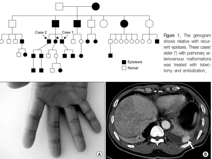
Figure 1. The genogram shows relative with recur- rent epistaxis. These cases' sister (*) with pulmonary ar- teriovenous malformations was treated with lobec- tomy and embolization.
Figure 2. Finger tips show sparse telangiectasia (A) and a computed tomographic scan (B) shows an arteriovenous malformation (white arrow).
강출혈이 있었으며, 누나가 폐동정맥기형으로 폐엽절제 술(lobectomy)과 폐혈관 색전술을 시행 받았다(Figure 1).
진찰 소견: 내원 당시 혈압 138/86 mmHg, 맥박수 110 회/분, 호흡수 20회/분, 체온 37.7oC이었고, 의식은 명료 하였다. 구강과 손가락 끝에 다발성 모세혈관확장증 소견 이 관찰되었다(Figure 2A). 청진상 좌측 폐에서 호흡음이 감소되었으나, 심음은 규칙적이었으며 심잡음은 없었다.
검사실 소견: 말초혈액검사에서 백혈구 14.760×10⁹/L (호중구 91.6%), 혈색소 80 g/L, 혈소판 212×109/L으로 백혈구 증가증이 있었다. 생화학 검사에서 총단백 64 g/L, 알부민 33 g/L, BUN 5.4 mmol/L, 크레아티닌 115 μmol/L, AST 0.83 μkat/L, ALT 0.51 μkat/L, 총빌리루빈 18.7 μmol/L, 프로트롬빈 시간 15.6초로 정상이었다.
방사선 소견: 가슴 엑스선 사진에서 폐 좌측 하부에 흉 수 소견이 있었으며, 흉부 전산화단층촬영에서는 양쪽 폐 야에 다발성 동정맥기형과 좌측 폐에 흉수가 관찰되었다
(Figure 2B).
치료 및 경과: 흉강천자는 동정맥기형 파열에 대한 위험 으로 환자가 거부하여 시행하지 못하였다. 임상적으로 부 폐렴성 흉수로 진단 후 항생제로 치료하였으며, 이후 흉부 전산화단층촬영에서 흉수가 감소되었다. 환자는 다시 입 원하여 폐동맥 혈관 조영술로 폐의 좌하엽 1곳, 우하엽 2곳, 우중엽 1곳에 색전술을 시행하였다(Figure 3). 이후 특별한 부작용이나 합병증 없이 외래 추적 관찰 중이다.
증례 2
환 자: 박○○, 64세, 남자(위 증례 환자의 형) 주 소: 흉부 전산화단층촬영에서 우연히 발견된 기관 (trachea) 내 결절
현병력: 폐동정맥기형에 대한 선별검사로 실시한 흉부 전산화단층촬영에서 기관에 결절이 관찰되어 기관지내시 경으로 확인하기 위해 내원하였다.
Figure 3. Pulmonary angiograms reveal multiple pulmo- nary arteriovenous malformations in both lungs.
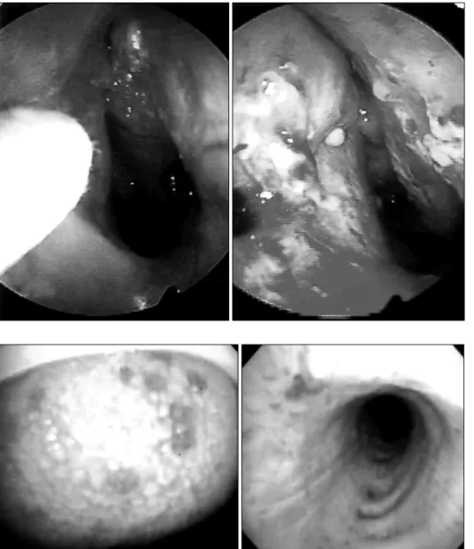
Figure 4. Nasal endos- copies show epistaxis and multiple telangiectasis on both nasal cavities.
Figure 5. Bronchoscopy re- veals multiple telangiectasis on tongue and bronchus.
과거력: 일상 생활 중에 반복적인 코출혈이 있어 6개월 전 타 병원에서 비중격성형술을 시행 받았으나, 이후에도 반복적인 코출혈로 본원 이비인후과 외래에서 수 차례 레 이저 절제(laser ablation)를 시행 받았다(Figure 4). 현기 증으로 신경과 외래에서 시행한 뇌 자기공명영상에서 우 중뇌동맥에 국소 협착만 있을 뿐 뇌농양 및 혈관기형은 없었다.
진찰 소견: 내원 당시 혈압 110/70 mmHg, 맥박수 80회/
분, 호흡수 20회/분, 체온 36.9oC이었고, 의식은 명료하였 다. 청진 시 심음은 규칙적이었으며 심잡음은 없었고 호 흡음도 깨끗하였다. 복부검사에서 이상소견은 없었다.
검사실 소견: 말초혈액검사에서 백혈구 4.170×10⁹/L (호중구 52.4%), 혈색소 101 g/L, 혈소판 367×109/L로 빈혈
소견이 있었다. 생화학 검사에서 총단백 65 g/L, 알부민 40 g/L, BUN 6.1 mmol/L, 크레아티닌 97.2 μmol/L, AST 0.44 μkat/L, ALT 0.46 μkat/L, 총빌리루빈 5.1 μmol/L, 프로트롬빈 시간 13.7초로 정상이었다.
방사선 소견: 가슴 엑스선 사진은 정상이었고 흉부 전 산화단층촬영에서 폐동정맥기형은 없었으나 기관 내에 6 mm 크기의 결절이 관찰되었다.
치료 및 경과: 기관지내시경에서 흉부 전산화단층촬영 에서 보이던 기관 내 결절은 분비물(secretion)로 생각되 었고, 기관 및 기관지에 다발성 모세혈관확장이 관찰되었 다(Figure 5). 퇴원 후 반복적인 코출혈로 국소적인 지혈 치료 중이다.
고 찰
유전출혈모세혈관확장증은 1864년 Sutton에 의해 비출 혈, 피부 모세혈관확장증 및 내부 출혈을 가진 환자로 처 음 소개된 이후 1896년 Rendu, 1901년 Osler, 1907년 Weber에 의해 각각 기술됨으로써 Osler-Weber-Rendu병 으로 명명되고 있다3. 상염색체 우성으로 유전되는 질환 으로 진단을 위해서는 반복적으로 나타나는 자발적 코출 혈, 입술, 구강, 코, 손가락 등의 다발성 모세혈관확장, 소 화기계의 모세혈관확장증 및 폐, 간, 뇌, 척수 등의 동정맥 기형으로 나타나는 내장기관 이상, 그리고 이에 상응하는 가족력으로, 이 4가지 중에서 적어도 3가지가 있으면 Osler-Weber-Renu병으로 진단할 수 있다1,4,5.
유전출혈모세혈관확장증은 염색체 9번의
Endoglin
유 전자와 염색체 12번의ALK-1
(activin receptor-like kin- ase-1) 유전자의 돌연변이와 연관성이 있는 것으로 알려 져 있다6. 1형과 2형, 두 가지 유형으로 나타나는데, 폐동 맥 기형의 빈도가 높은 1형에 비해 2형은 다소 경한 질병 의 경과를 나타낸다6.초기 혈관의 변화는 세정맥의 확장으로 시작되지만, 모 세혈관을 통한 동맥과의 연결은 유지되며, 혈관주위의 임 파구 침윤이 나타난다. 질환이 진행할수록 세정맥의 확장 이 더 두드러져 진피의 동정맥루나 단락과 같은 동정맥기 형을 만들고, 혈관주위의 임파구 침윤과 더불어 혈관벽에 탄성섬유의 소실이 일어난다7.
코출혈은 가장 흔한 증상으로 약 80%의 환자에서 나타 나며8, 10세 전후에 대부분 증상이 시작된다3. 코 점막의 모세혈관확장증의 자발적 출혈에 의해 발생하고, 경구 철 분제로 조절되는 경증에서 지속적인 수혈이 필요한 중증
의 출혈까지 다양한 범위로 나타난다5,8. 코출혈의 확립된 표준 치료는 없으나, 경증의 경우 국소 지혈, 혈관 결찰술, 혈관 색전술, 호르몬제 복용 등을 시행할 수 있으며, 심한 경우 수술적 치료를 고려해 볼 수 있다1. 전기 또는 화학 소작술(cauterization)은 코 점막에 손상을 주기 때문에 권 장되지 않는다1. 본 증례의 환자도 반복적인 코출혈로 수 차례 국소지혈을 하였고 빈혈을 보이고 있었다.
피부의 병변은 2 mm 정도의 황반성 모세혈관확장이 특징적으로 전체 환자의 75%에서 나타난다9. 주로 얼굴, 입술, 코, 혀, 귀, 손, 상체, 발 부분에 잘 생기며, 30대에 많이 발견되고 나이가 들면서 그 크기와 개수가 증가한다8. 특별한 치료가 필요치 않으며, 출혈이 반복되거나 미용상 의 이유로 레이저 절제나 국소 약품을 사용하기도 한다8. 폐동정맥기형은 유전출혈모세혈관확장증 환자의 15∼
33%에서 발견되며1,2, 호흡곤란, 피로, 청색증, 적혈구 증가 증이 나타날 수 있으나, 첫 증상은 보통 30∼40대에 우좌 단락(right to left shunt)에 의한 패혈성 색전이 뇌혈류로 유입되어 뇌경색, 뇌농양에 의한 신경학적 징후로 나타난 다5,10. 첫 번째 증례 환자도 50대에 뇌농양을 진단받고 수 술을 받은 과거력이 있었으나 이 당시 폐동정맥기형은 확 인할 수 없었다. 이처럼 어른에서 원인을 알 수 없는 뇌농 양이나 젊은 나이에 뇌경색이 발생하는 경우 폐동정맥기형 의 가능성을 염두에 두어야 한다11. 폐동정맥기형은 이와 같은 신경학적 합병증을 초래 할 수 있고, 이에 따른 사망 률이 높기 때문에 조기 진단과 치료가 중요시 되고 있다.
폐동정맥기형의 영양동맥(feeding artery)의 직경이 3 mm 이상이고, 다발성인 경우 신경학적 합병증의 발생 위험이 증가하므로 무증상의 경우라도 치료의 적응증이 된다12,13. 폐동정맥기형의 치료는 수술적으로 제거할 수도 있으나 최근에는 혈관색전술이 안전한 방법으로써 많이 시행되고 있다14.
유전출혈모세혈관확장증 환자의 10%에서 뇌동정맥기 형1과 점막의 모세혈관확장증에 의한 위장관 출혈이 있다1. 간의 침범은 10∼30% 정도의 환자에서 발견되며, 간동 맥과 정맥의 동정맥 기형으로 좌우단락(left to right shunt) 이 생성되면 울혈성 심부전의 증상이 나타날 수 있고8, 간 문맥과 간정맥의 단락으로 간성 혼수나 위장관 출혈이 발 생할 수도 있으며, 간동맥과 간문맥의 연결로 문맥고혈압 이나 그에 따른 식도 정맥류도 형성될 수 있다5. 간의 동정 맥기형은 가능한 고식적 치료가 원칙이며 최근 시도되는 혈관 색전술 또한 보고가 제한적이고 치명적인 간 괴사의 위험이 있어 간이식에 대한 고려가 병행 되야 한다15.
본 증례에서 형제를 포함한 3명의 남매가 모두 코출혈 의 과거력이 있었으며, 5대에 걸쳐 코출혈의 가족력이 있 는 전형적인 Osler-Weber-Renu병의 가계였다. 3남매 중 폐동정맥기형이 2명에서 발생하였으며 1명에서는 뇌농양 도 합병되었다. 따라서 반복적인 코출혈이나 모세혈관확 장증이 있는 환자에서는 유전출혈모세혈관확장증을 의심 해 보아야 하며, 자세한 병력 청취를 통한 가족력의 확인 이 진단에 도움이 될 수 있을 것이다. 또한 가족들을 대상 으로 적절한 선별 검사와 치료가 이루어져야 할 것이다.
요 약
저자들은 반복적인 코출혈이 있는 가족에서 폐동정맥기 형이 있는 동생과 점막의 모세혈관확장증을 가진 형을 경 험하고 희귀한 유전질환인 유전출혈모세혈관확장증의 가 계도를 확인하였기에 문헌고찰과 함께 보고하는 바이다.
참 고 문 헌
1. Begbie ME, Wallace GM, Shovlin CL. Hereditary haemor- rhagic telangiectasia (Osler-Weber-Rendu syndrome): a view from the 21st century. Postgrad Med J 2003;79:18-24.
2. Trembath RC, Thomson JR, Machado RD, Morgan NV, Atkinson C, Winship I, et al. Clinical and molecular ge- netic features of pulmonary hypertension in patients with hereditary hemorrhagic telangiectasia. N Engl J Med 2001;345:325-34.
3. Juares AJ, Dell'Aringa AR, Nardi JC, Kobari K, Gradim Moron Rodrigues VL, Perches Filho RM. Rendu-Osler- Weber syndrome: case report and literature review.
Braz J Otorhinolaryngol 2008;74:452-7.
4. Shovlin CL, Guttmacher AE, Buscarini E, Faughnan ME, Hyland RH, Westermann CJ, et al. Diagnostic criteria for hereditary hemorrhagic telangiectasia (Rendu-Osler- Weber syndrome). Am J Med Genet 2000;91:66-7.
5. Haitjema T, Westermann CJ, Overtoom TT, Timmer R, Disch F, Mauser H, et al. Hereditary hemorrhagic te- langiectasia (Osler-Weber-Rendu disease): new insights in pathogenesis, complications, and treatment. Arch
Intern Med 1996;156:714-9.
6. Sabba C, Pasculli G, Cirulli A, Gallitelli M, Virgilio G, Resta F, et al. Hereditary hemorrhagic teleangiectasia (Rendu-Osler-Weber disease). Minerva Cardioangiol 2002;50:221-38.
7. Braverman IM, Keh A, Jacobson BS. Ultrastructure and three-dimensional organization of the telangiectases of hereditary hemorrhagic telangiectasia. J Invest Dermatol 1990;95:422-7.
8. Guttmacher AE, Marchuk DA, White RI Jr. Hereditary hemorrhagic telangiectasia. N Engl J Med 1995;333:
918-24.
9. Plauchu H, de Chadarévian JP, Bideau A, Robert JM.
Age-related clinical profile of hereditary hemorrhagic te- langiectasia in an epidemiologically recruited population.
Am J Med Genet 1989;32:291-7.
10. Kjeldsen AD, Oxhoj H, Andersen PE, Elle B, Jacobsen JP, Vase P. Pulmonary arteriovenous malformations:
screening procedures and pulmonary angiography in patients with hereditary hemorrhagic telangiectasia.
Chest 1999;116:432-9.
11. Tabakow P, Jarmundowicz W, Czapiga B, Czapiga E.
Brain abscess as the first clinical manifestation of multi- ple pulmonary arteriovenous malformations in a patient with hereditary hemorrhagic telangiectasia (Rendu- Osler-Weber disease). Folia Neuropathol 2005;43:41-4.
12. Andersen PE, Kjeldsen AD, Oxhϕj H, Vase P, White RI Jr. Embolotherapy for pulmonary arteriovenous malfor- mations in patients with hereditary hemorrhagic te- langiectasia (Rendu-Osler-Weber syndrome). Acta Radiol 1998;39:723-6.
13. Cottin V, Plauchu H, Bayle JY, Barthelet M, Revel D, Cordier JF. Pulmonary arteriovenous malformations in patients with hereditary hemorrhagic telangiectasia.
Am J Respir Crit Care Med 2004;169:994-1000.
14. Dakeishi M, Shioya T, Wada Y, Shindo T, Otaka K, Manabe M, et al. Genetic epidemiology of hereditary hemorrhagic telangiectasia in a local community in the northern part of Japan. Hum Mutat 2002;19:140-8.
15. Garcia-Tsao G, Korzenik JR, Young L, Henderson KJ, Jain D, Byrd B, et al. Liver disease in patients with he- reditary hemorrhagic telangiectasia. N Engl J Med 2000;
343:931-6.