ISSN 0378-6471 (Print) ISSN 2092-9374 (Online) DOI : 10.3341/jkos.2010.51.1.120
= 증례보고 =
트리암시놀론의 망막색소상피세포 증식과 이동성 효과에 대한 일산화질소의 역할
김재우⋅이재형⋅이승희 대구가톨릭대학교 의과대학 안과학교실
목적: Triamcinolone acetonide (TA)은 망막색소상피세포 증식과 이동성에 미치는 영향과 이에 대한 일산화질소의 역할을 알아보고자 하였다.
대상과 방법: 망막색소상피세포주인 ARPE19 세포에 10 nM, 1 μM, 100 μM의 TA에 4일간 노출시켰으며 세포의 생존은 MTT assay로, 일산화질소의 생성은 Griess assay로 조사하였으며 세포의 활성에 미치는 영향을 알아보기 위해 이동성검사를 실시하였다. 이때 일산 화질소가 미치는 영향을 알아보기 위하여 N-acetyl cysteine에도 동시에 노출시켰다.
결과: TA는 1 μM 이하의 저 농도에서는 유의하게 망막색소상피세포의 증식을 증가시켰으나 고농도에서는 증식을 억제하였다. TA는 농도의 증가에 따라 유의하게 일산화질소의 생성을 억제시켰고 이동성을 저하시켰으며 이러한 효과는 N-acetyl cysteine에 의해 상쇄 되었다.
결론: TA는 농도에 따라 망막색소상피세포의 생존과 세포의 이동성에 영향을 미치며 그 기전으로 일산화질소가 관여할 수 있을 것으 로 생각된다.
<대한안과학회지 2010;51(1):120-125>
■ 접 수 일: 2009년 4월 3일 ■ 심사통과일: 2009년 9월 15일
■ 책 임 저 자: 김 재 우
대구시 남구 대명4동 3056-6 대구가톨릭대학교 의과대학 안과학교실 Tel: 053-650-4728, Fax: 053-627-0133 E-mail: [email protected]
Triamcinolone acetonide (TA)를 유리체강 내에 주입하 거나 결막하주사하여 미만성 당뇨병성 황반부종, 증식성 당 뇨망막증, 증식성 유리체망막증, 만성 포도막염, 망막부종 과 같은 여러 신생혈관성, 증식성 또는 부종성 질환의 치료 에 이용하고 있다.1-7TA는 결정성의 액체로써 항염증작용 과 항증식작용을 가지고 있으며 일반적인 부신피질호르몬 제의 효과가 하루정도 유지되는데 비해 수일간 지속적인 효과를 나타내며 안압상승을 유발하기도 한다.
망막색소상피세포는 망막의 광수용체와 맥락막 모세혈 관 사이에 있는 세포로서 생리적으로 광수용체의 영양공급 과 대사물질의 배출 및 세포탈락물의 탐식 등 시력의 유지 에 중요한 역할을 하며 산소의 소모량이 많아 산소유리기 의 생성도 많은 것으로 알려져 있으며8이러한 망막색소상 피의 산화적 손상은 연령관련성황반변성과도 밀접한 관련 이 있는 것으로 알려져 있다.9
일산화질소(Nitric oxide, NO)는 안구 내에서 다양한 조 직에 영향을 끼치며10생리적인 매개체일 뿐만 아니라 병리 적 역할을 나타내기도 한다.11NO는 세포의 종류에 따라 세
포의 증식 또는 세포고사를 유발할 수 있을 뿐만 아니라, 창상치유에도 영향을 끼치는 것으로 알려져 있으며12,13 세 포의 이동성에도 관여하는 것으로 알려져 있다.14 기존의 부신피질호르몬제와 NO의 상호작용에 관해서는 다양한 연 구가 되어 있으며 TA가 망막색소상피세포의 증식에 미치 는 영향은 알려져 있으나15-19세포의 이동성에 미치는 영 향과 이에 대한 일산화질소의 역할은 아직 자세히 알려져 있지 않다.
본 연구에서는 사람에서 유래하여 수립된 망막색소상피 세포주의 일종인 ARPE1920세포주를 이용하여 TA의 농도 에 따라 망막색소상피세포의 증식과 세포의 이동성에 미치 는 효과를 알아보고 그 기전에 반응성산소종의 일종인 NO 가 관여하는지를 알아보고자 하였다.
대상과 방법
세포배양
수립된 망막색소상피세포주(ARPE-19: ATCC No. CRL -2302)를 항생제(Gibco, Carlsbad, CA, USA)와 10% 우 태아혈청(Hyclone, Thermo Fisher Scientific, Waltham, MA, USA)이 포함된 Dulbecco’s modified Eagle’s medium 배지(DMEM, Gibco, Carlsbad, CA, USA)를 사용하여 5%
Figure 1. Effect of triamcinolone acetonide (TA) on the proliferation of ARPE19 cells exposed for 4 days. TA exhibited biphasic response and N-acetyl cysteine (NAC) increased proliferation (*, **; p<0.05).
Figure 2. Effect of triamcinolone acetonide (TA) on the migration of ARPE19 cells. TA inhibited cellular migration sighnificantly (*; p<0.05) and co-exposure of2 mM N-acetyl cysteine (NAC) abolished TA-induced inhibition of cellular migration (**; p<0.05).
CO2 배양기에서 배양하였다. 세포가 배양접시에 충만해지 면 10% 우태아혈청(Gibco, Carlsbad, CA, USA)을 포함한 배지로 1:4의 비율로 트립신 처리하여 계대 배양하였다.
약물처리
배양된 망막색소상피세포를 트립신 처리하여 24-well plate에 1×10⁴ cell/ml로 분주하여 하루 동안 부착시킨 후 0, 10 nM, 1 μM, 100 μM의 TA (Sigma, USA)에 4일간 노 출시켰으며 이때 NO 합성저해제인 NO 합성저해제인 Nω
-Nitro-L-arginine methyl ester (L-NAME, Sigma, USA) 0.5 nM과 항산화제인 N-acetylcysteine (NAC, Sigma, USA) 200 μM에도 동시에 노출시켜 동일한 실험을 반복하였다.
MTT assay와 Griess assay
TA가 망막색소상피세포의 생존에 미치는 영향을 알아보 기 위해 MTT assay를 시행하였다.21약물 처리하여 정치배 양한 후 4일째에 세포의 배지에 Methylthiazolyl-diphenyl -tetrazolium bromide (MTT, Sigma, USA)를 각 well당
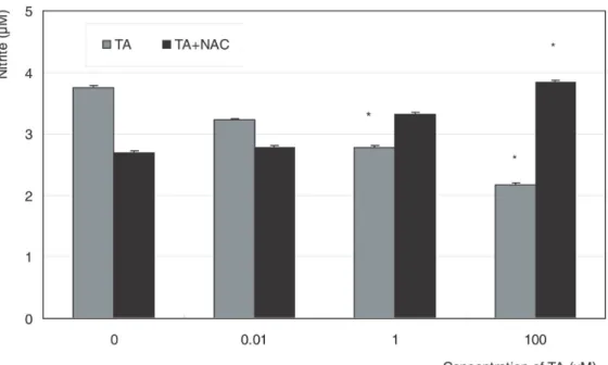
Figure 3. Effect of triamcinolone acetonide (TA) on the production of nitric oxide exposed for 4 days. Co-exposure of 200 μM N-acetyl cysteine (NAC) further abolished TA-induced inhibition of nitric oxide production (*; p<0.05).
100 μl씩 투여한 후 4시간 동안 정치배양한 다음 염류용액 으로 씻어낸 후 dimethylsulfoxide (Sigma, USA)를 각 well 당0.5 ml씩 넣어 10분 이상 흔든 다음 96-well plate에 200 μl씩 옮겨 Spectrophotometer (Sirio-S, Italy)로 570 nM에서 흡광도를 측정하였다. 이때 세포의 증식정도는 실 험군의 값을 약물처리를 하지 않은 대조군의 비로 나누어 백분율로 나타내었다. NO의 생성은 NO의 대사물인 아질산 염의 양을 측정하는 Griess assay를 이용하였는데22 약물 처리한 세포의 배지에 동량의 Griess 반응액(Sigma, USA) 을 섞은 후 96-well plate에 옮겨 Spectrophotometer로 540 nM에서 흡광도를 측정하였다. 이때 표준곡선을 구하 기 위해 sodium nitrite (Sigma, USA)를 단계적으로 희석 하여 사용하였다.
세포 이동성 실험(Migration assay)
TA가 망막색소상피세포의 이동성에 미치는 영향을 알아 보기 위해 wound migration assay를 변형하여 시행하였
다.23,24세포의 부착을 막기 위해 지름 10 mm의 원형 디스
크를 12-well plate의 바닥에 부착시킨 다음 트립신 처리 한 배양된 망막색소상피세포를 12-well plate에 다섯 시간 동안 분주하여 부착시킨 후 원형 디스크를 제거한 다음 신 선한 배지로 교환하고 나서 약물처리를 하여 정치 배양하 였다. 그 다음 이틀째에 눈금이 새겨진 격자판을 이용하여 제거된 원형 디스크 부위 안쪽으로 망막색소상피세포가 자 라 들어온 길이를 각각 네 군데에서 측정하여 평균치를 기
록하였다.
통계적 처리
모든 실험은 3계대에서 5계대 사이의 세포를 이용하였고 3회 이상 반복하여 시행하였다. 모든 실험에서 대조군은 약 물처리를 하지 않은 군으로 하였고 실험군과 대조군의 비 교는 unpaired t-test를 이용하였으며 유의수준은 0.05로 정하였다.
결 과
TA가 망막색소상피세포의 성장에 미치는 영향
TA에 노출한 후 4일째에 1 μM 이하의 농도에서는 약물 처리를 하지 않은 대조군에 비해 유의하게 세포가 증식하 였으나 농도가 증가함에 따라 세포의 생존이 오히려 유의 하게 감소하였다(p<0.05), (Fig. 1). 약물처리를 하지 않은 군과 NAC를 처리한 경우에는 유의한 차이를 보이지 않았 으나(p>0.05) TA에 노출한 경우에는 NAC를 처리한 군에 서는 유의하게 세포의 증식이 나타났다.
TA가 망막색소상피세포의 이동성에 미치는 영향 TA는 약물처리를 하지 않은 대조군에 비해 1 μM의 농도부 터 유의하게 망막색소상피세포의 이동을 억제하였다(p<0.05)
(Fig. 2). 그러나 NAC를 동시에 처치한 경우에는 세포의 이동성이 오히려 유의하게 증가되었다(p<0.05). 따라서 TA는 망막색소상피세포의 이동성을 억제하고 이러한 이동 성의 억제 효과에는 NO가 관여할 가능성이 있음을 알 수 있었다.
TA가 NO의 생성에 미치는 영향
고농도에서는 TA에 의한 세포의 생존 억제효과와 이동 성에 미치는 영향이 항산화제에 의해 상쇄되는 것으로 보 아 고농도의 TA에 의한 세포 증식 억제에 산화스트레스가 관여할 가능성이 있음을 알 수 있었으며 이를 확인하기 위 해 반응성산소종의 일종인 NO의 생성 변화를 조사하였다.
배양 후 이틀째에 약물처리를 하지 않은 군과 비교해서 L-NAME를 처리한 군에서 아질산염은 각각 1.81 μM과 1.11 μM로 유의한 차이를 보여 망막색소상피세포에서 아 질산염이 생성됨을 알 수 있었다(p<0.05). TA에 노출된 후 2일째에 1 μM 이상의 농도에 노출된 군에서 배지의 아 질산염 생성이 농도에 비례하여 약물처리를 하지 않은 대 조군에 비해 유의하게 감소하였으나(p<0.05) TA를 단독 으로 처리한 경우와 NAC를 동시에 처치한 경우에는 두 군 간의 아질산염 생성에 있어서 유의한 차이를 보이지 않았 으나 TA에 노출된 후 4일째에는 100 μM의 농도에서 TA 를 단독으로 처리한 경우와 NAC를 동시에 처치한 경우 두 군 간의 아질산염의 생성에 있어서 유의한 차이를 보였다 (p<0.05), (Fig. 3). 따라서 TA에 노출된 기간이 길어질수 록 배지에서 NO 생성이 더욱 억제됨을 알 수 있었으며 TA 에 의한 NO 생성감소는 항산화제인 NAC에 의해 상쇄됨을 알 수 있었다.
고 찰
TA는 저 농도에서는 배양된 망막색소상피세포의 생존을 촉진하였으나 농도가 증가하면 생존을 억제하는 이중적 반 응을 나타내었으며 농도의 증가에 따라 세포의 이동성을 감소시켰다. 이러한 TA의 증식과 이동성에 관한 효과는 NO의 생성과 관련 있는 것으로 나타났다.
부신피질호르몬제가 세포의 생존과 활성에 다양한 영향 을 끼친다는 것이 이미 잘 알려져 있으며 TA는 저 농도에 서는 세포의 생존이 증가하고 고농도에서는 세포의 생존이 감소하는 것으로 알려져 있는데 세포의 종류에 따라서 증 식을 억제시키는 농도가 각기 다른 것으로 보고되어 있
다.25-27망막색소상피세포주를 이용한 본 실험의 결과는 일
반적으로 부신피질호르몬제재에 노출시켰을 경우 저 농도
에서는 세포의 생존이 증가하고 고농도에서는 생존이 감소 하는 경향과 일치하는 것임을 알 수 있었으며 TA를 망막색 소상피세포에 노출시켰을 때 1 μM의 농도까지는 증식을 촉진하고 그 이상의 농도에서는 증식을 억제하는 기존의 보고와 일치한다.28이러한 결과는 TA의 농도에 따라 망막 에 독성을 나타낼 수 있다는 것을 보여주고 있는데 ARPE19 세포주를 이용한 실험결과에서는 임상적으로 사용된 농도보 다 훨씬 저 농도인 10 μg/ml (0.01 mg/ml)에서 세포독성을 나타낸다고 알려져 있으며26 그 기전으로 산화적 손상이 관 여하며 덱사메타존에 비해 독성이 심한 것으로 알려져 있 다.29따라서 비록 실험실내에서의 결과이지만 이러한 결과 는 임상적으로 TA를 사용할 경우 안압에 미치는 영향뿐만 아니라 망막에 미치는 독성을 고려하여 농도를 적절히 조 절할 필요가 있을 것이다.
NO는 자유유리기로서 안구 내에서도 다양한 조직에서 발현되며30 저 농도에서는 중요한 생리적 조절인자로 작용 하지만 고농도에서는 반응성산소종으로 전환되어 세포에 병적인 손상을 유발하기도 한다.30또한 NO는 세포의 생존 에 영향을 미칠 뿐만 아니라 창상치유과정에도 관여하여 섬유화에 중요한 매개체 역할을 하는 것으로 알려져 있
다.12,13 TA는 망막의 증식성, 신생혈관성 질환에서도 사용
되는데7 그 기전으로 산화스트레스가 관여하는 것으로 알 려져 있다.26,28,32본 연구의 결과에서 TA가 농도에 따라 망 막색소상피세포에서 세포의 생존과 증식에 영향을 줄 뿐만 아니라 세포의 이동성을 저하시켰으며, 이와 함께 NO의 생 성을 감소시킨 것으로 나타났다. 이러한 TA의 작용기전이 산화스트레스에 의한 것임을 고려하여 항산화제의 작용을 생각해보면 NO는 산화스트레스를 유발하는 superoxide 같 은 자유유리기와 결합하여 perox-ynitrite를 형성하여 여 러 가지 손상을 유발하게 되는데, 항산화제를 처치할 경우 자유유리기를 제거함으로써 결과적으로 NO의 생리적인 농 도를 유지하여 TA가 유발하는 증식과 이동성에 대한 효과 를 상쇄시킨 것으로 생각된다.
NO는 망막에서 신생혈관의 생성과 세포이동에 관여하는 것으로 알려져 있으므로33이러한 NO의 생성 억제가 TA에 의한 세포의 증식과 이동성에 영향을 미칠 수 있는 기전으 로 생각해 볼 수 있을 것이며 임상적으로는 망막의 반흔형 성 또는 섬유화를 촉진시킬 수 있을 것이다.
그러나 본 연구는 생체 내 실험이 아니며 향후 TA가 망 막색소상피세포에 미치는 구조적 또는 기능적 변화와 NO 이외의 다양한 반응성산소종의 관련 여부에 대한 보다 상 세한 연구가 향후 필요할 것으로 생각된다. 결론적으로 TA 는 농도에 따라 망막색소상피세포의 생존에 영향을 줄뿐만 아니라 세포의 이동성을 감소시켰으며 그 기전으로 반응성
산소종의 일종인 NO가 관여할 수 있을 것으로 생각된다.
참고문헌
1) Machemer R, Sugita G, Tano Y. Treatment of intraocular pro- liferations with intravitreal steroids. Trans Am Ophthalmic Soc 1979;77:171-80.
2) Penfold P, Gyory J, Hunyor A, Billson FA. Exudative macular degeneration and intravitreal triamcinolone: a pilot study. Aust N Z J Ophthalmol 1995;23:293-8.
3) Jonas JB, Hayler JK, Panda-Jonas S. Intravitreal injection of crystalline cortosine as adjunctive treatment of proliferative retinopathy. Br J Ophthalmol 2000;84:1064-7.
4) Danis RP, Ciulla TA, Pratt LM, Anliker W. Intravitreal triam- cinolone acetonide in exudative age-related macular degeneration.
Retina 2000;20:244-50.
5) Martidis A, Duker JS, Greenberg PB, et al. Intravitreal triamc- inolone for refractory diabetic macular edema. Ophthalmology 2002;109:920-7.
6) Jonas JB, Hayler JK, Söfker A, Panda-Jonas S. Jochen Intravitreal injection of crystalline cortisone as adjunctive treatment of proliferative diabetic retinopathy. Am J Ophthalmol 2001;131:
468-71.
7) Jonas JB, Kressig I, Degenring RF. Intravitreal triamcinolone acetonide for treatment of intraocular proliferative, exudative, and neovascular diseases. Prog Retin Eye Res 2005;24:587-611.
8) Schraermeyer U, Heimann K. Current understanding on the role of retinal pigment epithelium and its pigmentation. Pigment Cell Res 1999;12-9.
9) Proctor PH, Reynolds ES. Free radicals and disease in man.
Physiol Chem Phy Med NMR 1984;16:175-95.
10) Park GS, Kwon NS, Kim YM, Kim JC. The role of nitric oxide in ocular surface diseases. Korean J Ophthalmol 2001;15:59-66.
11) Becquet F, Courtois Y, Goureau O. Nitric oxide in the eye: Multi- faceted roles and diverse outcomes. Surv Ophthalmol 1997;42:
71-82.
12) Schäffer MR, Tantry U, Gross SS, et al. Nitric oxide regulates wound healing. J Surg Res 1996;63:237-40.
13) Schäffer MR, Tantry U, Van Wesep RA, Bardul A. Nitric oxide metabolism in wound. J Surg Res 1997;71:25-31.
14) Noiri E, Lee E, Testa J, et al. Podokinesis in endothelial cell migration: role of nitric oxide. Am J Phsiol 1998;274:C236-44.
15) Brodsky SV, Morrishow AM, Dharia N, et al. Glucose scavenging of nitric oxide. Am J Physiol Renal Physiol 2001;280:F480-6.
16) Johns DG, Dorrance AM, Tramontini NL, Webb RC.
Glucococorticoid inhibit tetrahydrobiopterin-dependent endoth- elial function. Exp Biol Med (Maywood) 2001;226:27-31.
17) Iuchi T, Akaike M, Mitsui T, Ohshima Y, et al. Glucocorticoid excess induces superoxide production in vascular endothelial cells and elicits vascular endothelial dysfunction. Circ Res 2003;92:
81-7.
18) Marinos RS, Zhang W, Wu G, et al. Tetrahydrobiopterin levels regulate endothelial cell proliferation. Am J Physiol Heart Cir Physiol 2001;281:H482-9.
19) Ishii M, Shimizu S, Yamamoto T, et al. Acceleration of oxidative stress-induced endothelial cell death by nitric oxide synthase dysfunction accompanied with decrease in tetrahydrobiopterin content. Life Sci 1997;61:739-47.
20) Dunn KC, Aotaki-Keen AE, Putkey FR, Hjelmeland LM.
ARPE19, a human retinal pigment epithelial cell line with diff- erentiated properties. Exp Eye Res 1996;62:155-69.
21) Mosmann T. Rapid colorimetric assay for cellular growth and survival: Application to proliferation and cytotoxicity assays. J Immunol Methods 1983;65:55-63.
22) Green LC, Wagner DA, Glogoski J, et al. Analysis of nitrate, nitrite and [15N]nitrate in biologic fluids. Anal Biochem 1982;
126:131-8.
23) Kahn AM, Allen JC, Seidel CL, Zhang S. Insulin inhibits migrat- ion of vascular smooth muscle cells with inducible nitric oxide synthase. Hypertension 2000;35:303-6.
24) Kiviluto T, Watanabe S, Hirose M, et al. Nitric oxide donors retard wound healing in cultured rabbit gastric epithelial cell monolayers. Am J Physiol Gastrointest Liver Physiol 2001;281:
G1151-7.
25) Blumenkranz MS, Claflin A, Hajek AS. Selection of therapeutic agents for intraocular proliferative disease. Arch Ophthalmol 1984;102:598-604.
26) Yeung CK, Chan KP, Chiang SW, et al. The toxic and stress res- ponses of cultured human retinal pigment epithelium (ARPE19) and human glial cells (SVG) in the presence of triamcinolone.
Invest Ophthalmol Vis Sci 2003;44:5293-300.
27) Spandau UH, Sauder G, Schubert U, et al. Effect of triamcinolone acetonide on proliferation of retinal endothelial cells in vitro and in vivo. Br J Ophthalmol 2005;89:745-7.
28) Matsuda S, Gomi F, Oshima Y, et al. Vascular endothelial growth factor reduced and connective tissue growth factor induced by triamcinolone in ARPE10 cells under oxidative stress. Invest Ophthalmol Vis Sci 2005;46:1062-8.
29) Chung H, Hwang JJ, Koh JY, et al. Triamcinolone acetonide- mediated oxidative injury in retinal cell culture: Comparison with dexamethasone. Invest Ophthalmol Vis Sci 2007;48:5742-9.
30) Nathanson JA, McKee M. Identification of an extensive system of nitric oxide-producing cells in the ciliary muscle and outflow pa- thway of the human eye. Invest Ophthalmol Vis Sci 1995;36:1765-73.
31) Moncada S, Palmer RMJ, Higgs EA. Nitric oxide: physiology, pa- thophysiology, and pharmacology. Pharmacol Rev 1991;43:109-42.
32) Kim KO, Yoon TJ, Choi GJ. Induction of angiogenic cytokines in cultured RPE by oxidative stress. J Korean Ophthalmol Soc 2004;45:1742-9.
33) Murohara T, Witzenbichler B, Spyridopoulos I, et al. Role of endothelial nitric oxide synthesis in endothelial cell migration.
Arterioscler Thromb Vasc Biol 1999;19:1156-61.
=ABSTRACT=
Role of Nitric Oxide in the Proliferative and Migratory Effect of Triamcinolone in RPE Cells
Jae Woo Kim, MD, PhD, Jae Hyung Lee, MD, Seung Hee Lee, MD
Department of Ophthalmology, Catholic University of Daegu College of Medicine, Daegu, Korea
Purpose: To investigate the role of nitric oxide (NO) on the proliferative and migratory effects of triamcinolone acetonide (TA) in retinal pigment epithelial cells.
Methods: After exposure to 10 nM, 1 μM, or 100 μM TA for four days, with or without co-exposure of antioxidant N-acetylcyteine, the proliferation and nitrite production of ARPE19 cells were assessed with MTT and Griess assays, respectively. Additionally, a cell migration assay was performed.
Results: Cellular survival increased after exposure to TA at low concentration but decreased at high concentration. TA decreased the production of NO and cellular migration significantly, and these effects were abolished by N-acetylcysteine.
Conclusions: TA showed a biphasic response on the proliferation and decreased cellular migration in ARPE19 cells, which may be mediated by nitric oxide.
J Korean Ophthalmol Soc 2010;51(1):120-125
Key Words: ARPE19, Migration, Nitric oxide, Triamcinolone acetonide
Address reprint requests to Jae Woo Kim, MD, PhD
Department of Ophthalmology, Catholic University of Daegu College of Medicine
#3056-6 Daemyeung-4-dong, Nam-gu, Daegu 705-718, Korea Tel: 82-53-650-4728, Fax: 82-53-627-0133, E-mail: [email protected]