DOI : 10.3341/jkos.2007.48.9.1227
망막중심정맥폐쇄는 흔한 망막 혈관 질환 중의 하나 이며, 망막의 사분역 모두에서 망막출혈과 정맥계의 확 장이 보이며 시신경유두와 망막의 부종이 관찰된다. 대 개 심한 시력저하를 보이는데 이는 황반을 덮는 망막출 혈, 허혈, 황반부종 등에 의해 나타난다.1
망막중심정맥폐쇄는 병리학적으로 망막중심정맥의 lamina cribrosa 부위나 동정맥 교차부위의 혈전형 성에 의해 발생한다.2 망막모세혈관의 기능적 및 구조 적 변화가 나타나며, 망막모세혈관 투과도가 증가하여 망막부종이 발생하게 된다. 망막모세혈관의 투과도 증 가와 이로 인한 망막부종은 망막내 혈액-망막장벽 (blood-retinal barrier)의 파괴로 인해 나타나며, 이는 VEGF (vascular endothelial growth factor)
로 매개된다.3 정상 망막에는 VEGF가 없거나 매우 적 게 존재하지만, 저산소 상태가 되면 VEGF의 생성이 증가되며,4 망막중심정맥폐쇄에서도 VEGF 수치가 증 가되어있는 것으로 알려져 있다.5
스테로이드는 이러한 VEGF 유전자의 발현을 감소 시키며, PDGF (platelet-derived growth factor) 와 platelet-activating factor 등과 같은 염증매개 물로부터 VEGF로 유도(induction) 되는 것을 막는 다.6,7 이러한 기전으로 유리체내 triamcinolone acetonide (TA) 주입이 시도되었으며, 이는 장액성 연령관련 황반변성, 당뇨성 황반부종, 심한 포도막염, 포도막염에 의한 낭포황반부종 등에서 효과가 있는 것 으로 알려져 있다.8-11
본 연구에서는 망막중심정맥폐쇄에 의한 황반부종을 보이는 환자에서 유리체내 트리암시놀론 주입술의 임상 적 효과를 허혈성 망막중심정맥폐쇄와 비허혈성 망막정 맥폐쇄로 나누어 비교하여 알아보고자 하였다.
대상과 방법
2004년 1월부터 2004년 10월까지 병원에서 망막중 심정맥폐쇄로 진단받은 70명의 환자를 대상으로 후향 적 의무기록 조사를 시행하였다. 모든 환자에게 스넬렌
망막중심정맥폐쇄환자에서 황반부종에 대한 유리체내 트리암시놀론주입술의 효과
강수연1․최문정1․유수진1․이태곤1․김순현2
건양대학교 김안과병원 안과학교실, 명곡 안연구소1, 누네병원2
목적 : 망막중심정맥폐쇄에 의한 황반부종 환자에서 유리체내 트리암시놀론 주입술의 효과를 알아보고자 하였다.
대상과 방법 : 황반부종을 동반한 망막중심정맥폐쇄 환자 70명 70안에 대해 유리체내 트리암시놀론(4 mg/0.1 cc) 주입군(36명 36안)과 비주입군(34명 34안) 사이의 시력변화, 황반부종의 두께변화를 비교하였다.
결과 : 허혈성 망막중심정맥폐쇄의 주입군과 비주입군 간에 초기와 주입 후 1주, 1, 3, 6개월째 시력은 통계적으로 유
의한 차이를 보이지 않았다(p>0.05). 비허혈성의 경우 초기와 주입 후 1주째 시력은 유의한 차이를 보이지 않았으나 (p>0.05), 1, 3개월째는 유의한 차이를 보였다(p=0.004, 0.001). 그러나 주입 후 6개월째는 두 군 간에 시력차이 를 보이지 않았다(p=0.345). 황반부종은 주입 전보다 유의한 감소를 보였고(p<0.001), 시력과의 상관관계는 허혈성 군에서는 유의하지 않았으나(p=0.510), 비허혈성 군에서는 상관관계를 보였다(p=0.024).
결론 : 유리체내 트리암시놀론 주입술은 망막중심정맥폐쇄로 인한 황반부종을 감소시키며, 비허혈성의 경우 일시적으
로 유의한 시력호전을 가져올 수 있다.
<한안지 48(9):1227-1233, 2007>
<접수일 : 2006년 7월 21일, 심사통과일 : 2007년 5월 29일>
통신저자 : 김 순 현
서울시 강남구 대치동 907-16 누네병원 안과
Tel: 02-2086-7770 Fax: 02-2086-7894 E-mail: [email protected]
* 본 논문의 요지는 2005년 대한안과학회 제93회 춘계학술대회 에서 구연으로 발표되었음.
시력표를 이용한 최대교정시력, 골드만안압계를 이용한 안압검사, 세극등 현미경검사 및 안저검사, 형광안저촬 영, 광간섭단층촬영(Optical coherent tomography 3; Carl Zeiss Ophthalmic Systems Inc, Dublin, Calif)을 시행하였다. 망막출혈과 확장되고 구부러진 망막정맥이 망막 사분역 모두에서 관찰되고 황반부종을 동반한 경우를 그 대상으로 하였다. 이중 형광안저촬영에서 비관류지역이 10 유두직경 면적(DA) 이상인 경우를 허혈성 망막중심정맥폐쇄라 하였고, 미 만인 경우를 비허혈성 망막중심정맥폐쇄라 분류하였다.
연구기간 중 비허혈성에서 허혈성으로 진행한 경우와 진단 당시 홍채나 전방각 신생혈관이 있는 경우는 연구 대상에서 제외하였다.
70명의 전체 환자 중 36명은 유리체내 트리암시놀론 주입술을 시행하였고, 주입술을 원하지 않았던 34명은 시행하지 않았다. 시술방법은 0.5% 프로파라케인 점안 제(proparacaine hydrochloride)를 점안한 후 5%
povidone iodine으로 안검과 결막낭을 소독하였다.
개검기로 눈을 벌리고 각막윤부에서 3.5 mm 떨어진 하비측 부위의 평면부에 triamcinolone acetonide 4 mg (0.1 cc)를 30-gauge 바늘을 사용하여 주사하 였다. 추적관찰 중 황반부종이 재발하여 재주입술을 시 행한 경우는 본 연구대상에서 제외하였다.
주입 후 1주, 이후 매1개월마다 외래경과 관찰하였 고, 주입 후 1주, 1개월, 3개월, 6개월째 시력 및 안압 검사, 세극등현미경검사, 안저검사, 형광안저촬영, 광 간섭단층촬영을 시행하였다. 황반부종의 두께는 광간섭 단층촬영의 axial scan상 중심와에서 내경계막과 망 막색소상피층 사이의 거리를 측정한 망막두께와 정상망 막두께의 차이로 정의하였다. 중심와의정상 망막 두께
는 193 µm를 기준으로 하였다.12
최대교정시력은 logMAR (logarithm of the minimal angle of resolution)시력으로 전환하였으 며 통계적인 분석은 Chi-square, paired t-test, Fisher’s exact test를 사용하였다. 황반부종의 두께 와 시력과의 상관관계는 Pearson’s correlation을 이용하였으며, 통계학적 유의성은 P값 0.05를 기준으 로 평가하였다.
결 과
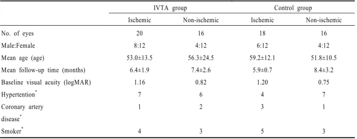
대상 환자 70안 중에서 유리체내 트리암시놀론 주입 술을 시행받은 경우는 36안이었으며, 이중 허혈성 망막 중심정맥폐쇄 환자는 20명, 비허혈성 망막중심정맥폐 쇄 환자는 16명이었다. 남자가 12명(33.3%), 여자가 24명(66.7%)이었다. 평균 나이는 허혈성 군은 53.0
±13.5세, 비허혈성 군은 56.3±24.5세였으며, 경과 관찰 기간은 허혈성 군은 평균 6.4개월, 비허혈성 군은 7.4개월이었다
환자가 원치않았던 경우는 유리체내 트리암시놀론 주입술을 시행하지 않았다. 유리체내 트리암시놀론 주 입술을 시행받지 않은 대조군은 총 34안이었으며, 허혈 성 망막중심정맥폐쇄로 분류된 경우는 18안, 비허혈성 망막중심정맥폐쇄의 경우는 16안이었다. 후향적 연구 이므로 주입군과 성비, 초기 평균시력, 기저질환 등을 고려하여 대조군 선정에 유의하였으며, 이들 중 남자가 10명(29.4%), 여자가 24명(70.6%)이었다. 평균 나 이는 허혈성 군은 59.2±12.1세, 비허혈성 군은 51.8
±10.5세였으며, 경과관찰 기간은 허혈성 군은 평균 5.9개월, 비허혈성군은 8.4개월이었다(Table 1).
Table 1. Baseline characteristics
IVTA group Control group
Ischemic Non-ischemic Ischemic Non-ischemic
No. of eyes Male:Female Mean age (age)
Mean follow-up time (months) Baseline visual acuity (logMAR) Hypertention*
Coronary artery disease*
Smoker*
20 8:12 53.0±13.5
6.4±1.9 1.16
7 1
4
16 4:12 56.3±24.5
7.4±2.6 0.82
6 2
3
18 6:12 59.2±12.1
5.9±0.7 1.20
4 3
5
16 4:12 51.8±10.5
8.4±3.2 0.75
7 1
3
*: No statistically significant with all characteristics.
허혈성 망막중심정맥폐쇄 환자에서 초기 평균시력은 logMAR scale 최대교정시력으로 주입군은 1.16, 대 조군은 1.20이며, 주입 후 1주째, 1, 3, 6개월째 평균 시력은 주입군은 1.15, 1.13, 1.08, 1.08이었으며, 대 조군은 1.22, 1.18, 1.16, 1.15이었다. 주입군과 대조 군 사이에 시술 전 시력은 통계적으로 유의한 차이를 보이지 않았다(p=0.625). 이후 두 군 간에 시술 1주 째, 1, 3, 6개월째 시력은 각각에서 모두 유의한 차이 를 보이지 않았다(p=0.460, 0.532, 0.489, 0.470) (Fig. 1).
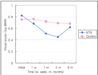
비허혈성 망막중심정맥폐쇄 환자에서는 초기 평균시 력은 주입군에서 0.82, 대조군은 0.75이었으며, 시술 후 1주째, 1, 3, 6개월째 평균시력은 주입군에서 0.68, 0.51, 0.45, 0.62이었으며, 대조군에서는 0.76, 0.72, 0.69, 0.68이었다. 주입군과 대조군 사이에 시술 전 시 력(p=0.221)과 시술 1주째 시력(p=0.188)은 통계 적으로 유의한 차이를 보이지 않았다. 그러나 허혈성 망막중심정맥폐쇄 군에서와 달리, 시술 후 1개월째 (p=0.004)와 3개월째(p=0.001) 대조군에 비해 주 입군의 최종시력이 유의하게 호전되었다. 그러나 시술 후 6개월째(p=0.345) 주입군과 대조군의 시력차이는
통계적으로 유의하지 않았다(Fig. 2).
시술 후 2줄 이상의 최종시력의 호전을 보인 경우는 주입군에서 총 18안(허혈성 5안, 25%, 비허혈성 13 안, 81%)이었고, 대조군에서 16안(허혈성 4안, 22%, 비허혈성 12안, 75%)이었다. 시력변화가 없거나 1줄 이하의 변화를 보인 경우는 주입군에서 총 8안(허혈성 6안, 30%, 비허혈성 2안, 13%)이었으며, 대조군에서 8안(허혈성 5안, 28%, 비허혈성 3안, 19%)이었다.
또한 시력이 2줄 이상 감소한 경우는 주입군에서 총 10 안(허혈성 9안, 45%, 비허혈성 1안, 6%)이었고, 대조 군에서는 10안(허혈성 9안, 50%, 비허혈성 1안, 6%) 이었다. 주입술 시행여부에 따른 허혈성 망막중심정맥 폐쇄군과 비허혈성 망막중심정맥폐쇄군 각각에서 시력 변화 분포는 통계적으로 유의한 차이를 보이지 않았다 (p=0.970, Table 2).
황반부종의 두께는 허혈성 망막중심정맥폐쇄군에서 주입 시술 전 310±43 µm에 비하여 시술 후 1주, 1, 3, 6개월째 각각 209±52 µm, 60±92 µm, 18±59 µm, 43±87 µm로 모두 시술 전보다 통계적으로 유의 하게 감소하였다(p<0.001). 마찬가지로 비허혈성 망막 중심정맥폐쇄군에서 주입시술 전 307±51 µm에 비하 Figure 1. Average visual acuity in ischemic central retinal
vein occlusion group.
0 0.2 0.4 0.6 0.8 1 1.2 1.4
Initial 1 w 1 m 3 m 6 m
Time (w: week. m: months)
Visual acuity (log MAR) .
IVTA Control
Table 2. Improvement of final visual acuity
IVTA group (%) Control group (%)
Ischemic Non-ischemic Ischemic Non-ischemic
Improved* Unchanged Worsen
5/20 (25) 6/20 (30) 9/20 (45)
13/16 (81 )† 2/16 (13)
1/16 (6)
4/18 (22) 5/18 (28) 9/18 (50)
12/16 (75) † 3/16 (19)
1/16 (6)
*: Improved at least two lines with Snellen chart.
†: No statistically significant difference (P>0.05) in two groups by chi-square test.
Figure 2. Average visual acuity in non-ischemic central retinal vein occlusion group.
0 0.2 0.4 0.6 0.8 1
Initial 1 w 1 m 3 m 6 m
Time (w: week. m: months)
Visual acuity (log MAR) .
IVTA Control
여 시술 후 1주, 1, 3, 6개월째 각각 187±69 µm, 23
±88 µm, 4±47 µm, 15±78 µm로 모두 시술 전보다 유의하게 감소하였다(p<0.001, Table 3).
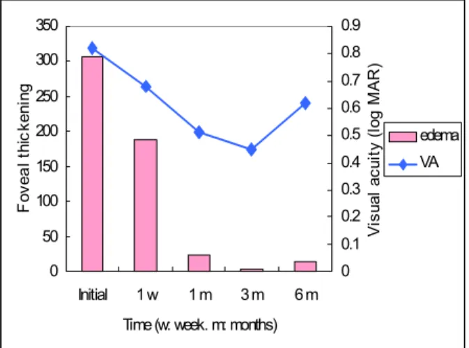
주입군에서 시력호전과 황반두께와의 상관관계는 허 혈성 망막중심정맥폐쇄 군에서 통계적으로 유의한 상관 관계를 보이지 않았으나(p=0.510, Fig. 3), 비허혈 성 망막중심정맥폐 군에서는 통계적으로 유의한 상관관 계를 보였다(p=0.024, Fig. 4). 대조군에서는 추적 검사시 광간섭단층촬영을 시행하지 않았다.
36안의 유리체내 트리암시놀론 주입군 모두에서 치 명적 합병증(망막박리, 백내장, 유리체 출혈, 안내염 등)없이 안전하게 시술이 진행되었으며, 21 mmHg를 초과하는 안압상승은 허혈성과 비허혈성 군에서 각각 1 안에서 있었다. 2안 모두 주입 후 3개월째 안압상승이 나타났으나 약물치료로 안압이 조절되었다.
경과관찰 기간 중 홍채신생혈관이나 신생혈관녹내장
이 발생한 경우는 주입군 중 허혈성 망막중심정맥폐쇄 군에서만 3안(15%)이었고, 대조군에서는 허혈성 망막 중심정맥폐쇄 군 4안(22%), 비허혈성 망막중심정맥폐 쇄 군에서 1안(6%)이었다. 주입군과 대조군 간에 신생 혈관 발생은 통계적으로 유의한 차이를 보이지 않았다 (p=0.472, Table 4).
고 찰
망막중심정맥폐쇄에 의한 황반부종의 치료는 제한적 이다. Central Vein Occlusion Study의 연구에 따 르면 격자레이저치료가 망막중심정맥폐쇄에 의한 황반 부종을 유의하게 감소시키지만 시력의 호전을 가져오지 는 않았다.13 따라서 망막중심정맥폐쇄에 의한 망막 의 순환상태를 개선시키려는 다양한 노력들이 시도되 고 있다.
Table 3. Changes of foveal thickness (µm)
Foveal thickening†: mean±SD (µm)
Ischemic (n=20)* Non-ischemic (n=16)* Initial
1 week 1 month 3 month 6 month
310±43 209±52 60±92 18±59 43±87
307±51 187±69 23±88 4±47 15±78
*: Statistically significant difference from initial value (P<0.05) at all time points by paired t-test.
†: (measured-normal) foveal thickness.
No statistically significant difference (P=0.510) by Pearson’s correlation.
Figure 3. Correlation of macular edema and visual acuity in IVTA group of ischemic central retinal vein occlusion patients.
0 50 100 150 200 250 300 350
Initial 1 w 1 m 3 m 6 m Time (w: week. m: months)
Foveal thickening
1.04 1.06 1.08 1.1 1.12 1.14 1.16 1.18
Visual acuity (log MAR)
edema VA
Statistically significant difference (P=0.024) by Pearson’s correlation.
Figure 4. Correlation of macular edema and visual acuity in IVTA group of non-ischemic central retinal vein occlusion patients.
0 50 100 150 200 250 300 350
Initial 1 w 1 m 3 m 6 m Time (w: week. m: months)
Foveal thickening
0 0.1 0.2 0.3 0.4 0.5 0.6 0.7 0.8 0.9
Visual acuity (log MAR)
edema VA
유리체내 tissue plasminogen activator 주입 술, radial optic neurotomy, 레이저를 이용한 망막 맥락막 측부순환의 생성 등이 이에 속하나, 아직은 보 다 많은 연구가 필요한 실정이다.14-17
그러나 유리체내 트리암시놀론 주입술은 황반부종을 효과적으로 줄여주는 것으로 보고되었고,18-20 비허혈성 망막중심정맥폐쇄의 경우 주입술 후 황반부종의 해부학 적 호전 및 시력의 호전을 보이는 반면, 허혈성 망막중 심정맥폐쇄의 경우는 황반부종의 감소는 나타나나, 시 력의 호전은 유의하지 않은 것으로 알려져 있다.1,21
본 연구에서는 허혈성과 비허혈성 망막중심정맥폐쇄 로 나누어 유리체내 트리암시놀론 주입 후 황반부종과 시력의 변화를 대조군과 비교하여 알아보았다. 기존의 연구에서와 마찬가지로 비허혈성 망막중심정맥폐쇄에 서는 주입술 후 황반부종과 시력의 호전을 보였지만, 허혈성 망막중심정맥폐쇄에서는 황반부종은 유의하게 감소하였지만 시력호전은 유의하지 않았다. 심한 허혈 이 발생하면 망막내층에 비가역적인 손상이 오고 이로 인해 황반부종이 호전되더라도 시력회복이 어려울 수 있다.1 따라서 이러한 이유로 허혈성 망막중심정맥폐쇄 의 경우 트리암시놀론 주입 후 황반부종의 해부학적 호 전에도 불구하고 시력호전이 유의하지 않으리라 생각 된다.
그러나 유리체내 트리암시놀론 주입술은 시간이 지 날수록 그 효과가 감소되어 황반부종이 재발하는 한계 점이 있다.9 본 연구에서는 재발과 이에 따른 재주입술 여부에 관해서는 포함되지 않아서, 이에 대한 추가적인 연구가 필요하리라 생각된다. 또한 최근 당뇨황반부종 환자에서 유리체내 트리암시놀론 주입술 이후 황반부종 이 감소된 시기에 격자레이저치료를 시행하여 황반부종 의 재발을 막는 시도도 보고된 바 있다.22 마찬가지로 망막중심정맥폐쇄에서도 이와 같은 치료를 시행해 볼 수 있으리라 생각된다.
본 연구에서는 경과관찰 기간 동안 홍채신생혈관, 신 생혈관녹내장이 주입군의 3안에서 발생하였고 3안 모 두 허혈성 망막중심정맥폐쇄 환자였다. 대조군에서는 허혈성 망막중심정맥폐쇄 환자 4안, 비허혈성 망막중심 정맥폐쇄 환자 1안에서 발생하였다. 환자수가 적어 통
계적으로 유의하지는 않았으나, 대조군에서 신생혈관 발생률이 높았다. 트리암시놀론은 신생혈관 생성에 관 여하는 VEGF의 발현을 막는 것으로 알려져 있다.1 따 라서 주입군에서 유리체내 트리암시놀론이 VEGF의 발현을 막음으로써 망막과 홍채의 신생혈관을 감소시킨 다고 생각할 수 있다.
유리체내 트리암시놀론 주입 후 발생할 수 있는 합병 증으로는 안압상승, 백내장, 망막박리, 유리체출혈, 안 내염 등이 있으나 그 빈도는 매우 낮은 것으로 알려져
있고,9,10,23 본 연구에서도 안압상승이 2안에서 발생한
것 외에 시력에 치명적 감소를 초래할 만한 심각한 합 병증은 발생하지 않았다. 안압상승이 발생한 2안 모두 약물치료로 안압이 조절되었다.
결론적으로 유리체내 트리암시놀론 주입술은 망막중 심정맥폐쇄로 인한 황반부종의 해부학적 호전을 가져올 수 있는 효과적 치료라 생각된다. 특히 비허혈성 망막 중심정맥폐쇄에서는 일시적으로 시력의 호전도 기대할 수 있어서 환자의 삶의 질을 향상시킬 수 있을 것으로 생각된다.
그러나 본 연구가 적은 환자수와 후향적 연구라는 제 한점이 있어, 차후 많은 환자를 대상으로 긴 추적관찰 을 통해 유리체내 트리암시놀론 주입술의 합병증에 대 한 체계적인 연구, 황반부종의 재발에 따른 재주입술의 결과에 대한 연구 또한 필요하리라 생각된다.
참고문헌
1) Bashshur ZF, Ma’luf RN, Allam S, et al. Intravitreal triamcinolone for the management of macular edema due to nonischemic central retinal vein occlusion. Arch Ophthalmol 2004;122:1137-40.
2) Green WR, Chan CC, Hutchins GM, Terry JM. Central retinal vein occlusion: a prospective histopathologic study of 29 eyes in 28 cases. Trans Am Ophthalmol Soc 1981;79:371-422.
3) Aiello LP, Bursell SE. Clermont A, et al. Vascular endothelial growth factor-induced retinal permeability is mediated by protein kinase C in vivo and suppressed by an orally effective beta-isoform-selective inhibitor. Diabetes 1997;46:1473-80.
4) Vinores SA. Youssri AO, Luna JD, et al. Upregulation of vascular endothelial growth factor in ischemic and Table 4. Incidence of neovascularization
IVTA group (%) Control group (%)
Ischemic Non-ischemic Ischemic Non-ischemic
NVI/NVG† (%)* 3 (15) 0 (0) 4 (22) 1 (6)
*: Statistically not significant difference with IVTA and control group by Fisher’s exact test.
NVI/NVG†: neovascularization on iris/neovascular glaucoma.
non-ischemic human and experimental retinal disease. Histol Histopathol 1997;12:99-109.
5) Pe’er J, Folberg R, Itin A, et al. Vascular endothelial growth factor upregulation in human central retinal vein occlusion.
Ophthalmology 1998;105:412-6.
6) Nauck M, Karakiulakis G, Perruchoud AP, et al.
Corticosteroids inhibit the expression of the vascular endothelial growth factor gene in human vascular smooth muscle cells. Eur J Pharmacol 1998;341:309-15.
7) Nauck M, Roth M, Tamm M, et al. Induction of vascular endothelial growth factor by platelet-activating factor and platelet-derived growth factor is downregulated by corticosteroids. Am J Respir Cell Mol Biol 1997;16:398-406.
8) Jonas JB, Jreissig I, Sofker A, Degenring RF. Intravitreal injection of triamcinolone for diffuse diabetic macular edema.
Arch Ophthalmol 2003;121:57-61.
9) Martidis A, Duker JS, Greenberg PB, et al. Intravitreal triamcinolone for refractory diabetic macular edema.
Ophthalmology 2002;109:920-7.
10) Young S, Larkin G, Braniey M, Lightman S. Safety and efficacy of intravitreal triamcinolone for cystoid macular oedema in uveitis. Clin Experiment Ophthalmol 2001;29:2-6.
11) Benhamou N, Massin P, Haouchine B, et al. Intravitreal triamcinolone for refractory pseudophakic macular edema. Am J Ophthalmol 2003;135:246-9.
12) Jung HJ, Hyun JH, Kim YI, Yun IH. Normal macular thickness measured macular mapping of OCT3. J Korean Ophthalmol Soc 2004;45:962-8.
13) The Central Vein Occlusion Study Group. Evaluation of grid pattern photocoagulation for macular edema in central vein occlusion. The Central Vein Occlusion Study Group M report.
Ophthalmology 1995;102:1425-33.
14) Elman MJ, Raden RZ, Carrigan A. Intravitreal injection of tissue pasminogen activator for central retinal vein occlusion.
Trans Am Ophthalmol Soc 2001;99:219-21.
15) Opremcak EM, Bruce RA, Lomeo M, et al. Radial optic neurotomy for central retinal vein occlusion. Retina 2001;21:
408-15.
16) Weiss JN, Bynoe LA. Injection of tissue plasminogen activator into a branch retinal vein in eyes with central retinal vein occlusion. Ophthalmology 2001;108:2249-57.
17) McAllister IL, Douglas JP, Constable IJ, Yu DY.
Laser-induced chorioretinal venous anastomosis for nonischemic central retinal vein occlusion. Am J Ophthalmol 1998;126:
219-29.
18) Ip MS, Kumar KS. Intravitreous triamcinolone acetonide as treatment for macular edema from central retinal vein occlusion. Arch Ophthalmol 2002;120:1217-9.
19) Greenberg PB, Martidis A, Rogers AH, et al. Intravitreal triamcinolone acetonide for macular oedema due to central retinal vein occlusion. Br J Ophthalmol 2002;86:247-8.
20) Jonas JB, Kreissig I, Degenring RF. Intravitreal triamcinolone acetonide as treatment of macular edema in central retinal vein occlusion. Graefes Arch Clin Experiment Ophthalmol 2002;240:782-3.
21) Ip MS, Gottlieb JL, Kahana A, et al. Intravitreal triamcinolone for the treatment of macular edema associated with central retinal vein occlusion. Arch Ophthalmol 2004;122:1131-6.
22) Kang SW, Sa HS, Cho HY, Kim JI. Macular grid photocoagulation after intravitreal triamcinolone acetonide for diffuse diabetic macular edema. Arch Ophthalmol 2006;124:
653-8.
23) Benz MS, Murray TG, Dubovy SR, et al. Endophthalmitis caused by mycobacterium chelonae abscessus after intravitreal injection of triamcinolone. Arch Ophthalmol 2003;121:271-3.
=ABSTRACT=
The Efficacy of Intravitreal Triamcinolone Acetonide Injection for Macular Edema in Central Retinal Vein Occlusion
Su Yeon Kang, M.D.1, Moon Jeong Choi, M.D.1, Su Jin Yoo, M.D.1, Tae Gon Lee, M.D.1, Soon Hyun Kim, M.D., PhD.2
Myung-Gok Eye Research Institute, Department of Ophthalmology, Konyang University, Kim's Eye Hospital1, Seoul, Korea Nune Eye Hospital2, Seoul, Korea
Purpose: To investigate the effect of intravitreal injection of triamcinolone acetonide (IVTA) for the treatment of macular edema in central retinal vein occlusion (CRVO).
Methods: Seventy patients with CRVO and persistent macular edema were included in this retrospective study. Thirty-six eyes of 36 patients received an intravitreal injection of 4 mg (0.1 cc) triamcinolone and 34 eyes of 34 control patients didnot receive an injection. Any differences in visual acuity and foveal thickness were measured and compared between the two groups. The correlation between the change in foveal thickness and visual acuity was also evaluated.
Results: In ischemic CRVO group, the mean difference of visual acuity between the injection and the control groups was not statistically significant (p<0.05) at the initial measurement and at one week, one, three, and six months after the injection. However, in the non-ischemic CRVO group it was not clinically significant (p<0.05) at the initial time, one week or six months after the injection. But it was clinically significant at one months and three months after receiving the injection (p=0.004 and 0.001). The mean difference in foveal thickness was clinically significant (p<0.001). The visual acuity and foveal thickness were correlated in non-ischemic group but not in the ischemic group.
Conclusions: The IVTA appears to be an effective treatment in patients with macular edema associated with CRVO. The visual acuity of patients with non-ischemic CRVO improved temporally after the IVTA injection, but there was no significant improvement in ischemic CRVO group.
J Korean Ophthalmol Soc 48(9):1227-1233, 2007
Key Words: Central retinal vein occlusion, Intravitreal injection, Triamcinolone acetonide, Macular edema
Address reprint requests to Soon Hyun Kim, M.D., Ph.D.
Nune Eye Hospital
#907-16 Daechi-dong, Gangnam-gu, Seoul 135-841, Korea
Tel: 82-2-2086-7770, Fax: 82-2-2086-7894, E-mail: [email protected]