DOI : 10.3341/jkos.2009.50.8.1190
= 증례보고 = 접수번호 : 50-08-12-24
당뇨황반부종의 치료에서 유리체강내 트리암시놀론과 베바시주맙 주입술의 효과 비교
오세범⋅문준웅⋅김형찬 건국대학교 의학전문대학원 안과학교실
목적: 당뇨황반부종 치료에서 유리체강내 트리암시놀론 주입술과 베바시주맙 주입술의 효과에 대해 알아보고자 하였다.
대상과 방법: 당뇨황반부종의 치료로 유리체강내 트리암시놀론 주입술(Intravitreal Triamcinolone Acetonide, IVTA) 또는 베바시주맙 주입술(Intravitreal Bevacizumab, IVB)을 시행 받은 환자들에 대하여 후향적 의무기록조사를 통하여 시술 전, 시술 후 1주, 1개월, 2개월, 3개월, 6개월에 최대교정시력, 중심황반두께, 총황반부피를 조사하였다. 또한, 안압상승, 백내장 진행 등의 부작용에 대해서도 조사하였다.
결과: 대상안은 총 72명 72안(IVTA군: 40안, IVB군: 32안)이었다. 최대교정시력은 IVTA군에서는 시술 후 1주일부터 3개월까지 유의한 시력호전을 보였고, IVB군에서는 시술 후 1주일부터 2개월까지 유의한 시력호전을 보였다. 중심황반두께와 총황반부피는 IVTA군에서 시술 후 1주일부터 3개월까지 유의하게 감소하였으며, IVB군에서는 시술 후 1주일부터 2개월까지 유의하게 감소하였다. IVTA군에서 IVB군에 비해 1개월부터 3개월까지 유의한 시력호전과 중심황반두께 및 총황반부피의 감소를 보였다. IVTA군에서 경과관찰 중 5안 (12.5%)에서 안압이 25 mmHg 이상으로 상승하였다.
결론: 당뇨황반부종에서 유리체강내 트리암시놀론과 베바시주맙 주입술 모두 단기간 기능적, 해부학적 호전을 보인다. 하지만 트리암 시놀론 치료군에서 더 좋은 치료효과를 보였고, 그 지속기간도 더 길었다.
<대한안과학회지 2009:50(8):1190-1196>
■ 접 수 일: 2008년 12월 31일 ■ 심사통과일: 2009년 5월 12일
■ 책 임 저 자: 김 형 찬
서울특별시 광진구 화양동 4-12번지 건국대학교병원 안과
Tel: 02-2030-5270, Fax: 02-2030-5273 E-mail: [email protected]
* 본 논문의 요지는 2008년 대한안과학회 제99회 추계학술대회에서 구연으로 발표되었음.
* 이 논문은 2008년도 건국대학교 학술진흥연구비 지원에 의한 논문임.
당뇨황반부종은 당뇨망막병증 환자에서 시력저하를 초래 하는 가장 흔한 원인으로 모든 당뇨환자의 10%, 증식당뇨 망막병증 환자의 70%, 당뇨 유병기간이 20년 이상 된 환자 의 29%에서 발생한다.1Early Treatment Diabetic Retino- pathy Study (ETDRS)는 당뇨황반부종 환자에서 국소레이 저 치료가 중등도 시력손상의 위험을 반으로 줄일 수 있고, 특히 유의한 황반부종에서 그 효과가 더 크다고 하였다. 그 러나, 3년 후 치료받은 환자의 12%에서만 중등도 미만의 시력저하가 있었고, 시력이 호전된 경우는 3% 이하에 불과 하였다.2
당뇨황반부종의 정확한 발생기전에 대해서는 아직 알려진 바 없으나, 프로스타글란딘 및 성장인자들의 분비에 의한 투과성 항진, 혈액망막장벽의 파괴, 미세혈관류의 누출, 망막 색소상피의 이상, 유리체망막견인 등이 관여하는 것으로 알 려져 있다.3
유리체강내 트리암시놀론 주입술(Intravitreal Triam- cinolone Acetonide, IVTA)은 당뇨황반부종 발생기전에 관여하는 혈액망막장벽을 안정화시키고,4 프로스타글란딘, 인터루킨 등과 같은 염증매개물질을 억제하고,5,6혈관 투과 성을 증가시키는 혈관내피성장인자를 억제함으로써 황반부 종을 감소시키며 시력을 개선시킨다.7,8 Larsson et al9은 당 뇨황반부종 환자에서 IVTA 후 3개월까지 시력개선 및 황 반두께의 유의한 감소가 있었다고 하였고, Selim et al10 은 IVTA 후 3개월, 6개월 째 각각 68%, 29%에서 2줄 이상의 시력개선이 있었다고 하였다. 당뇨황반부종의 치료로서 IVTA는 안압 상승 등의 여러 부작용 및 당뇨황반부종의 재발 가능성에도 불구하고 그 효과가 인정되어 널리 쓰이고 있 다.11-16
최근에 당뇨황반부종 환자에서 인터루킨(Interleukin-6) 및 혈관내피성장인자(vascular endothelial growth factor, VEGF)의 유리체강내 농도가 증가되어 있다는 사실이 밝혀
졌고,17,18 항혈관내피성장인자인 베바시주맙의 유리체강내
주입술(Intravitreal Bevacizumab, IVB)이 당뇨황반부종의 치료에 효과적이었다는 보고가 있다.19-23
따라서 본 연구에서는 당뇨황반부종의 치료로 쓰이고 있는 IVTA와 IVB 각각의 치료효과 및 합병증에 대해 비교해 보 고자 하였다.
대상과 방법
2007년 8월부터 2008년 2월까지 당뇨황반부종으로 진단 받은 18세 이상의 환자들 중 빛간섭단층촬영(Optical co- herence tomography (OCT), Carl Zeiss Meditec, Dublin, CA, USA)에서 중심황반두께 300 µm 이상, 최대교정시력 (logMAR) 0.4 이하인 환자를 대상으로 IVTA 혹은 IVB를 시행한 후 6개월 이상 경과관찰이 가능하였던 환자 72명, 72안의 의무기록을 후향적으로 조사하였다.
시력저하를 일으킬 수 있는 백내장 혹은 각막혼탁, OCT 에서 유리체망막견인이 있거나, 최근 3개월 이내에 황반부종 치료를 받았거나, 이전에 범안저광응고술 혹은 국소레이저 치료를 받은 경우, 다른 안내 수술을 받은 적이 있는 경우, 녹내장이 있는 경우 등은 제외하였다.
경과관찰 중 OCT에서 망막내 체액의 증가로 시력이 감 소된 경우에는 의사의 판단에 따라 재치료를 시행하였다.
IVB에서 두 번째 치료는 16안에서 평균 12주, 세 번째 치 료는 7 안에서 평균 17주에 시행하였고, IVTA에서 두 번째 치료는 11안에서 평균 17주에 시행하였다.
모든 환자의 연령, 당뇨 유병기간 등을 조사하였으며, 시술 전 최대교정시력 및 안압을 측정하였고, 세극등현미경검사, 안저검사, 형광안저혈관조영술, OCT를 시행하였다.
시술 후 1주, 1개월, 2개월, 3개월, 6개월째 최대교정시 력(logMAR), OCT로 측정한 중심황반두께(central foveal thickness, CMT) 및 총황반부피(total macular volume, TMV)를 조사하였다. 한 줄 미만의 안정된 시력을 보이는 환자 및 한 줄에서 세 줄 사이의 중등도 시력변화를 보이는 환자의 비율을 조사하였다. 또한 안전성을 알아보기 위해 안압 변화, 백내장 진행유무, 염증 유무, 전신 혈전증 유무 등에 대해 알아보았다.
CMT와 TMV는 빛간섭단층촬영(Stratus OCT model 3000, Carl Zeiss, Meditec Inc. USA)을 이용하여 중심와 에서 직경 1 mm 이내의 평균두께인 CMT를 구했고, 황반 두께를 volume data (cubic millimeters)로 변환하여 TMV를 계산하였다.
모든 대상안에 대해 proparacaine hydrochloride 0.5%
(AlcaineⓇAlcon)로 점안 마취 후 10% 베타딘 용액을 이 용하여 안검 소독을 하였고, 결막낭에 5% 베타딘 용액을 점안하였다. 개검기를 사용해 눈을 벌린 후 30 게이지 주사 바늘을 이용하여 triamcinolone acetonide 4.0 mg/0.1ml 또 는 bevacizumab 1.25 mg/0.05ml를 유수정체안에서는 각막 윤부에서 3.5 mm, 무수정체 또는 위수정체안에서는 각막 윤부에서 3.0 mm 떨어진 상이측 또는 하이측 섬모체평면부를 통해 유리체강 내로 주입하였다. 항생제(Ofloxacin, TarividⓇ),
점안 후 도상검안경을 통해 중심망막동맥의 박동을 확인하 였다.
시력은 소수점시력으로 측정한 후 통계분석을 위해 logMAR 시력으로 전환하였으며, 안전수지 50 cm, 안전수지 5 cm, 안 전수동의 시력은 각각 20/2000, 20/20000, 20/40000으로 환산하였다.24 LogMAR 시력에서 0.1 logMAR의 차이는 ETDRS 시력표에서 한 줄의 시력변화를 나타낸다.
통계적인 분석은 SPSS 12.0 (SPSS, Inc., Chicago, IL)를 이용하였다. 두 치료군 간 환자들의 특성 비교는 Mann- Whitney test, 주사 전과 후의 시력, 황반두께 및 황반부피의 변화 중 군내 비교는 Wilcox sign rank test, 군 간 비교는 Mann-Whitney test로 분석하였다. 그리고 유리체강내 주 입술 후 3개월, 6 개월에 한 줄 이내의 안정된 시력변화를 보이는 환자 및 세 줄 이내의 중등도 시력변화를 보이는 환 자의 비율은 chi-square test로 평가하였다. 통계적인 유의 성은 p값 0.05 미만으로 하였다.
결 과
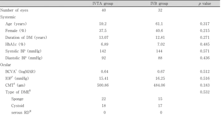
IVTA군은 총 40명, 40안으로 평균연령은 59.2±13.32 세이었고, 남자 25명, 여자 15명이었다. OCT로 분류한 당 뇨황반부종의 형태는 스폰지성 황반부종이 22안, 낭포성 황반부종이 18안이었다. IVB군은 총 32명, 32안으로 평균 연령은 61.1±11.76세이었고, 남자 19명, 여자 13명이었다.
황반부종의 형태는 스폰지성 15안, 낭포성 17안이었다. 두 군 간의 평균연령, 성비, 당뇨유병기간, 최대교정시력, 안 압, CMT, 황반부종의 형태 비교에서 통계적으로 유의한 차 이는 없었다(Table 1).
IVTA군 및 IVB군에서 치료 전 최대교정시력(logMAR±
SD)은 각각 0.64±0.11, 0.67±0.13이었고, CMT는 각각 500.86±62.37 µm, 481.06±54.06 µm, TMV는 각각 10.60±
0.83 mm3, 10.86±0.93 mm3, 그리고 안압은 15.41±0.77 mmHg, 16.25±0.49 mmHg이었다.
최대교정시력은 IVTA군에서 주사 후 1주, 1개월, 2개월, 3개월에 각각 0.55±0.15, 0.49±0.14, 0.48±0.12, 0.50±
0.13으로 치료 전과 비교하여 유의하게 호전되었고(p<0.05), IVB군에서도 주사 후 1주, 1개월, 2개월에 각각 0.58±0.15, 0.54±0.11, 0.55±0.14로 치료 전과 비교하여 유의하게 호 전되었다(p<0.05). 두 치료군 간 비교에서는 IVTA군에서 1, 2, 3개월에 IVB군보다 유의한 시력호전을 보였다(p<0.05, Fig. 1).
CMT는 IVTA군에서 주사 후 1주, 1개월, 2개월, 3개월 에 각각 392.54±66.73 µm, 267.96±58.67 µm, 275.32±
61.08 µm, 335.45±30.45 µm으로 치료 전과 비교하여 유의
Table 1. Baseline characteristics
IVTA group IVB group pvalue
Number of eyes 40 32
Systemic
Age (years) 59.2 61.1 0.317
Female (%) 37.5 40.6 0.215
Duration of DM (years) 13.07 12.81 0.271
HbA1c (%) 6.89 7.02 0.485
Systolic BP (mmHg) 142 144 0.571
Diastolic BP (mmHg) 92 88 0.436
Ocular
BCVA* (logMAR) 0.64 0.67 0.512
IOP† (mmHg) 15.41 16.25 0.516
CMT‡(µm) 500.86 484.06 0.183
Type of DME§ 0.532
Sponge 22 15
Cystoid 18 17
serous RD∏ 0 0
*BCVA=best corrected visual acuity; †IOP=intraocular pressure; ‡CMT=central macular thickness; §DME=diabetic macular edema; ∏RD=retinal detachment.
Figure 2. Change in central macular thickness (CMT, µm) after intravitreal bevacizumab (IVB) and triam- cinolone injections (IVTA) at follow-up visits. *denotes statistically significant change from baseline at each visit within group (p<0.05). **denotes statistically sig- nificant difference between two groups at each follow- up visit (p<0.05).
Figure 1. Change in best corrected visual acuity (BCVA, logMAR) after intravitreal bevacizumab (IVB) and tri- amcinolone injections (IVTA)at follow-up visits. *denotes statistically significant change from baseline at each visit within group (p<0.05). **denotes statistically significant difference between two groups at each follow-up visit (p<0.05).
하게 호전되었고(p<0.05), IVB군에서는 주사 후 1주, 1개월, 2개월에 각각 402.12±61.72 µm, 334.56±59.47 µm, 342.09±64.93 µm으로 치료 전과 비교하여 유의하게 호전 되었다(p<0.05). 두 치료군 간 비교에서는 IVTA군에서 1, 2, 3개월에 IVB군보다 유의한 해부학적 호전을 보였다 (Fig. 2).
TMV는 IVTA군에서 주사 후 1주, 1개월, 2개월, 3개월에
각각 8.52±0.87 mm3, 8.27±0.92 mm3, 8.35±0.89 mm3, 8.97±0.94 mm3으로 치료 전과 비교하여 유의하게 호전되 었고(p<0.05), IVB군에서도 주사 후 1주, 1개월에 각각 9.22±0.89 mm3, 9.17±0.97 mm3으로 치료 전과 비교하여 유의하게 호전되었다(p<0.05). 두 치료군 간 비교에서는 IVTA군에서 1, 2, 3개월에 IVB군보다 유의한 해부학적 호 전을 보였다(Fig. 3).
Figure 3.Change in total macular volume (TMV, mm3) after intravitreal bevacizumab (IVB) and triamcinolone injections (IVTA) at follow-up visits. *denotes sta- tistically significant change from baseline at each visit within-group (p<0.05). **denotes statistically signi- ficant difference between two groups at each follow-up visit (p<0.05).
Figure 4. The proportion of patients losing (-3 lines
~-1 line), maintaining (-1 line~+1 line), or gaining (+1~+3) best corrected visual acuity (BCVA) at 3 months. *indicates a statistically significant difference between the two groups.
Figure 5. The proportion of patients losing (-3 lines
~-1 line), maintaining (-1 line~+1 line), or gaining (+1~+3) best corrected visual acuity (BCVA) at 6 months. There was no statistically significant difference between the two groups.
Figure 6.Change in intraocular pressure (IOP, mm Hg) after intravitreal bevacizumab (IVB) and triamcinolone injections (IVTA) at follow-up visits. *denotes statis- tically significant change from baseline at each visit within group (p<0.05). **denotes statistically significant difference between the two groups at each visit (p<0.05).
시술 후 3개월에 한 줄 이내의 안정된 시력변화를 보인 경우는 IVTA군과 IVB군에서 각각 50%, 66%로서 차이가 없었으나, 한 줄에서 세 줄 사이의 중등도 미만의 시력저하를 보이는 환자의 비율은 각각 3%, 17%로서 IVB군에서 많았다.
중등도 미만의 시력상승을 보인 경우는 각각 47%, 17%로서 IVTA군에서 유의하게 더 많았다. 그러나 시술 후 6개월에 두 치료군 사이에는 유의한 차이가 없었다(Fig. 4, 5).
안압은 IVTA군에서 시술 후 1, 2개월에 17.35±0.95 mmHg, 17.87±0.60 mmHg로 치료 전에 비해 유의하게 상승 되었고(p<0.05), IVB군에서는 시술 후 치료 전과 비교해 유의한 차이를 보이지 않았다(Fig. 6). IVTA군에서 경과관찰 기간 중 5안(12.5%)에서 안압이 25 mmHg 이상으로 상승
되었으나, 점안 안압하강제를 사용한 후 안압은 잘 조절되 었다. IVTA군에서 2안(5%)에서 백내장이 진행하였으나, IVB군에서는 백내장 진행이 없었다. 그리고 안구내 염증 및 전신적 혈전증은 두 군 모두에서 발생하지 않았다.
고 찰
당뇨황반부종의 발생기전은 아직 확실하게 밝혀져 있지 않지만 프로스타글란딘, 인터루킨 등과 같은 염증매개물질 에 의한 투과성 항진이 중요한 역할을 하는 것으로 알려져 있으며, 이미 여러 연구에서 이러한 염증반응을 억제할 수 있는 스테로이드인 트리암시놀론을 유리체강내 주사하여 황반부종을 감소시키고 시력을 향상시킬 수 있었다.4-8 그
러나, 유리체강내 트리암시놀론 주입술은 대부분의 경우 황 반부종이 재발하며, 일부 환자들에서는 안압상승, 백내장의 진행 등이 발생하였다.
VEGF는 혈관생성인자로서 내피세포를 증식시키고 혈관 투과성을 항진시킨다.25,26당뇨황반부종 환자의 유리체강내 VEGF 농도가 증가되어 있고, 증가된 VEGF 농도는 황반부종 의 심한 정도와 밀접하다고 알려져 있으며, 항 VEGF 항체인 bevacizumab 유리체강내 주입술은 당뇨황반부종의 치료로 쓰이고 있다.7,8그러나 IVB의 지속효과는 길지 않아서 Shimura et al27은 당뇨황반부종 환자에서 IVB 시술 후 유의한 시력 호전을 보이나 3개월 이후에는 치료 전 시력으로 돌아간다 고 하였다. 본 연구에서는 시술 후 한 줄에서 세 줄 사이의 중등도 미만의 시력호전을 보인 환자의 비율은 3개월째 IVTA군에서 47%로 IVB군에서 보다 유의하게 높았으나, 시술 후 6개월째에는 IVTA군에서17%, IVB군 8%로 시술 후 3개월에 비해 낮은 비율을 보였으며, 두 치료군 사이에 는 유의한 차이가 없었다. 이는 IVTA 혹은 IVB 모두 시술 3개월 이후에는 그 효과가 점점 감소한다는 기존의 연구결 과와 일치한다.9
Arevallo et al28은 당뇨황반부종 환자에서 IVB 시술 후 필요하면 재치료를 하였고 6개월 경과관찰 중 55%에서 2줄 이상의 시력호전을 보였다고 하였는데, 본 연구에서는 시술 후 6개월에 대부분 안정된 시력을 보였으나 시력호전을 보인 환자의 비율은 이전 연구 결과와 비교해 낮았다. 그 원인으로 생각할 수 있는 것은 본 연구에서 시술 전 중심망막두께가 500 µm 정도의 심한 황반부종 환자가 상대적으로 더 많았고, 필요한 정도보다 주사 횟수가 상대적으로 적었기 때문일 것 으로 생각된다.
그러나 본 연구와 IVTA 및 IVB로 2개월 동안의 단기간 치료효과를 보고한 IBEME study29와 Shimura et al27의 연 구들과 비교했을 때, 세 연구 모두에서 비슷한 시력호전 양상을 보였으며, 세 연구 모두에서 IVTA가 IVB보다 유의 한 시력호전을 보였다.
다른 연구에서와 같이 본 연구에서도 IVTA가 IVB보다 더 좋은 효과를 보였는데 그 이유로 생각할 수 있는 것은 다음과 같다. 당뇨황반부종의 발생기전은 크게 두 가지로 나누어 생각할 수 있는데, 첫째는 망막혈관의 투과성 항진 이며, 둘째는 혈관으로부터 조직 내로의 수분 유입(water flux)의 증가이다.30 VEGF는 혈관의 투과성을 항진시키는 작용을 하는 것으로 알려져 있으나 조직 내로의 수분 유입을 증가시킨다는 가설은 아직 입증된 바 없다. 이에 비해 스테 로이드인 트리암시놀론은 VEGF의 표현(expression)을 감 소시켜서 세포외 공간에 수분이 축적되는 것을 예방하며, 또한 스테로이드 특유의 항염증작용에 의해 부종을 감소시
킨다.5,6 이러한 관점에서 보면, IVB는 유리체강 내에 존재 하는 자유로운 VEGF의 양을 감소시키는 작용만 있는데 비해 트리암시놀론은 여러 가지 작용기전을 가진 다목적 약제라 고 할 수 있으며, 이러한 여러 가지 작용에 의해 IVTA가 IVB보다 더 좋은 효과를 보였다고 추정할 수 있다.
IVTA의 가장 흔한 합병증은 안압상승이며, 그 외에도 백 내장의 진행, 유리체출혈, 망막박리, 안내염 등과 같은 합병 증이 올 수 있다.31-34본 연구에서는 IVTA 후 시력에 유의 한 영향을 미칠 부작용이 발생한 예는 없었으나, 40안 중 5안에서 일시적인 안압상승이 관찰되었으며 점안 안압하강 제를 통하여 잘 조절되었다. IVB 후 백내장의 진행, 포도막염, 급성 망막색소상피파열, 유리체출혈, 안내염, 뇌혈관에 대 한 부작용 등과 같은 합병증에 대한 보고가 있었으나,35-37 본 연구에서는 어떠한 합병증도 없었다.
이번 연구의 제한점으로 후향적 연구이고, 재치료를 한 환자가 포함되어 있으며, 경과관찰 기간이 짧았다는 점을 들 수 있다. 시술 3개월 이후에 당뇨황반부종이 재발하여, CMT가 증가하거나 시력저하를 호소하는 환자들에게 재치 료를 권유하지만, 환자의 순응도 등으로 인하여 실제 주사 시행 여부는 이에 미치지 못하는 경우가 일부 있었다. 또한, 재치료를 시행한 증례들이 포함되었기 때문에 두 가지 약 제의 단일 주입술 효과를 비교할 수 없었다.
결론적으로, 당뇨황반부종에서 유리체강내 트리암시놀론 혹은 베바시주맙 주입술 모두 단기간 기능적, 해부학적 호 전을 보였지만 트리암시놀론 치료군에서 그 효과가 상대적 으로 현저하게 나타나며 지속기간도 길었다. 하지만 시술 후 3개월부터 6개월까지 두 치료군 모두 효과가 점점 감소 하는 양상을 보였다. 그리고 베바시주맙 주입술은 트리암시 놀론 주입술보다 안압이 더 안정적이었다.
앞으로 당뇨황반부종 환자에서 유리체강내 주입술의 장 기적인 치료 효과 및 예후인자를 알아보기 위한 전향적인 연구가 필요할 것으로 사료된다.
참고문헌
1) Klein R, Klein BE, Moss SE, et al. The Wisconsin Epidemiologic Study of Diabetic Retinopathy. IV. Diabetic macular edema.
Ophthalmology 1984;91:1464-74.
2) Early Treatment Diabetic Retinopathy Study Research Group.
Photocoagulation for diabetic macular edema. Early Treatment Diabetic Retinopathy Study report number 1. Arch Ophthalmol 1985;103:1796-806.
3) Ryan SJ. Nonproliferative diabetic retinopathy. In: Chew EY, Ferris FL III, eds. Retina, 4th ed. New York: Mosby, Elsevier Inc., 2006; v. 2. chap. 67.
4) Wilson C, Berkowitz BA, Sato Y, et al. Treatment with intra- vitreal steroid reduces Blood-retinal barrier breakdown due to
retinal photocoagulation. Arch Ophthalmol 1992;110:1155-9.
5) Funatsu H, Yamashita H, Noma H, et al. Increased levels of vascular endothelial Growth factor and interleukin-6 in the aqueous humor of diabetics with macular edema. Am J Ophthalmol 2002;
133:70-7.
6) Antcliff RJ, Marshall J. The pathogenesis of edema in diabetic maculopathy. Semin Ophthalmol 1999;14:223-32.
7) Fischer S, Renz D, Schaper W, Karliczek GF. In vitro effects of dexamethasone on hypoxia- induced hyperpermeability and expression of vascular endothelial growth factor. Eur J Phamacol 2001;411:231-43.
8) Nauck M, Karakiulakis G, Perruchoud AP, et al. Corticosteroids inhibit the expression of vascular endothelial growth factor gene in human vascular smooth muscle cells. Eur J Phamacol 1998;341:
309-15.
9) Larsson J, Zhu M, Sutter F, et al. Relation between reduction of foveal thickness and visual acuity in diabetic macular edema treated with intravitreal triamcinolone. Am J Ophthalmol 2005;
139:802-6.
10) Selim Kocabora M, Kucuksahin H, Gulkilik G, et al. Treatment of diabetic macular edema with intravitreal triamcinolone acetonide injection: functional and anatomical outcomes. J Fr Ophthalmol 2007;30:32-8.
11) Martidis A, Duker JS, Greenberg PB, et al. Intravitreal triam- cinolone for refractory diabetic macular edema. Ophthalmology 2002;109:920-7.
12) Tano Y, Chandler D, Machemer R. Treatment of intraocular proliferation with intravitreal injection of triamcinolone acetonide.
Am J Ophthalmol 1980;90:810-6.
13) Jonas JB, Kreissig I, Sofker A, et al. Intravitreal injection of triam- cinolone for diffuse diabetic macular edema. Arch Ophthalmol 2003;121:57-61.
14) Sutter FK, Simpson JM, Grilles MC. Intravitreal triamcinolone for diabetic edema that persists after laser treatment: three month efficacy and safety results of a prospective, randomized, double- masked, placebo-controlled clinical trial. Ophthalmology 2004;
111:2044-9.
15) Massin P, Andren F, Haouchine B, et al. Intravitreal triamcino- lone acetonide for diabetic macular edema-Preliminary results of a prospective controlled trial. Ophthalmology 2004;111:218-24.
16) Verma LK, Vivek MB, Kumar A, et al. A prospective controlled trial to evaluate the adjunctive role of posterior subtenon triam- cinolone in the treatment of diffuse diabetic macular edema. J Ocular Pharmacol Ther 2004;20:277-84.
17) Funatsu H, Yamashita H, Ikeda T, et al. Vitreous levels of interleukin-6 and vascular endothelial growth factor are related to diabetic macular edema. Ophthalmology 2003;110:1690-6.
18) Funstsu H, Yamashita H, Sakata K, et al. Vitreous levels of vascular endothelial growth factor and intracellular adhesion molecule 1 are related to diabetic macular edema. Ophthalmology 2005;112:806- 16.
19) Boulton M, Foreman D, Williams G, et al. VEGF localization in diabetic retinopathy. Br J Ophthalmol 1998;82:561-8.
20) Adamis AP, Miller JW, Bernal MT, et al. Increase vascular endo- thelial growth factor level in the vitreous of eyes with proliferative diabetic retinopathy. Am J Ophthalmol 1994;118:445-50.
21) Macugen Diabetic Retinopathy Study Group. A phase II rando- mized double-marked trial of pegaptanib, an anti-vascular endo- thelial growth factor aptamer, for diabetic macularedema. Oph- thalmology 2005;112:1747-57.
22) Rosenfeld PJ, Fung AE, Puliafito CA. Optical coherence tomo- graphy findings after an intravitreal injection of bevacizumab (Avastin) for macular edema from central retinal vein occlusion.
Ophthalmic Surg Lasers Imaging 2005;36:336-9.
23) Haritoglou C, Kook D, Neubauer A, et al. Intravitreal bevacizu- mab (Avastin) therapy for persistent diffuse diabetic macular edema. Retina 2006;26:999-1005.
24) Hijikata K, Masuda K. Visual prognosis in Behet’s disease: effects of cyclophosphamide and colchicines. Jpn J Ophthalmol 1978;
22:506-19.
25) Strom C, Sander B, Klemp K, et al. Effect of ruboxistaurin on blood-retinal barrier permeability in relation to severity of leakage in diabetic macular edema. Invest Ophthalmol Vis Sci 2005;46:
3855-8.
26) Ferrara N. Vascular endothelial growth factor: basic science and clinical progress. Endocr Rev 2004;25:581-611.
27) Shimura M, Nakazawa T, Yasuda K, et al. Comparative therapy evaluation of intravitreal bevacizumab and triamcinolone acetonide on persistent diffuse diabetic macular edema. Am J Ophthalmol 2008;145:854-61.
28) Arevallo JF, Fromow-Guerra J, Quiroz-Mercado H, et al. Primary bevacizumab (Avastin) for diabetic macular edema: results from the Pan-American Collaborative Retina Study Group at 6-month flollow-up. Ophthalmology 2007;114:743-50.
29) Paccola L, Costa RA, Folgosa MS, et al. Intravitreal triamcinolone versus bevacizumab for treatment of refractory diabetic macular edema (IBEME study). Br J Ophthalmol 2008;92:76-80.
30) Stefansson E. Ocular oxygenation and the treatment of diabetic retinopathy. Surv Ophthalmol 2006;51:364-80.
31) McCuen B II, Bessler M, Tano Y, et al. The lack of toxicity of intravitreally administered triamcinolone acetonide. Am J Ophthalmol 1981:91:785-8.
32) Moshfeghi D, Kaiser P, Scot I, et al. Acute endophthalmitis following Intravitreal triamcinolone acetonide injection. Am J Ophthalmol 2003;136:791-6.
33) Nelson M, Tennant M, Sivalingam A, et al. Infectious and pre- sumed noninfectious endophthalmitis after intravitreal triamcino- lone acetonide injection. Retina 2003;23:686-91.
34) Roth D, Chieh J, Spirn M, et al. Noninfectious endophthalmitis associated with intravitreal triamcinolone injection. Arch Oph- thalmol 2003;121:1279-82.
35) Jonas JB, Spandau UH, Schlichtenbrede F. Short-term compli- cations of intravitreal injections of triamcinolone and bevacizumab.
Eye 2008;22:590-1.
36) Pieramici DJ, Avery RL, Castellarin AA, et al. Case of anterior uveitis after intravitreal injection of bevacizumab. Retina 2006;
26:841-2.
37) Meyer CH, Mennel S, Schmidt JC, Kroll P. Acute retinal pigment epithelial tear following intravitreal bevacizumab (Avastin) injection for occult choroidal neovascularization seconday to age related macular degeneration. Br J Ophthalmol 2006;90:1207-8.
=ABSTRACT=
Comparison of Effects of Intravitreal Triamcinolone and Bevacizumab in the Treatment of Diabetic Macular Edema
Se Beum Oh, MD, Jun Woong Moon, MD, Hyung Chan Kim, MD, PhD
Department of Ophthalmology, College of Medicine, Konkuk University, Seoul, Korea
Purpose: To compare the effect of an intravitreal injection of triamcinolone acetonide with bevacizumab in the treatment of diabetic macular edema (DME).
Methods: For this study, the medical records of patients with diabetic macular edema, who received intravitreal triamcinolone injection (IVTA) or intravitreal bevacizumab injection (IVB), were reviewed. Best corrected visual acuity (BCVA), central macular thickness (CMT) and total macular volume (TMV) were evaluated before injection and at 1 week, 1 month, 2 months, 3 months, and 6 months after injection. The adverse events, such as increased intraocular pressure, and progression of cataract, were also reviewed.
Results: A total of 72 eyes from 72 patients, (IVTA: 40 eyes, IVB: 32 eyes) were included in this study. In the IVTA group, BCVA improved significantly at 1 week after injection and was maintained until 3 months after injection. In the IVB group, BCVA improved significantly at 1 week after injection and was maintained until 2 months after injection. In the IVTA group, CMT and TMV decreased significantly at 1 week after injection and were maintained until 3 months after injection, while in the IVB group CMT and TMV were maintained until 2 months after injection. The IVTA group showed significantly better results in visual improvement, CMT and TMV reduction compared to the results of the IVB group, from 1 month to 3 months after injection. In the IVTA group, intraocular pressure increased to more than 25 mmHg in 12.5% of patients during the follow-up period.
Conclusions: While the functional and anatomical improvements are achieved by both IVTA and IVB for diabetic macular edema, the effect of IVTA is more prominent with longer duration than IVB.
J Korean Ophthalmol Soc 2009;50(8):1190-1196
Key Words: Diabetic macular edema, Intravitreal bevacizumab, Intravitreal triamcinolone acetonide
Address reprint requests to Hyung Chan Kim, MD
Department of Ophthalmology, Konkuk University Medical Center, Konkuk University School of Medicine
#4-12 Hwayang-dong, Gwangjin-gu, Seoul 143-729, Korea
Tel: 82-2-2030-5270, Fax: 82-2-2030-5273, E-mail: [email protected]