ISSN 0378-6471 (Print) ISSN 2092-9374 (Online) DOI : 10.3341/jkos.2010.51.1.35
= 증례보고 =
신생혈관성 나이관련황반변성에서 베바시주맙 주입과 광역학요법을 병합한 일차치료 효과
유영주1⋅박혜진1⋅이태곤2⋅이동원1⋅조성원1⋅이재흥1
건양대학교 김안과병원 안과학교실, 명곡 안연구소1, 건양대학교 의과대학 안과학교실2
목적: 나이관련황반변성에 의한 맥락막신생혈관에서 bevacizumab 단독치료와 광역학요법과의 병합치료 효과를 알아보고자 하였다.
대상과 방법: 72명 72안을 대상으로 27안에서는 bevacizumab만을 주사하였고 45안에서는 광역학요법을 병행하여 6개월 뒤에 최대교 정시력, 중심황반두께와 재 치료 여부를 비교하였다.
결과: 단독치료군의 경우 평균시력(LogMAR) 0.62±0.34에서 0.56±0.33 (p=0.03)으로, 병합치료군은 0.61±0.33에서 0.48±0.21 (p=0.001) 로 유의한 향상을 보였으며, 치료 6개월 뒤 병합치료군에서 더 시력이 향상되었다(p=0.049). 중심황반두께는 모두 시술 전에 비해 유의하게 감소하였으나(p=0.001), 두 군 간의 차이는 없었다. 재 치료는 단독치료군이 10안(37.0%)으로, 병합치료군 12안(26.7%)보다 유의하게 높은 비율로 나타났다(p=0.02).
결론: 나이관련황반변성에 의한 맥락막신생혈관환자에서 bevacizumab 단독치료 및 광역학요법과의 병합치료 모두 시술 후 6개월까 지 중심황반두께를 감소시키고 시력이 호전되었으며, 병합치료의 경우 최대교정시력과 재 치료율에서 더 좋은 결과를 보여주었다.
<대한안과학회지 2010;51(1):35-41>
■ 접 수 일: 2009년 4월 29일 ■ 심사통과일: 2009년 10월 13일
■ 책 임 저 자: 이 태 곤
대전시 서구 가수원동 685 건양대학교병원 안과
Tel: 042-600-9258, Fax: 042-600-9176 E-mail: [email protected]
* 본 논문의 요지는 2008년 대한안과학회 제99회 춘계학술대회에서 구연 으로 발표 되었음.
* 본 논문의 요지는 2008년 제8회 European Vitreoretinal Surgery congress 에서 구연으로 발표되었음.
맥락막신생혈관은 실명의 흔한 원인이며 그 중 90% 이 상이 나이관련황반변성으로 인해 생기게 되고 그 유병율도 증가하는 추세이다.1치료 방법으로는 레이저광응고술을 비 롯하여 광감작물질인 verteporfin을 이용한 광역학요법과 스테로이드 유리체강 내 주사, 항혈관내피증식인자의 유리 체강내 주사 등이 있다.2-4
현재까지 널리 이용된 광역학요법은 광감작물질과 689 nm 파장의 광선을 이용하여 신생혈관의 응고와 폐쇄 효과 를 가져 올 수 있어 중등도 이상의 시력 손실을 막는데 효 과적이나 영구적 반흔의 형성과 이로 인한 시력저하의 가 능성이 있다.5
여러 연구 결과 혈관내피증식인자는 나이관련황반변성 에서 맥락막혈관신생의 발생에 중요한 역할을 하는 것으로 알려져 있으며, 최근 개발되는 여러 종류의 혈관내피증식인 자의 항체들은 맥락막혈관신생의 진행을 억제할 것으로 기
대되고 있다.6-8
최근 이 두 가지 치료 효과의 상승 작용을 기대하여 두 치료를 병합하여 치료를 시행하고 있는데, 병합치료의 장점 으로는 항혈관내피증식인자의 주입으로 광역학요법 후 일 시적으로 증가한 혈관내피증식인자를 억제할 수 있으며, 광 역학요법의 효과로 안내 주사의 횟수를 줄이며, 좀 더 영구 적인 효과를 가져올 것으로 기대할 수 있으나, 단점으로는 광역학요법으로 인한 망막과 모세혈관의 손상 및 반흔 형 성의 문제가 있다.5,9
이에 본 연구에서는 bevacizumab (Avastin®, Genetech, San Francisco, CA, USA)을 이용한 항혈관내피증식인자 의 단독치료와 광역학요법과의 병합치료의 효과를 비교하 고 최대교정시력의 변화, 재치료율 및 예후인자를 조사하여 좀 더 효과적이고 안전한 치료 방법을 알아보고자 하였다.
대상과 방법
2007년 1월부터 2008년 3월까지 본원에서 나이관련황 반변성과 동반된 중심와밑 맥락막신생혈관으로 진단 받아 verteporfin을 이용한 광역학요법 및 유리체강내 bevaci- zumab 주입술을 받은 환자 중에서 치료 종료 후 6개월 뒤 경과 관찰이 가능하였던 72명 72안을 대상으로 후향적으로 조사 하였다. 이 중 광역학요법과 유리체강내 bevacizumab 주입술을 동시에 받은 병합치료군 45안이었으며, 유리체강내
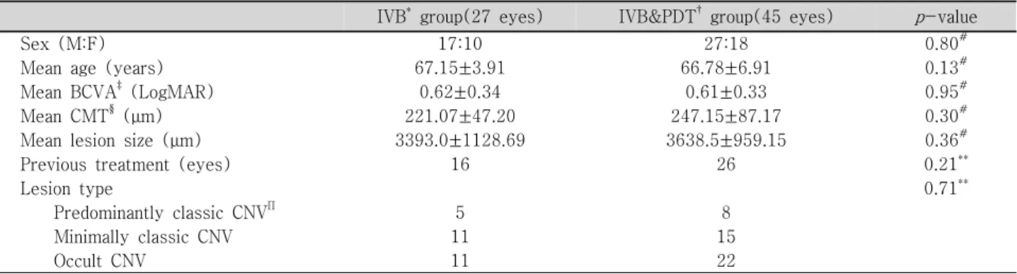
Table 1. Baseline demographics for patients
IVB* group(27 eyes) IVB&PDT† group(45 eyes) p-value
Sex (M:F) 17:10 27:18 0.80#
Mean age (years) 67.15±3.91 66.78±6.91 0.13#
Mean BCVA‡(LogMAR) 0.62±0.34 0.61±0.33 0.95#
Mean CMT§ (μm) 221.07±47.20 247.15±87.17 0.30#
Mean lesion size (μm) 3393.0±1128.69 3638.5±959.15 0.36#
Previous treatment (eyes) 16 26 0.21**
Lesion type 0.71**
Predominantly classic CNVΠ 5 8
Minimally classic CNV 11 15
Occult CNV 11 22
*IVB=Intravitreal bevacizumab; †PDT=Photodynamic therapy; ‡BCVA=Best corrected visual acuity; §CMT=Central macular thickness; ΠCNV=Choroidal neovascularization; #p-value by independent t-test; **p-value by Chi-square test.
Table 2. Changes in best corrected visual acuity
IVB* group(27 eyes) IVB & PDT† group(45 eyes) p-value
Pretreatment BCVA‡(LogMAR) 0.62±0.34 0.61±0.33 0.95§
BCVA at 6 months (LogMAR) 0.56±0.33 0.48±0.21 0.049§
p-value 0.03Π 0.001Π
*IVB=Intravitreal bevacizumab; †PDT=Photodynamic therapy; ‡BCVA=Best corrected visual acuity. §p-value by independent t-test; Πp-value by paired t-test.
bevacizumab 주입술만 받은 단독치료군 27안이었다.
50세 이상의 나이관련황반변성으로 인한 중심와밑 맥락 막신생혈관이 있는 환자를 대상으로 하였으며, 최대 교정시 력이 스넬렌 시력표 20/400 이상, 형광안저조영에서 전체 병변의 크기가 5400 μm 이하인 경우로 하였다. 이 외에 결 절맥락막혈관병증, 변성근시, 망막혈관종성증식 및 그 외 이차적 맥락막 신생혈관은 제외하였으며, 심한 백내장 혹은 매질 혼탁, 포도막염, 최근 3개월 이내에 안내 수술을 받은 경우와 최근 6개월 이내에 맥락막신생혈관에 대한 치료를 받은 병력이 있는 경우는 제외하였다.
치료를 받은 모든 환자에서 치료 전 최대교정시력을 측 정하였으며 전안부검사, 도상검안경검사, 90D 렌즈를 이 용한 황반부검사, 형광안저조영술, 인도시아닌그린형광안 저조영술 및 빛간섭단층촬영을 이용한 황반두께검사를 시 행하였다. 광역학 치료는 TAP (Treatment of Age-related macular degeneration with Photodynamic Therapy) 연구 방법에 따라 시행하였다.3 유리체강내 bevacizumab 주입 술은 1.25 mg/0.05 cc를 유리체강내 주입하였으며, 광역 학요법과 동반하여 시행한 경우에는 광역학요법치료 1주 이내 주입 후 6주 간격으로 총 3회 주입하거나, 단독치료 의 경우에는 6주 간격으로 3회 주입하였다. 치료 종료 후 1개월에서 3개월 간격으로 시력 측정 및 안저검사를 시행 하였으며 형광안저조영술과 빛간섭단층촬영을 이용한 황 반두께검사 및 인도시아닌그린형광안저조영술을 시행하 였다. 추적 관찰 도중 최대교정시력의 저하, 빛간섭단층촬영
에서 황반두께가 감소하지 않은 경우, 형광안저혈관조영술과 인도시아닌그린형광안저조영술에서 지속적인 혈관 누출 또 는 새로운 병변이 발견될 경우, 유리체강내 bevacizumab 혹 은 ranibizumab의 단독주입 또는 광역합요법과의 병합치 료를 치료자의 판단에 따라 추가적으로 시행하였다.
일차 결과로는 병합치료군과 단독치료군의 시술 종료 6 개월 뒤 최대교정시력의 변화, 황반두께의 변화와 재치료 여부를 확인하였으며, 이차 결과로는 두 군에서 각각 최대 교정시력의 악화가 없었던 경우에 요인 분석을 SPSS 통계 프로그램을 이용하여 시행하였다.
결 과
대상 환자 72명(72안)의 평균 연령은 66.9세 이였으며, 단독치료를 받은 경우가 27안, 병합치료는 45안이었다. 성 별, 평균 연령, 치료 전 최대교정시력, 중심황반두께, 병변 의 크기, 이전의 치료 병력 및 TAP 연구에 따른 병변의 종 류는 두 치료군 간에 유의한 차이는 없었다(Table 1).
일차 연구 결과로 치료 종료 후 6개월에 최대교정시력, 중심황반두께 및 재 치료 여부를 비교하였다. 최대교정시력 은 단독치료군은 0.62±0.34에서 0.56±0.33 (p=0.03)으로, 병합치료군은 0.61±0.33에서 0.48±0.21 (p=0.001)로 모 두 치료 전에 비하여 유의한 시력호전이 있었으며, 치료 전 두 군 간의 최대교정시력에는 통계적으로 유의한 차이는 없 었으나, 치료 6개월 뒤 최대교정시력은 병합치료를 시행한 경
Table 3. Changes in central macular thickness
IVB*group (27 eyes) IVB & PDT† group (45 eyes) p-value
Pretreatment CMT‡(μm) 221.07±47.20 247.15±87.17 0.30§
CMT at 6 months (μm) 153.29±19.94 175.39±59.79 0.19§
p-value 0.001Π 0.001Π
*IVB=Intravitreal bevacizumab; †PDT=Photodynamic therapy; ‡CMT=Central macular thickness. §p-value by independent t-test; Πp-value by paired t-test.
Table 4. Retreatment outcomes
IVB group (27 eyes) IVB & PDT group (45 eyes) p-value
Previous treatment 16 (59.3%) 26 (53.3%) 0.21Π
PDT* 0 4
IVB† 14 16
PDT & IVTA‡ 2 6
Retreatment at 6 months 10 (37.0%) 12 (26.7%) 0.02Π
IVB 4 8
IVR§ 0 4
IVB & PDT 6 0
*PDT=Photodynamic therapy; †IVB=Intravitreal bevacizumab; ‡IVTA=Intravitreal triamcinolone acetonide; §IVR=Intravitreal ranibizumab. Π p-value by Chi-square test.
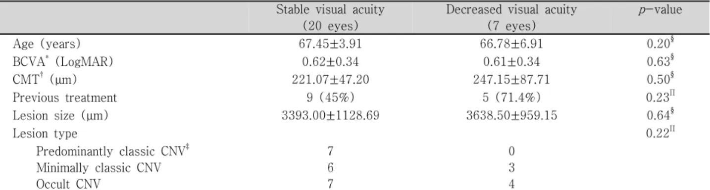
Table 5. Baseline factors related to visual prognosis in intravitreal bevacizumab group Stable visual acuity
(20 eyes)
Decreased visual acuity (7 eyes)
p-value
Age (years) 67.45±3.91 66.78±6.91 0.20§
BCVA* (LogMAR) 0.62±0.34 0.61±0.34 0.63§
CMT†(μm) 221.07±47.20 247.15±87.71 0.50§
Previous treatment 9 (45%) 5 (71.4%) 0.23Π
Lesion size (μm) 3393.00±1128.69 3638.50±959.15 0.64§
Lesion type 0.22Π
Predominantly classic CNV‡ 7 0
Minimally classic CNV 6 3
Occult CNV 7 4
*BCVA=Best corrected visual acuity; †CMT=Central macular thickness; ‡CNV=Choroidal neovascularization. §p-value by independent t-test; Πp-value by Chi-square test.
Table 6. Baseline factors related to visual prognosis in IVB & PDT group Stable visual acuity
(38 eyes)
Decreased visual acuity (7 eyes)
p-value
Age (years) 66.08±6.91 70.43±5.44 0.12§
BCVA* (LogMAR) 0.60±0.33 0.69±0.41 0.53§
CMT†(μm) 249.32±89.84 251.00±71.01 0.98§
Previous treatment 14 (36.7%) 2 (28.6%) 0.21Π
Lesion size (μm) 3477.30±912.75 4398.43±848.43 0.02§
Lesion type 0.03Π
Predominantly classic CNV‡ 8 0
Minimally classic CNV 10 5
Occult CNV 20 2
*BCVA=Best corrected visual acuity; †CMT=Central macular thickness; ‡CNV=Choroidal neovascularization. §p-value by independent t-test; Πp-value by Chi-square test.
우에서 통계적으로 유의하게 시력이 좋았다(Table 2). 빛간 섭단층촬영을 이용한 중심황반두께는 단독치료군은 221.07
±47.20에서 153.29±19.94 (p=0.001), 병합 치료군은 247.15
±87.17에서 175.39±59.79 (p=0.001)로 두 군에서 모두 치 료 6개월 뒤 통계적으로 유의하게 감소하였으나, 두 군 간 의 차이는 없었다(Table 3). 치료 6개월 뒤 재 치료를 받은
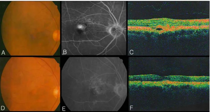
Figure 1. A 70-year–old woman with visual loss in her left eye was diagnosed with neovascular age-related macular degeneration. Her visual acuity was 5/200 in the left eye. Fundus photography showed subretinal fluids with subretinal hemorrhages on the macula (A). Fluorescein angiography demonstrated hyperfluorescence corresponding to leakage (B). Optical coherence tomography showed a moderate thickened retina with subretinal fluids and cystic spaces (C). The patient was offered three consecutive intravitreal injections of bevacizumab. Six months after injection, the visual acuity in the left eye was 20/70. Fundus photography showed decreased of subretinal fluids and subretinal hemorrhages (D). Fluorescein angiography showed decreased hyperfluorescein without leakage (E).
Optical coherence tomography showed a significant resolution of subretinal fluids and cystic spaces (F).
경우는 단독치료군이 10안(37.0%)으로, 병합치료군 12안 (26.7%)보다 유의하게 높은 비율로 나타났다(Table 4).
이차 연구 결과로는 최대교정시력이 치료 전과 비교하여 변화가 없거나 1줄 이상의 향상을 보인 경우를 악화되지 않 고 안정된 경우로 보고 치료 전 병변의 특징에 따른 관련인 자를 보았다. 단독치료군의 경우 시력의 안정을 나타내었던 20안에서 시력이 악화되었던 경우와 비교하여 치료 전 병 변의 특징에 따른 차이가 없었으나, 병합치료를 시행한 군 은 시력의 안정이 있었던 38안에서 시력이 악화되었던 7안 에 비해 통계적으로 유의하게 병변의 크기가 작았고, 전형 및 잠복 맥락막신생혈관이 많았다(Table 5, 6).
본 연구에서는 대상 환자 72안 중 시술 종료 후 6개월까 지의 경과 관찰 기간 동안 안내염, 망막색소상피박리, 주사 로 인한 백내장 형성, 망막박리 등 특이할 만한 부작용은 나타나지 않았다.
고 찰
맥락막신생혈관은 망막하 또는 망막색소상피하에서 발
생하는 비정상적인 신생혈관으로 이로부터 누출된 삼출물, 혈액 또는 이차적으로 발생하는 섬유혈관조직과 허혈 등에 의해 망막 손상을 일으키는 것으로 알려져 있으며, 황반부 에 발생하는 경우 심한 시력 저하를 유발한다.10,11
최근 맥락막신생혈관의 새로운 치료제로 주목 받고 있는 항혈관내피증식인자의 유리체강내 주입술은 시력의 회복, 중심황반두께의 감소와 병변의 안정을 보여 광역학요법 단 독치료보다 더 좋은 결과를 얻었다.12-14그러나 정기적으로 안내 주사를 시행해야 하며, 이로 인한 안내염의 발생이 Fintak et al15에 의하면 약 4500회의 주사 중 1회(0.02%), Mason et al16에 의하면 5233회의 주사 중 1회(0.019%)의 낮은 빈도로 보고되고 있다. 또한 반복적인 주사 요법 이후 에 생긴 비감염성 안내염도 보고되고 있으며, 이러한 안내 염이 통증과 함께 심각한 시력 저하를 야기하면 수술적 치 료까지 필요할 수 있다.17 따라서 치료회수를 줄여보기 위 해 광역학요법과 항혈관내피증식인자의 유리체강내 주입술 을 병합한 다양한 방법의 치료가 시도 되고 있으며, 대부분 좋은 결과를 보여주었으나 아직 더 우수한 치료 방침을 정 하기 위한 노력이 계속 되고 있다.8,18-20
Figure 2.A 71-year–old-man with visual loss in his right eye was diagnosed with neovascular age-related macular degeneration. His visual acuity was 20/70 in the right eye. Fundus photography showed subretinal fluids with subretinal hemorrhages on the macula (A). Fluorescein angiography demonstrated hyperfluorescence corresponding to leakage (B). A pigment epithelial detachment and a retinal thickening were showed by optical coherence tomography (C). The patient was offered a photodynamic therapy and three consecutive intravitreal injections of bevacizumab. Six months after injection, the visual acuity in the left eye was 20/25. Fundus photography showed complete resolution of a pigment epithelial detachment and subretinal hemorrhages (D). Fluorescein angiography showed decreased hyperfluorescein without leakage (E). Optical coherence tomography showed a complete resolution of a pigment epithelial detachment and a foveal depression (F).
본 연구에서는 나이관련황반변성과 동반된 맥락막신생 혈관환자 72명 72안 중 bevacizumab 만 6주 간격으로 3회 주사한 27안과 광역학요법을 1회 시행하고 bevacizumab을 3회 시행한 45안을 대상으로 하였다. 두 군 모두 시술 전에 비해 최대교정시력과 중심황반두께의 유의한 호전이 관찰 되었다. 치료 후 3줄 미만의 시력저하를 보인 경우는 병합 치료를 한 경우 41안(91.1%), 단독치료를 한 경우 25안 (92.6%)으로 전형 맥락막신생혈관에서 광역학요법과 ra- nibizumab을 매달 주입한 기존의 FOCUS 연구의 90.5% 및 전형 맥락막신생혈관에 ranibizumab을 매달 주입한 ANCHOR 연구의 96.4%와 비교하여 비슷한 결과를 보였다.18,21그러 나 본 연구의 경우 약 10개월의 연구 기간 동안 평균 3.3회 의 주입술을 시행하였으며, 1년간 13회의 주입술을 받은 FOCUS 연구나 ANCHOR 연구와 비교하여 비록 연구 방법 이나 대상 환자 수는 적었지만, 더 적은 시술로도 비슷한 결과를 얻을 수 있음을 알 수 있었다. 본 연구와 마찬가지 로 고정된 치료 간격보다 빛간섭단층촬영의 중심황반두께 를 참고로 ranibizumab 재치료 여부를 결정하였던 PrONTO
연구의 경우, 12개월의 연구 기간 동안 3줄 미만의 시력저 하가 95%의 대상 안에서 관찰되었으며, 평균 5.6회의 주입 술을 시행하였다.22 또한 국내의 한 연구에서는 31안을 대 상으로 bevacizumab을 6주 간격 3회 시행 후 12개월간 경 과 관찰을 하였는데, 3줄 미만의 시력저하가 있었던 경우는 29안(93.5%)이었다.23
본 연구에서 경우 중심황반두께는 시술 후 6개월 뒤 두 군 모두 시술 전에 비해 유의한 감소를 보여주었으며, 평균 69.8 μm의 감소가 있었다. Ranibizumab을 이용하여 전형 맥락막신생혈관환자를 대상으로 한 PrONTO 연구의 경우 12개월 후 평균 177.8 μm의 중심황반두께감소가 관찰되었 는데,22 시술 전 중심황반두께가 PrONTO 연구의 경우 평 균 393.9 μm 이었던 데 반해 본 연구에서는 평균 234.1 μm 로 시술 전 중심황반두께가 비교적 얇았기 때문에 중심황 반두께감소의 정도가 작았던 것으로 보여진다. 다른 연구에 서의 중심황반두께 감소를 보면 Ranibizumab을 이용하여 비전형 맥락막신생혈관을 대상으로 한 MARINA 연구에서 는 12개월 후 평균 123 μm의 감소가 있었다.24또한 beva-
cizumab을 6주 간격으로 3회 주입 후 12개월간 경과 관찰 한 국내 연구에서 Oh et al23은 평균 102 μm, Kim et al25은 평균 114.8 μm의 중심황반두께 감소가 있었다고 보고하였다.
본 연구에서, 최대교정시력은 bevacizumab 단독치료군 의 평균 0.56±0.33에 비해 병합치료를 받은 군에서 평균 0.48±0.21로 시술 후 6개월 뒤 통계적으로 유의하게 시력 이 좋았다. 재치료율도 단독치료군이 10안(37.0%)으로 병 합치료군 12안(26.7%)보다 유의하게 높은 비율로 나타났 다. 따라서 광역학요법을 병합하는 것이 bevacizumab 단독 치료보다 재치료율을 낮추고 치료 6개월 뒤의 시력도 더 좋 은 것으로 볼 수 있다. 또한 광역학요법을 병합한 군에서는 시력 이 안정된 경우 시술 전 평균 병변의 크기가 3477.30±912.75 μm로시력이 악화되었던 7안의 평균 4398.43±848.43 μm 보다 유의하게 치료 전 병변의 크기가 작았다. 시력이 안정 된 경우와 시력이 저하된 경우를 병변의 특징에 따라 비교 하면 전형 맥락막신생혈관이 있었던 경우가 각각 8안과 0 안, 잠복 맥락막신생혈관이 있었던 경우가 각각 20안, 2안 으로 병변의 특징에 따른 분포에 유의한 차이가 있었다. 반 면, bevacizumab 단독치료만 시행한 군에서는, 시력이 안 정된 경우와 시력이 저하된 경우에 시술 전 요인에 따른 유 의한 차이는 없었다. 기존에 시행되었던 TAP 연구에 의하 면 전형 맥락막신생혈관이 있는 중심와밑 병변에서 광역학 요법은 플라시보에 비하여 증등도 또는 심한 시력 상실의 위험을 감소시킨다고 보고 하였으며, VIP 연구에서는 전형 맥락막신생혈관이 보이지 않는 맥락막신생혈관에서도 병변 의 크기가 작거나 초기 시력이 안 좋을 경우 광역학요법이 효과가 있음을 발표하였다.3,26따라서 광역학 요법과 병합 치료에서 좋은 결과를 나타낸 본 연구의 경우에도 기존의 연구 결과와 비슷한 시술 전 요인이 있었음을 알 수 있었다 (Fig. 1, 2).
이러한 결과를 종합하여 볼 때 bevacizumab 단독 치료와 광역학요법과의 병합 치료 모두 시력 호전 및 중심황반두 께의 감소에 유의하게 좋은 결과를 나타내고 재치료율도 비교적 적었다. 특히 병합치료에서 더 우수한 결과를 얻을 수 있는 경우로는 전형 맥락막신생혈관과 잠복 맥락막신생 혈관이 있거나 초기 병변의 크기가 작은 경우임을 알 수 있 었는데, 이 결과는 추후 대상환자를 선정할 때 고려 사항이 될 수 있을 것이다.
참고문헌
1) Polito A, Isola M, Lanzetta P, et al. The natural history of occult choroidal neovascularization associated with age-related macular degeneration: a systemic review. Ann Acad Med Singapore 2006;
35:145-50.
2) Macular Photocoagulation Study group. Subfoveal neovascular lesions in age related macular degeneration: guidelines for evaluat- ion and treatment in the macular photocoagulation study. Arch Ophthalmol 1991;109:1242-57.
3) Treatment of Age related macular degeneration with Photod- ynamic Therapy (TAP) Study Group. Photodynamic therapy of subfoveal choroidal neovascularization in age related macular degeneration with verteporfin: two-year results of 2 randomized clinical trials-TAP report 2. Arch Ophthalmol 2001;119:198-207.
4) Emerson MV, Lauder AK, Flaxel CJ et al. Intravitreal bevacizu- mab(Avastin) treatment of neovascular age-related macular degen- eration. Retina 2007;27:439-44.
5) Bashshur ZF, Schakal A, Hamam RN, et al. Intravitreal bevacizu- mab vs. verteporfin photodynamic therapy for neovascular age-related macular degeneration. Arch Ophthalmol 2007;125:1357-61.
6) Ferrara N. Vascular endothelial growth factor: basic science and clinical progress. Endocr Rev 2004;25:581-611.
7) Adamis AP, Shima DT. The role of vascular endothelial growth factor in ocular health and disease. Retina 2005;25:111-8.
8) Lazic R, Gabric N. Verteporfin therapy and intravitreal bevacizu- mab combined and alone in choroidal neovascularization due to age-related macular degeneration. Ophthalmology 2007;114:1179-85.
9) Ladas ID, Kotsolis AI, Papakostas TD, et al. Intravitreal bevaci- zumab combined with photodynamic therapy for the treatment of occult choroidal neovascularization associated with serous pig- ment epithelium detachment in age-related macular degeneration.
Retina 2007;27:891-6.
10) Bressler SB, Bressler NM, Fine SL, et al. Natural course of choro- idal neovascular membranes within the foveal avascular zone in senile macular degeneration. Am J Ophthalmol 1982;93:157-63.
11) Guyer DR, Fine SL, Maguine MG, et al. Subfoveal choroidal ne- ovascular membranes in age-related macular degeneration. Visual prognosis in eyes with relatively good visual acuity. Arch Oph- thalmol 1986;104:702-5.
12) Emerson MV, Lauder AK, Flaxel CJ, et al. Intravitreal bevacizu- mab(Avastin) treatment of neovascular age-related macular degen- eration. Retina 2007;27:439-44.
13) Lynch SS, Cheng CM. Bevacizumab for neovascular ocular dise- ase. Ann Pharmacother 2007;41:614-25.
14) Bressler NM, Chang TS, Fine JT, et al. Improved vision-related function after ranibizumab vs. photodynamic therapy: a randomi- zed clinical trial. Arch Ophthalmol 2009;127:13-21.
15) Fintak DR, Shah GK, Blinder KJ, et al. Incidence of endopht- hal- mitis related to intravitreal injection of bevacizumab and rani- bizumab. Retina 2008 ;28:1395-9.
16) Mason JO 3rd, White MF, Feist RM, et al. Incidence of acute on- set endophthalmitis following intravitreal bevacizumab (Avastin) injection. Retina 2008;28:564-7.
17) Yenerel NM, Dinc UA, Gorgun E. A case of sterile endophtha- lmitis after repeated intravitreal bevacizumab injection. J Ocul Pharmacol Ther 2008;24:362-3.
18) Heier JS, Boyer DS, Ciulla TA, et al. Ranibizumab combined with verteporfin photodynamic therapy in neovascular age-re- lated ma- cular degeneration. Arch Ophthalmol 2006;124:1532-42.
19) Augustin AJ, Puls S, Offermann I. Triple therapy for choroidal neovascularization due to age-related macular degeneration: Vert- eporfin PDT, Bevacizumab and Dexamethasone. Retina 2007;27:
133-40.
=ABSTRACT=
Primary Combined Photodynamic Therapy and Intravitreal Bevacizumab Injection for Neovascular
Age-related Macular Degeneration
Young Ju Lew, MD, PhD1, Hae Jin Park, MD1, Tae Gon Lee, MD2, Dong Won Lee, MD, PhD1, Sung Won Cho, MD, PhD1, Jae Heung Lee, MD, PhD1
MyungGok Eye Research Institute, Konyang University, Kim’s Eye Hospital1, Seoul, Korea, Department of Ophthalmology, College of Medicine, Konyang University2, Deajeon, Korea
Purpose: To investigate the efficacy of the combined treatment of photodynamic therapy (PDT) with verteporfin and intravitreal bevacizumab in patients with neovascular age-related macular degeneration.
Methods: Forty-five eyes received a single session of PDT following three intravitreal bevacizumab (1.25 mg) injections at six-week intervals (IVB+PDT group). Twenty-seven eyes received three intravitreal bevacizuamb (1.25 mg) injections only at six-week intervals (IVB group).
Results: After the six months of follow up, the mean BCVA (LogMAR) changed significantly from 0.62±0.34 to 0.56±0.33 in the IVB group (p=0.03) and from 0.61±0.33 to 0.48±0.21 in the IVB+PDT group (p=0.001). The mean BCVA was more improved in the IVB+PDT group (p=0.049). The mean CMT changes were significant in both groups immediately after treatment, but there was no significant difference at six months between the two groups. Ten eyes (37.0%) in the IVB group and 12 eyes (26.7%) in the IVB+PDT group required retreatments during the six months of follow-up (p=0.02).
Conclusions: Significant improvement in visual acuity and reduction in central macular thickness over a six-months period were observed in both groups. The mean visual acuity was significantly better and the rate of reinjections after the initial treatment schedule was lower in the IVB+PDT group.
J Korean Ophthalmol Soc 2010;51(1):35-41
Key Words: Age-related macular degeneration, Bevacizumab, Choroidal neovascularization, Photodynamic therapy
Address reprint requests to Tae Gon Lee, MD
Department of Ophthalmology, Konyang University, College of Medicine
#685 Gasuwon-dong, Seo-gu, Daejon 302-718, Korea
Tel: 82-42-600-9258, Fax: 82-42-600-9176, E-mail: [email protected] 20) Ladewig MS, Karl SE, Hamelmann V, et al. Combined intra- vitreal bevacizumab and photodynamic therapy for neovascular age-related macular degeneration. Graefes Arch Clin Exp Ophth- almol 2008; 246:17-25.
21) Brown DM, Kaiser PK, MIchels M, et al. ANCHOR study group.
Ranibizumab versus verteporfin for neovascular age-related macu- lar degeneration. N Eng J Med 2006;335:1432-44.
22) Fung AE, Lalwani GA, Rosenfeld PJ, et al. An optical coherence tomography-guided, variable dosing regimen with intravitreal ranibizumab (Lucentis) for neovascular age-related macular deg- eneration. Am J Ophthalmol 2007;143:566-83.
23) Oh SB, Cho WB, Moon JW, Kim HC. Effect and prognostic factors of intravitreal bevacizumab on choroidal neovascularization from age-related macular degeneration. J Korean Ophthalmol Soc 2009;
50:202-10.
24) Kaiser PK, Blodi BA, Shapiro H, et al. Angiographic and optical coherence tomographic results of the MARINA study of ranib- izumab in neovascular age-related macular degeneration. Opht- halmology 2007;114:1868-75.
25) Kim YH, Kim ES, Yu SY, Kwak HW. Long-term effect of in- travitreal bevacizumab for CNV secondary to age-related macular degeneration. J Korean Ophthalmol Soc 2008;49:1935-40.
26) Treatment of age-related macular degeneration with photodyn- amic therapy and Verteporfin in photodynamic therapy study groups. Effect of lesion size, visual acuity, and lesion composition on visual acuity change with and without Verteporfin therapy for choroidal neovascularization secondary to age-related macular degeneration: TAP and VIP Report No.1 Am J Ophthalmol 2003;
136:407-18.