Received January 26, 2011, Revised August 31, 2011, Accepted for publication September 16, 2011
Corresponding author: Jade Wititsuwannakul, Division of Dermatology, Department of Medicine, Faculty of Medicine, Chulalongkorn Uni- versity and King Chulalongkorn Memorial Hospital, Thai Red Cross Society, Bangkok 10330, Thailand. Tel: 66-2-2564253, Fax: 66-2-256- 4253 (ext102), E-mail: [email protected]
This is an Open Access article distributed under the terms of the Creative Commons Attribution Non-Commercial License (http://
creativecommons.org/licenses/by-nc/3.0) which permits unrestricted non-commercial use, distribution, and reproduction in any medium, provided the original work is properly cited.
CASE REPORT
Fig. 1. Pedigree of family with generalized Dowling-Degos disease (DDD). Proband (P), his first son (case 2), and his second daughter (case 3) had generalized DDD.
Generalized Dowling-Degos Disease: Case Reports
Jade Wititsuwannakul, Nopadon Noppakun
Division of Dermatology, Department of Medicine, Faculty of Medicine, Chulalongkorn University and King Chulalongkorn Memorial Hosptial, Thai Red Cross Society, Bangkok, Thailand
Dowling-Degos disease (DDD) is a rare autosomal dominant trait characterized by numerous, symmetrical, progressive and pigmented macules over the axillae, groins, face, neck, arms and trunk as well as scattered comedo-like lesions (dark dot, follicles) and pitted acneiform scars. Histopathology is diagnostic testing using a distinctive form of acanthosis, characterized by an irregular elongation of thin branching rete ridges, with a concentration of melanin at the tips. We report cases of generalized DDD in a single family with autosomal dominant penetrance. DDD can be presented in a generalized form with hypopigmented lesions instead of reticulate hyperpigmentation confined to the flexor areas.
This form can be differentiated from DUH by histopathology.
(Ann Dermatol 25(3) 360∼364, 2013) -Keywords-
Dowling-Degos disease, Pigmentary disorder, Reticulate
INTRODUCTION
Dowling-Degos disease (DDD) is a rare autosomal domi- nant trait characterized by numerous, symmetrical, progre- ssive and pigmented macules over the axillae, groins, face, neck, arms and trunk as well as scattered comedo- like lesions (dark dot follicles) and pitted acneiform scars1-3.
The disorder usually appears and/or worsens after puberty.
DDD, dyschromatosis symmetrica hereditaria (DSH), dy- schromatosis universalis hereditaria (DUH) and reticulate acropigmentation of Kitamura (RAPK) share clinical fea- tures with each other; yet, they have different pathology findings4-7. Histopathology is diagnostic testing using a distinctive form of acanthosis, characterized by an irre- gular elongation of thin branching rete ridges, with a concentration of melanin at the tips8,9. Recently, the loss- of-function in the keratin 5 (KRT5) gene and a gene locus mapping to chromosome 17p13.3 have been described recently in cases of DDD10,11. In this study, we report a family with autosomal dominant inheritance (Fig. 1) of a skin disorder with clinical features of generalized DDD (Fig. 2~4).
CASE REPORT
Case 1
The proband was an 82-year-old man who was presented with progressively generalized skin lesions since adole- scence. He began to develop areas of hyperpigmentation, initially on the face, but then soon affecting the neck, axillae, groin, trunk and limbs (Fig. 2). The eruption has continued to become more extensive throughout adult-
Fig. 4. Daughter of proband, 50-year-old woman. (A) Reticulate pigmented macules, multiple pits and comedo-like lesions distributed over the face, perioral area and neck. (B, C) Reticulate hyperpigmentation on forearms.
Fig. 3. Son of proband, 58-year-old man. (A) Reticulate pig- mented macules, multiple pits and comedo-like lesions distributed over the face, perioral area and neck. (B) Reticulate hyperpigmentation on axilla.
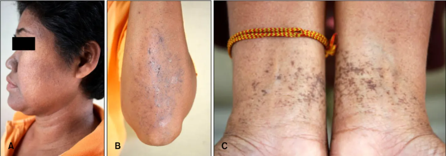
Fig. 2. Skin lesion of proband, 82-year-old man. (A, B) Reticulate hyperpigmentation, multiple pits and comedo-like lesions distributed over the face, perioral area and neck. (C) Reticulate hyperpigmentation with symmetrically distributed hypopigmented macules on shins. (D) Comedo-like lesions distributed over the back.
hood leading to generalized pruritus. The patient was born of a non-consanguineous marriage. All of his off- spring were affected with the same disease. His general physical examination was normal. Skin examination revealed a symmetrical reticulate hyperpigmentation pre- dominantly affecting the lower face, skin folds, major flexures, back and legs. Symmetrically distributed reticu- late hypopigmentation was also seen in the legs. Multiple pits varying in size from 1∼3 mm and open comedone- like lesions were distributed over the cheeks, perioral area, neck and back. No breaks in the epidermal ridge pattern on the palms and soles were seen. The mucosa, nails, teeth and hair were normal. On investigation, com- plete blood count, blood chemistry, liver and renal function tests were all within normal limits.
A biopsy was conducted from a hyperpigmented lesion
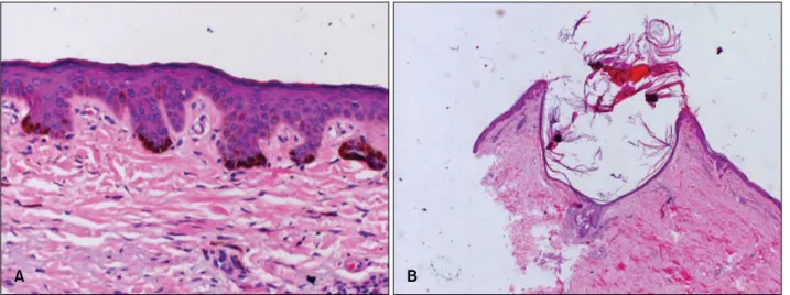
Fig. 5. Skin biopsy specimens from proband of hyperigmented macule (A) and comedo-like lesion (B). (A) Epidermal hyperkeratosis, acanthosis, irregular elongated thin branching rete ridges growing down into the dermis and increased melanin pigment in the lower part of rete pegs (H&E, ×200). (B) Keratin-filled cysts resembling comedones with irregular elongated thin branching rete ridges growing down into the dermis and increased melanin pigment in the lower part of rete pegs (H&E, ×40).
over the shin, and histopathological examination revealed hyperkeratosis, acanthosis and irregular elongated thin branching rete ridges growing down into the dermis and increased melanin pigment in the lower part of the rete pegs (Fig. 5). Another skin biopsy from comedo-like lesions on the back shows keratin-filled cysts resembling comedones (Fig. 5). These features were entirely consis- tent with the clinical diagnosis of DDD8,9.
Case 2
A 58-year-old man (first son of the proband) was presented with reticulate hyperpigmented skin lesions over the face, chest, back, axillae and flexures since adolescence. Initi- ally, the lesions, which first started over the face, were raised above the skin surface and were small. Gradually, these lesions progressed to form pits filled with blackish material. He developed similar lesions over the back and chest over the next year. There was no history of other skin diseases. His father, all his siblings and their offspring were affected with the same disease. His general physical examination was within normal limits. Skin examination revealed open comedone-like lesions and multiple pits of varying sizes distributed over the face, neck and limbs.
The face and neck were more severely affected when compared to the trunk and limbs. There was diffuse reti- culate pigmentation over the face, axillae, inguinal region and back. The rest of the skin examination was un- remarkable.
Case 3
A 50-year-old woman, the younger sister of the patient in
case 2, had progressively generalized skin lesions after puberty. Her skin lesions were very similar to his brother’s lesions, with generalized reticulate hyperpigmentation, open comedo-like lesions and multiple pits of varying sizes on the face, perioral area, flexures and axillae. There was no history of inflammatory skin diseases prior to the onset of the lesion. There was no significant past medical, surgical and gynecological history. Skin examination revealed multiple pits of varying sizes distributed over the face, upper back, upper chest, arms, cubital fossa and over forearms. There was diffuse reticulate pigmentation over the cubital fossa, axillae, inguinal region and face. The rest of the skin examination was unremarkable. Her general physical examination and all relevant investigations were within the normal limit. We did not perform a genetic study in our patients due to their religious belief.
DISCUSSION
DDD has characteristics of clinical and histopathological features. The diagnosis is very simple when classical fea- tures are present, as observed in our cases. We had considered the differential diagnosis of familial dyske- ratotic comedones, DSH, DUH and RAPK, which share some clinical features with each other but have different pathology findings4-7. In our cases, the proband, his off- spring and niece had generalized DDD. The generalized DDD has the typical feature of classic DDD (reticulate hyperpigmentation at flexural area) and generalized reticulate hyperpigmentation at the trunk, limbs and other cutaneous findings, including reticulate hypopigmented
macules, facial pits, comedo-like papules and palmar pits.
Histopathology is a diagnostic testing with a distinctive form of acanthosis, characterized by downward elon- gations of thin rete ridges with reticulated or fenestrated patterns, with a concentration of melanin at the tips and occasional follicular plugging and horn cysts8,9.
Due to the overlap of clinical features, there is debate as to whether DDD, DUH and RAPK are separate entities or represent a spectrum of a single disorder. RAPK is charac- terized by atrophic pigmented spots on the back of the hands and feet and palmar pits. These lesions may or may not be observed in patients with DDD12-15. These findings were not present in our patients. DUH (a generalized disorder) and DSH (a localized disorder) are characterized by diffuse symmetrically distributed hypopigmented ma- cules or papules mixed with hyperpigmentation. In DSH, the pigmentary changes are confined to the back aspects of the hands and feet16.
The genetic defect of DDD has not yet been well defined.
A recently reported series described the loss of-function mutations in the KRT5 gene10,11. These data confirm that haploinsufficiency for K5 engenders an excess of unpair- ed, soluble K14 that is then responsible for DDD and points to a prominent role for the keratin intermediate filament cytoskeleton within the basal keratinocytes in epidermal pigment biology17-19. Another genetic defect of DDD has been reported in the gene locus mapping to chromosome 17p13.3 and chromosome 1q21 with patho- genic mutations located in the DSRAD gene11.
In order to categorize these different disorders with over- lapping clinical or histopathologic features, it seems that there are two major groups20. The first is DDD, which has the characteristic histologic findings of elongated rete ridges, horn cyst formation and hyperpigmented tips. The distribution may be reticulate in flexural areas (classic DDD) or generalized with hypopigmented papules (gene- ralized DDD), and it may be distinguished by the pre- sence of acantholytic cells (Galli-Galli disease). The se- cond group is dyschromatosis, including DUH and DSH.
Clinically, both have hyperpigmented and hypopigmented lesions and histopathologic findings which are different from those of the DDD group.
In conclusion, we report cases of generalized DDD in a single family with autosomal dominant penetrance. DDD can be presented in a generalized form with hypopig- mented lesions instead of reticulate hyperpigmentation confined to the flexor areas. This form can be differenti- ated from DUH by histopathology.
REFERENCES
1. Crovato F, Nazzari G, Rebora A. Dowling-Degos disease (reticulate pigmented anomaly of the flexures) is an auto- somal dominant condition. Br J Dermatol 1983;108:473- 476.
2. Rebora A, Crovato F. The spectrum of Dowling-Degos disease. Br J Dermatol 1984;110:627-630.
3. Kim YC, Davis MD, Schanbacher CF, Su WP. Dowling- Degos disease (reticulate pigmented anomaly of the flexu- res): a clinical and histopathologic study of 6 cases. J Am Acad Dermatol 1999;40:462-467.
4. Lestringant GG, Masouyé I, Frossard PM, Adeghate E, Galadari IH. Co-existence of leukoderma with features of Dowling-Degos disease: reticulate acropigmentation of Ki- tamura spectrum in five unrelated patients. Dermatology 1997;195:337-343.
5. Ostlere L, Holden CA. Dowling-Degos disease associated with Kitamura's reticulate acropigmentation. Clin Exp Der- matol 1994;19:492-495.
6. Sandhu K, Saraswat A, Kanwar AJ. Dowling-Degos disease with dyschromatosis universalis hereditaria-like pigmenta- tion in a family. J Eur Acad Dermatol Venereol 2004;18:
702-704.
7. Thami GP, Jaswal R, Kanwar AJ, Radotra BD, Singh IP.
Overlap of reticulate acropigmentation of Kitamura, acro- pigmentation of Dohi and Dowling-Degos disease in four generations. Dermatology 1998;196:350-351.
8. Harper JI, Trembath RC. Genetics and genodermatoses. In:
Rook AJ, Burns T, editors. Rook's textbook of dermatology.
7th ed. Malden, Mass: Blackwell Science, 2004;15.11-15.13.
9. Howell JB, Freeman RG. Reticular pigmented anomaly of the flexures. Arch Dermatol 1978;114:400-403.
10. Betz RC, Planko L, Eigelshoven S, Hanneken S, Pasternack SM, Bussow H, et al. Loss-of-function mutations in the keratin 5 gene lead to Dowling-Degos disease. Am J Hum Genet 2006;78:510-519.
11. Li CR, Xing QH, Li M, Qin W, Yue XZ, Zhang XJ, et al. A gene locus responsible for reticulate pigmented anomaly of the flexures maps to chromosome 17p13.3. J Invest Der- matol 2006;126:1297-1301.
12. Crovato F, Desirello G, Rebora A. Is Dowling-Degos disease the same disease as Kitamura's reticulate acropig- mentation? Br J Dermatol 1983;109:105-110.
13. Berth-Jones J, Graham-Brown RA. A family with Dowling Degos disease showing features of Kitamura's reticulate acropigmentation. Br J Dermatol 1989;120:463-466.
14. Cox NH, Long E. Dowling-Degos disease and Kitamura's reticulate acropigmentation: support for the concept of a single disease. Br J Dermatol 1991;125:169-171.
15. Al Hawsawi K, Al Aboud K, Alfadley A, Al Aboud D.
Reticulate acropigmentation of Kitamura-Dowling Degos disease overlap: a case report. Int J Dermatol 2002;41:
518-520.
16. Oyama M, Shimizu H, Ohata Y, Tajima S, Nishikawa T.
Dyschromatosis symmetrica hereditaria (reticulate acropig- mentation of Dohi): report of a Japanese family with the
condition and a literature review of 185 cases. Br J Der- matol 1999;140:491-496.
17. Bonifas JM, Rothman AL, Epstein EH Jr. Epidermolysis bullosa simplex: evidence in two families for keratin gene abnormalities. Science 1991;254:1202-1205.
18. Bonifas JM, Bare JW, Lynch ED, Lebo RV, Epstein EH Jr.
Regional assignment of the human keratin 5 (KRT5) gene to chromosome 12q near D12S14 by PCR analysis of somatic cell hybrids and multicolor in situ hybridization. Genomics
1992;13:452-454.
19. Stephens K, Ehrlich P, Weaver M, Le R, Spencer A, Sybert VP. Primers for exon-specific amplification of the KRT5 gene: identification of novel and recurrent mutations in epidermolysis bullosa simplex patients. J Invest Dermatol 1997;108:349-353.
20. Wu YH, Lin YC. Generalized Dowling-Degos disease. J Am Acad Dermatol 2007;57:327-334.