Copyright © 2011 Journal of Korean Neurotraumatology Society
47
CASE REPORTJ Kor Neurotraumatol Soc 2011;7:47-49 ISSN 1738-8708
Received: March 15, 2011 / Revised: March 17, 2011 Accepted: April 13, 2011
Address for correspondence: Sung Ho Choi, MD
Department of Neurosurgery, Gachon University of Medical &
Science, Gil Medical Center, 1198 Guwol-dong, Namdong-gu, Incheon 405-760, Korea
Tel: +82-32-460-3304, Fax: +82-32-460-3899 E-mail: phoenix3070@naver.com
외상성 급성 경막하 혈종의 조기 자연 감소
가천의과학대학교 길병원 신경외과학교실
최성호 .박찬우 .김재명 .김은영 .유찬종 .김영보 .이상구 .박철완 .김우경 .이 언
Rapid Spontaneous Resolution of Traumatic Acute Subdural Hematoma
Sung Ho Choi, MD, Chan Woo Park, MD, Jae Myung Kim, MD, Eun Young Kim, MD, Chan Jong Yoo, MD, Young Bo Kim, MD, Sang Gu Lee, MD,
Cheol Wan Park, MD, Woo Kyung Kim, MD and Uhn Lee, MD
Department of Neurosurgery, Gachon University of Medical & Science, Gil Medical Center, Incheon, Korea
Rapid spontaneous resolution of traumatic acute subdural hematoma (SDH) is a rare and unexpectable phenomenon. A 67-year-old man presented with confused mental state after falling down. Initial brain computed tomography (CT) scan revealed an acute SDH with thickness of approximately 10mm, which was managed conservatively. Six days after con- servative management, follow-up CT showed complete resolution of the hematoma. The other case of 61-year-old woman was an acute traumatic SDH after falling out of wheelchair that reduced spontaneously. We report two cases of traumatic acute SDH with spontaneous reduction and possible mechanism of spontaneous resolution of acute SDH with relevant re- view of the literature. (J Kor Neurotraumatol Soc 2011;7:47-49)
KEY WORDS: Rapid resolution ㆍAcute subdural hematoma.
서 론
급성 경막하 혈종은 일반적으로 심한 두부 손상으로 유 발되며 높은 사망률을 보이고 대체로 조기에 시행되는 수 술이 예후를 향상시킬 수 있다고 알려져 있어 빠른 수술 적 치료가 권유된다.
2,9,11,14)그러나 외상성 급성 경막하 혈 종의 조기 자연 소실된 경우들이 드물게 보고되었다.
3,5-8,11-13,15-17)
저자들은 외상성 급성 경막하 혈종의 조기 흡수
에 대한 가능한 기전(mechanism)을 추정하고 더불어 문 헌 고찰을 하였다.
증 례
증 례 1
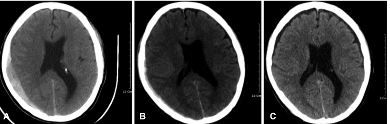
2년 전 간질 병력이 있는 67세 남자가 경련하면서 넘어 져 머리를 다쳐 응급실에 내원하였다. 의식은 혼돈 상태 (Glasgow Coma Scale: GCS 14)였으며, 내원 당시 시행 한 뇌 전산화단층촬영에서 우측 측두-두정부에 10 mm 두께의 경막하 혈종이 있었고 (Figure 1A), 중심선의 반대 편 이동은 없었다. 3시간 동안 경과관찰 후 뇌 전산화단 층촬영을 다시 하였고, 경막하 혈종은 소량만 관찰되었다 (Figure 1B). 내원 6일 뒤 시행한 뇌 전산화단층촬영에서 경막하 혈종은 더 이상 보이지 않았으며, 양측의 전두, 측두 및 두정부에서 경미한 경막하 수종만이 관찰되었다 (Fig - ure 1C). 보존적 치료를 받은 환자는 의식이 정상상태로 호전되어 퇴원하였다.
증 례 2
당뇨 신부전증으로 1년 전부터 혈액투석 중인 61세 여
online©MLComm
48
J Kor Neurotraumatol Soc 2011;7:47-49 Rapid Resolution of SDH자가 휠체어를 타고 가다 떨어져 응급실에 내원하였다.
의식은 기면상태 (GCS 13)였으며, 양측 동공은 우측 3 mm, 좌측 2 mm 크기로 양측 모두 빛에 반응이 있었다.
내원 당시 시행한 뇌 전산화단층촬영에서 약 17 mm 두 께의 급성 경막하 혈종이 우측 대뇌 반구에서 관찰되었 고, 약 18 mm의 중심선 좌측 이동이 있었다. 응급수술 을 시행하려고 하였으나, 보호자가 수술을 원하지 않아 신장 내과로 전과 후 보존적 치료를 하였다. 의식상태가 점 차 호전이 되었고, 내원 11일째 촬영한 뇌 전산화단층 영 상에서 경막하 공간에 있던 혈종은 거의 사라지고, 중심선 이동도 보이지 않았다 (Figure 2). 환자 의식이 비교적 조 기에 회복된 점을 고려하여 급성 경막하 혈종의 조기 자연 감소라고 판단하였다.
고 찰
급성 경막하 혈종은 사망률이 높고 조기 수술이 예후 를 향상시킨다고 알려져 있어 빠른 수술적 치료가 권유된
다.
2,9,11,14)그러나 몇몇 학자들은 경막하 혈종의 자연 흡수
로 인하여 수술적 조치를 취하지 않아도 좋은 경과를 보 이는 경우에 대하여 증례보고를 하였고 급성 경막하 혈종 의 조기 흡수에 대한 많은 가능한 기전을 제시하였다.
3,5-8,11-13,15-17)
Sato 등
16)은 1985년 이후 보고된 외상성 급성 경
막하 혈종 33예를 분석하고 가능한 기전을 제시하였다.
첫 번째, 가능한 기전은 경막하 혈종과 함께 지주막이 찢 어지고 이로 인해 혈종이 뇌척수액에 노출, 희석되어 씻겨 지며(wash-out) 소실되는 것이며, 두 번째 기전은 급성 경막하 혈종이 급성 뇌부종으로 인해 발생한 압력으로 혈 종이 재분배(redistribution)되어 자연소실이 발생한다는
것이다.
5-7,11,12,15,16)그리고 뇌실질의 위축이 동반되면 지주
막하 공간이 넓어 발생한 혈종의 재분배가 원활하여 경 막하 혈종의 소실이 용이하다고 주장하였다.
13)또한, 뇌 경 막하 혈종 발생 이후 척추에 경막하 혈종이 동반되면서 뇌 경막하 혈종이 소실된 경우가 보고되면서 두개외로 혈종 재분배(extracranial redistribution) 가능성이 제시되 었다.
1,10,18)증례 1의 경우, 초기 뇌 전산화단층 영상을 바탕으로 판 단하면 기존 뇌실질 위축이 있는 환자로 급성 혈종이 주
FIGURE 1. Brain CT images of case 1. A: Initial brain CT scan on admission shows an acute SDH with a maximal thickness of ap- proximately 10 mm. B: A follow-up brain CT scan 3 hours after the initial scan shows a resolution of the SDH. C: 6 days later after admission, a follow-up brain CT image revealed a complete resolution of acute SDH. SDH: subdural hematoma.A B C
FIGURE 2. Brain CT images of case 2. A: Initial brain CT scan revealed an acute SDH with a maximal thickness of approxi- mately 17 mm in the right con- vexity and a midline shift of 18 mm. B: A follow-up brain CT im- age 11days after the initial scan shows a resolution of SDH.
SDH: subdural hematoma.
A B
www.neurotrauma.or.kr
49
Sung Ho Choi, et al.
위 뇌부종에 의한 압력으로 혈종 재분배가 일어난 것으 로 판단된다.
증례 2의 경우, 초기 뇌 전산화단층촬영이후 11일째 추 적 검사 한 것이므로 혈종의 조기 소실이라고 단정할 수 는 없으나 조기 신경학적 회복이 있었고, 급성 뇌부종에 의한 압력 및 재분배와 함께 뇌척수액의 희석 등이 경막하 혈종의 자연 감소에 기여하였다고 추정된다. 또한 초기 뇌 전산화단층촬영에서 많은 양의 경막하 혈종이 뇌척수액과 혼합된 양상으로 보아 상기 추정을 뒷받침한다고 볼 수 있 다. Kato 등
6)은 외상성 급성 경막하 혈종이 조기 소실된 28예를 분석하고 21예에서 초기 두부 전산화단층촬영상 혈종부위에 낮은 밀도 띠(low-density band)가 보였으 며 이러한 것은 혈종이 뇌척수액과 혼합된 양상으로 혈종 소실 및 재분배에 중요하다고 주장했다. Imai
4)는 경천막 헤르니아(transtentorial herniation)를 보이는 다량의 외상성 급성 경막하 혈종의 조기 소실을 보고하면서 혈종 이 뇌척수액과 혼합되어 있는바 이는 지주막이 찢어지고 이로 인해 혈종이 뇌척수액에 노출되면서 희석되어 씻겨 진 것으로 추정하면서, 외상성 급성 경막하 혈종에서 수 술적 치료가 절실하지만 환자의 전신상태로 수술적 치료 가 어려울 경우 보존적 치료가 대안이 될 수 있음을 강조 하였다.
결 론
수술적 치료가 필요하다고 판단되는 외상성 급성 경막 하 출혈이 조기에 자연 소실된 두 예를 경험하였으며, 그 기전으로는 뇌부종으로 두개내 압력증가에 의한 혈종 재 분배 및 뇌척수액에 의한 혈종 희석에 의한 것으로 추정 된다.
중심 단어: 조기소실·급성 경막하 혈종.
■ The authors have no financial conflicts of interest.
REFERENCES
1) Bortolotti C, Wang H, Fraser K, Lanzino G. Subacute spinal sub- dural hematoma after spontaneous resolution of cranial subdural hematoma: causal relationship or coincidence? Case report. J Neu-
rosurg 100:372-374, 2004
2) Calhoun JM, Boop F. Spontaneous spinal subdural hematoma:
case report and review of the literature. Neurosurgery 29:133-134, 3) Cohen JE, Eger K, Montero A, Israel Z. Rapid spontaneous reso-1991 lution of acute subdural hematoma and HIV related cerebral atro- phy: case report. Surg Neurol 50:241-244, 1998
4) Imai K. Rapid spontaneous resolution of signs of intracranial her- niation due to subdural hematoma--case report. Neurol Med Chir (Tokyo) 43:125-129, 2003
5) Inamasu J, Nakamura Y, Saito R, Kuroshima Y, Mayanagi K, Ohba S, et al. Rapid resolution of traumatic acute subdural hematoma by redistribution. Am J Emerg Med 20:376-377, 2002
6) Kato N, Tsunoda T, Matsumura A, Yanaka K, Nose T. Rapid spon- taneous resolution of acute subdural hematoma occurs by redistri- bution--Two case reports. Neurol Med Chir (Tokyo) 41:140-143, 7) Kuroiwa T, Tanabe H, Takatsuka H, Arai M, Sakai N, Nagasawa S, 2001 et al. Rapid spontaneous resolution of acute extradural and sub- dural hematomas. Case report. J Neurosurg 78:126-128, 1993 8) Kundra SN, Kundra R. Extracranial redistribution causing rapid
spontaneous resolution of acute subdural hematoma. Neurol In- dia 53:124, 2005
9) Langmayr JJ, Ortler M, Dessl A, Twerdy K, Aichner F, Felber S.
Management of spontaneous extramedullary spinal haematomas:
results in eight patients after MRI diagnosis and surgical decom- pression. J Neurol Neurosurg Psychiatry 59:442-447, 1995 10) Lecouvet FE, Annet L, Duprez TP, Cosnard G, Scordidis V, Mal-
ghem J. Uncommon magnetic resonance imaging observation of lumbar subdural hematoma with cranial origin. J Comput Assist Tomogr 27:530-533, 2003
11) Matsuyama T, Shimomura T, Okumura Y, Sakaki T. Rapid resolu- tion of symptomatic acute subdural hematoma: case report. Surg Neurol 48:193-196, 1997
12) Nagao T, Aoki N, Mizutani H, Kitamura K. Acute subdural hema- toma with rapid resolution in infancy: case report. Neurosurgery 19:465-467, 1986
13) Niikawa S, Sugimoto S, Hattori T, Ohkuma A, Kimura T, Shino- da J, et al. Rapid resolution of acute subdural hematoma--report of four cases. Neurol Med Chir (Tokyo) 29:820-824, 1989 14) Osborn AG. Diagnostic Neuroradiology. St. Louis, MO: Mosby,
pp205-207, 1994
15) Polman CH, Gijsbers CJ, Heimans JJ, Ponssen H, Valk J. Rapid spontaneous resolution of an acute subdural hematoma. Neuro- surgery 19:446-448, 1986
16) Sato M, Nakano M, Sasanuma J, Asari J, Watanabe K. Rapid res- olution of traumatic acute subdural haematoma in the elderly. Br J Neurosurg 19:58-61, 2005
17) Suzuki Y, Kawamata T, Matsumoto H, Kunii N, Matsumoto K.
[A resolving sign of acute subdural hematoma: from report of two cases]. No Shinkei Geka 26:1025-1029, 1998
18) Yamaguchi S, Kurisu K, Arita K, Takeda M, Tani I, Araki O. Si- multaneous cranial and spinal subdural hematoma. Neurol Med Chir (Tokyo) 45:645-649, 2005