CASE REPORT
선천적 등쪽 췌장 발육 부전증을 동반한 다비장 증후군 환자에서 나타난 급성 췌장염 1예와 내시경 초음파의 역할
정재훈, 김광하, 송근암, 이동건, 문지윤, 정재훈, 김 석1
부산대학교 의학전문대학원 내과학교실, 영상의학교실1
Polysplenia Syndrome with Congenital Agenesis of Dorsal Pancreas Presenting as Acute Pancreatitis and the Role of Endoscopic Ultrasonography in Its Diagnosis
Jae Hoon Jeong, Gwang Ha Kim, Geun Am Song, Dong Gun Lee, Ji Yoon Moon, Jae Hoon Cheong and Suk Kim1 Departments of Internal Medicine and Radiology1, Pusan National University School of Medicine, Busan, Korea
A 49-year-old female was admitted to our hospital for acute pancreatitis. The abdomen CT scan incidentally showed midline liver with hepatomegaly, centrally located gallbladder, pancreas truncation, right sided small bowel, left sided large bowel, interruption of the inferior vena cava with azygos continuation, preduodenal portal vein, and multiple spleens in the left upper quadrant. In MRCP, the head of pancreas was enlarged and short main pancreatic duct without accessory duct was showed.
EUS revealed enlarged ventral pancreas with a main pancreatic duct of normal caliber, absence of the accessory pancreatic duct and the dorsal pancreas. She was diagnosed as polysplenia syndrome with agenesis of dorsal pancreas. It is a rare congenital anomaly frequently associated with various visceral anomalies including multiple spleens, impaired visceral lateraliza- tion, congenital heart diseases, gastrointestinal abnormalities and azygos continuation of the inferior vena cava. We report a case of polysplenia syndrome with agenesis of dorsal pancreas presenting acute pancreatitis. (Korean J Gastroenterol 2012;60:47-51)
Key Words: Polysplenia; Heterotaxy syndrome; Pancreatitis; Preduodenal portal vein; Agenesis of the dorsal pancreas
Received February 11, 2011. Revised May 6, 2011. Accepted May 12, 2011.
CC This is an open access article distributed under the terms of the Creative Commons Attribution Non-Commercial License (http://creativecommons.org/licenses/
by-nc/3.0) which permits unrestricted non-commercial use, distribution, and reproduction in any medium, provided the original work is properly cited.
교신저자: 김광하, 602-739, 부산시 서구 구덕로 179, 부산대학교 의학전문대학원 내과학교실
Correspondence to: Gwang Ha Kim, Department of Internal Medicine, Pusan National University School of Medicine, Pusan National University Hospital, 179 Gudeok-ro, Seo-gu, Busan 602-739, Korea. Tel: +82-51-240-7869, Fax: +82-51-244-8180, E-mail: [email protected]
Financial support: None. Conflict of interest: None.
INTRODUCTION
Heterotaxy is a rare condition wherein the orderly arrange- ment of organs and blood vessels, as seen in situs solitus and situs inversus, is lost.1 In addition, it has multiple congenital anomalies. Polysplenia is a subtype of heterotaxy (asplenia is another) characterized by cardiac malformations (both at- ria displaying the morphology of the left atrium), bilobed lung (each lung having the morphology of a left lung), visceral mal- rotation, multiple spleens, interruption of the inferior vena cava with azygos or hemizygos continuation, and preduo-
denal portal vein.2,3 Patients with polysplenia sometimes have short pancreas or agenesis of dorsal pancreas, which is related to the possibility of increased incidence of pancreatitis.4,5
EUS provides high-resolution images of the entire pancre- atic parenchyma and allows direct visualization of the ductal system.6 Therefore, EUS may be helpful in the diagnosis of polysplenia syndrome with agenesis of the dorsal pancreas when CT scans and MRI were inconclusive for the pancreatic lesion. Herein, we report a case of agenesis of the dorsal pan- creas in association with polysplenia syndrome that man-
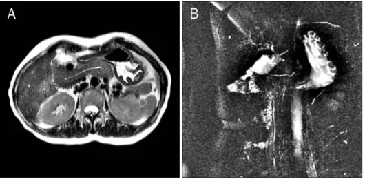
Fig. 1. Abdomen CT scans showing a midline liver, a centrally located gallbladder, multiple spleens in the left upper quadrant (arrowhead), a preduodenal portal vein (arrow), a short pancreas (A), an inferior vena cava interruption with an azygos continuation (arrow) (B), and a right-sided small bowel and left-sided large bowel (C).
Fig. 2. MRI scan (A) and MRCP (B) showing the main pancreatic duct and no accessory duct.
A 49-year-old female was referred to our hospital for acute persistent epigastric pain for 1 week prior to admission. She had no other illnesses and no remarkable family medical history. She denied any history of drug or alcohol abuse. The laboratory findings were normal except for elevated of serum amylase (2,023 IU/L, normal range 36-128 IU/L) and lipase (1,312 U/L, normal range 22-51 U/L) levels. Therefore, she was diagnosed with acute pancreatitis. Additional examina- tions were done to find the cause of pancreatitis. The serum calcium level was 8.6 mg/dL and triglyceride level was 52 mg/dL. The serum IgG4 level was 25 mg/dL. Abdomen CT scan revealed a midline liver with hepatomegaly, a centrally located gallbladder, multiple spleens in the left upper quad- rant behind the stomach, an inferior vena cava interruption
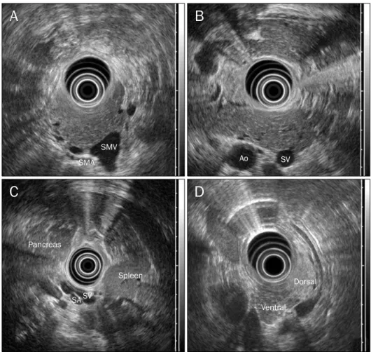
evident. MRI demonstrated that main pancreatic duct was short and the accessory duct was absent (Fig. 2). EUS re- vealed a hypertrophied pancreatic head with a main pancre- atic duct of normal caliber, absence of the accessory pancre- atic duct, homogenous echogenicity throughout the pancre- atic parenchyme, splenic vessels that were contact with the posterior wall of the stomach, and a preduodenal portal vein crossing over the pancreatic head. In addition, there were no sludges or stones in the gallbladder and bile duct and no pan- creatic divisum (Fig. 3). Endoscopy revealed a normal major papilla but the absence of the minor papilla. All these findings led to the diagnosis of dorsal pancreatic agenesis with poly- splenia syndrome. After fasting and conservative treatment, the patient’s symptoms improved, and her serum amylase and lipase levels were normalized. No evidence of relapse
Fig. 3. EUS showing homogenous echogenicity throughout the pancrea- tic parenchyme (A), a hypertrophied pancreatic head, the absence of the accessory pancreatic duct (B), and splenic vessels contacting the pos- terior wall of the stomach (C). EUS showing different echogenicity of the ventral and dorsal pancreas in normal subject (D). SMV, superior mesenteric vein; SMA, superior mesenteric artery; Ao, aorta; SV, splenic vein; SA, splenic artery.
was noted in the 12-month follow-up.
DISCUSSION
Polysplenia syndrome is a rare anomaly that occurs in ap- proximately 4 of every million live births.7 It is complex set of various visceral anomalies including multiple spleens, im- paired visceral lateralization, cardiovascular anomalies, gas- trointestinal abnormalities, and short pancreas.1 Rarely, genitourinary anomalies such as double ureters, renal agen- esis or hypoplastic kidney are reported as part of the poly- splenia syndrome.8 However, this syndrome does not have fixed set of characteristics that are present in all cases.1
The unique sign of the polysplenia syndrome is the pres- ence of multiple spleens, mostly located throughout the greater curvature of the stomach, which is to the right of the midline in more than 60% of patients.9
Cardiac anomaly is frequently associated with polysplenia syndromes in children but this is far less frequent in adults because of early decease in children with this anomaly.2
Cardiovascular anomalies associated with polysplenia syn- drome include the absence or hypoplasia of the suprarenal inferior vena cava (with or without azygos or hemiazygos con- tinuation), dextrocardia, ventricular septal defects, and mor- phologic left ventricular outflow obstruction, and these anomalies lead patients to death with congestive heart fail- ure, hypoplastic left ventricle or morbidity of cardiac oper- ation before 5 years old.2 Our patient did not have specific past history about heart disease, in addition, electrocardio- graphy and echocardiography was normal.
Preduodenal portal vein is a portal vein located in front of the pancreatic head at CT image. It passes ventral to the duo- denum and the head of the pancreas. Preduodenal portal vein might interfere mechanically with pancreatic develop- ment, so it is associated with annular pancreas and malrotation.3 This vascular anomaly has importance in points of the prevention of accident during pancreaticobiliary operation.10
Abdominal complaints are the most common symptoms in adult, and sometimes CT scan shows intestinal malrotation.
creatitis with complete agenesis of dorsal pancreas can cause abdominal pain.5 Sempere et al.12 reviewed 14 cases of complete agenesis of dorsal pancreas and reported ab- dominal pain (13 cases, 92.9%) and pancreatitis (7 cases, 50%). Rakesh et al.5 showed that 4 cases with complete agenesis of dorsal pancreas had pancreatitis and abdominal pain. In complete agenesis of dorsal pancreas, pancreatitis may be related with sphincter of Oddi dysfunction or higher intrapancreatic duct pressures due to hypertrophy of the ven- tral gland.13,14 In our cases, there were no other causes of pancreatitis. However, hyperplasia of ventral pancreas was noted at EUS and might cause increased intrapancreatic duct pressure or hypersecretion of pancreatic enzyme result- ing in pancreatitis.
Embryologically, the pancreas arises from ventral and dor- sal buds. While ventral bud forms the uncinate process and pancreatic head, the dorsal bud becomes the body and tail.
The dorsal pancreas and spleen develop in the dorsal meso- gastrium. Therefore, concomitant anomalies of both organs can be expected in patient with polysplenia syndrome.4 Short pancreas in polysplenia syndrome means partial agenesis or hypoplasia of the dorsal pancreas.15 In partial agenesis, the minor papilla with a remnant of the accessory pancreatic duct and the body of the pancreas are present but the minor papilla, accessory pancreatic duct, body and tail of the pan- creas are absent in complete agenesis.15,16 Partial agenesis of the pancreas can remain asymptomatic because of the functional reserves of the exocrine and the endocrine pancreas.17 The clinical significance in complete agenesis is a possible relationship to early or late onset diabetes melli- tus, probably because most of the islet cells are located in the pancreatic body and tail.18,19 However, our patient did not have diabetes and fasting sugar was 99 mg/dL.
We confirmed polysplenia syndrome with CT scans and MRI/MRCP but confused whether she had partial agenesis of dorsal pancreas or complete agenesis of dorsal pancreas with hypertrophied ventral pancreas. ERCP is considered the gold standard for the diagnosis of agenesis of the dorsal pancreas.16 However, ERCP is an invasive method, and it is also technically difficult to identify the minor papilla and to cannulate a catheter into it.16 CT scans, MRCP and ERCP have
was helpful in the identification of agenesis of the dorsal pan- creas when other imaging modalities were inconclusive. EUS can directly visualize the body and tail of the pancreas and preduodenal portal vein during the retraction the duodenum to the gastric antrum (called as “stripping”).12 With the EUS positioned in the gastric antrum, the body and the tail of the pancreas are visualized between the posterior wall of the stomach and the splenic vessels.6 Agenesis of the dorsal pancreas may be considered, as in our patient, if the ac- cessory pancreatic duct, and the body and tail of pancreas (without any structure between the stomach and the splenic vessels) are not visualized.12 If the pancreatic parenchyma showed homogenous echogenicity during stripping, we could suspect hypertrophied ventral pancreas because the dorsal and ventral pancreas had the different echogenicity (Fig.
3D).20 As a result, we confirmed the diagnosis of complete agenesis of the dorsal pancreas associated with poly- psplenia syndrome.
In conclusion, polysplenia syndrome is a rare disease en- tity in adult and associated with agenesis of dorsal pancreas presenting with pancreatitis. EUS is helpful in the diagnosis of dorsal pancreatic agenesis in polysplenia syndrome when CT scans and MRI are inconclusive.
REFERENCES
1. Fulcher AS, Turner MA. Abdominal manifestations of situs anomalies in adults. Radiographics 2002;22:1439-1456.
2. Peoples WM, Moller JH, Edwards JE. Polysplenia: a review of 146 cases. Pediatr Cardiol 1983;4:129-137.
3. Li CS, Tu HY, Chen RC, Yang MT, Ting CC. Polysplenia syndrome associated with preduodenal portal vein and short pancreas:
Incidental findings in a case of CBD adenocarcinoma. Chin J Radiol 2001;26:269-274.
4. Maier M, Wiesner W, Mengiardi B. Annular pancreas and agen- esis of the dorsal pancreas in a patient with polysplenia syndrome. AJR Am J Roentgenol 2007;188:W150-W153.
5. Rakesh K, Choung OW, Reddy DN. Agenesis of the dorsal pan- creas (ADP) and pancreatitis - is there an association? Indian J Gastroenterol 2006;25:35-36.
6. Palazzo L. Echoendoscopy of the pancreas. Gastroenterol Hepatol 2002;25:26-34.
7. Tsutsumi R, Nagata Y, Enjoji A, Ohno Y, Kamito H, Kanematsu T.
Situs ambiguous with gastric cancer: report of a case. Surg Today 2007;37:676-679.
8. Hadar H, Gadoth N, Herskovitz P, Heifetz M. Short pancreas in polysplenia syndrome. Acta Radiol 1991;32:299-301.
9. Gayer G, Apter S, Jonas T, et al. Polysplenia syndrome detected in adulthood: report of eight cases and review of the literature.
Abdom Imaging 1999;24:178-184.
10. Semb BK, Halvorsen JF. Repair of preduodenal portal vein injury occurring during biliary surgery. Acta Chir Scand 1973;139:312- 313.
11. Bobba RK, Arsura EL, Naseem M, Ashraf N. Polysplenia in an eld- erly male: diagnostic approaches and review of the literature.
Eur J Intern Med 2005;16:608-609.
12. Sempere L, Aparicio JR, Martínez J, Casellas JA, de Madaria E, Pérez-Mateo M. Role of endoscopic ultrasound in the diagnosis of agenesis of the dorsal pancreas. JOP 2006;7:411-416.
13. Nishimori I, Okazaki K, Morita M, et al. Congenital hypoplasia of the dorsal pancreas: with special reference to duodenal papil- lary dysfunction. Am J Gastroenterol 1990;85:1029-1033.
14. Gold RP. Agenesis and pseudo-agenesis of the dorsal pancreas.
Abdom Imaging 1993;18:141-144.
15. Suda K, Matsumoto Y, Fujii H, Miura K, Nobukawa B. Clinico- pathologic differentiation of atrophy of the pancreatic body and tail aplasia. Int J Pancreatol 1998;24:227-235.
16. Schnedl WJ, Reisinger EC, Schreiber F, Pieber TR, Lipp RW, Krejs GJ. Complete and partial agenesis of the dorsal pancreas within one family. Gastrointest Endosc 1995;42:485-487.
17. Schnedl WJ, Piswanger-Soelkner C, Wallner SJ, et al. Agenesis of the dorsal pancreas and associated diseases. Dig Dis Sci 2009;54:481-487.
18. Fukuoka K, Ajiki T, Yamamoto M, et al. Complete agenesis of the dorsal pancreas. J Hepatobiliary Pancreat Surg 1999;6:94-97.
19. Wildling R, Schnedl WJ, Reisinger EC, et al. Agenesis of the dorsal pancreas in a woman with diabetes mellitus and in both of her sons. Gastroenterology 1993;104:1182-1186.
20. Mallery S, Gupta K. Diagnosis and staging of solid pancreatic neoplasms. In: Gress F, Savides T, eds. Endoscopic ultraso- nography. 2nd ed. West Sussex: Wiley-Blackwell, 2009:111.