Angiokeratoma circumscriptum of the buccal mucosa:
a case report and literature review
Young-Hoon Kang*, June-Ho Byun*, Bong-Wook Park
Department of Oral and Maxillofacial Surgery, Gyeongsang National University School of Medicine, Jinju, Korea
Abstract(J Korean Assoc Oral Maxillofac Surg 2014;40:240-245)
Angiokeratoma is a benign cutaneous lesion of the capillaries, presenting as dilated vessels in the upper part of the dermis. Although this disorder is classified into various types and has been occasionally reported in the skin of the scrotum or extremities, the involvement of the oral cavity mucosa has been rarely reported. The present study reports a case of angiokeratoma circumscriptum in the buccal mucosa. The expression of vascular endothelial growth factor (VEGF) and both of its receptors (VEGFR-1 and VEGFR-2) was demonstrated by immunohistochemistry in the endothelial cells lining the dilated vessels. The expression of VEGFR-2 was higher than that of VEGFR-1 in the endothelial cells in the lesion, indicating an increased rate of endothelial cell proliferation within the lesion. Interestingly, some of the endothelial cells co-expressed VEGF and its two receptors. These results sug- gest that endothelial cells in the pathologically dilated vessels possess VEGF autocrine growth activity involved in vasculogenesis and maintenance in angiokeratoma lesions. To our knowledge, this is the second report published on isolated oral angiokeratoma confined to the buccal mucosa and the first case report on angiokeratoma circumscriptum involving the buccal mucosa.
Key words: Angiokeratoma circumscriptum, Buccal mucosa
[paper submitted 2014. 6. 18 / accepted 2014. 7. 12]
and (5) a multiple papular and plaque-like form (angiokera- toma circumscriptum)2,3. Although the clinical characteristics are different, common histological features include hyper- keratosis of the epidermis and dilated vascular spaces with or without organizing thrombi in the papillary dermis of cutane- ous lesions4,5. Among the five types of angiokeratomas, soli- tary angiokeratoma is the most common, and angiokeratoma circumscriptum is the least frequent2.
Most of the cases of angiokeratoma in the oral cavity have presented in conjunction with generalized systemic condi- tions (Fabry disease and fucosidosis). Isolated oral angiokera- toma unrelated to any systemic disease or cutaneous lesion has been rarely reported. To the best of our knowledge, only ten cases of isolated angiokeratoma in the oral cavity have been reported in the literature3-12.(Table 1) The majority of re- ported oral angiokeratoma cases occurred on the tongue, but one case involved the buccal mucosa.
In this report, we describe an additional case of angiokera- toma circumscriptum in the oral cavity that involved the buc- cal mucosa without accompanying systemic disease or skin lesion. To characterize this unusual oral lesion, the surgical specimen was analyzed by immunohistochemistry for the
I. Introduction
Angiokeratoma is a rare cutaneous vascular disorder of the papillary dermis that is characterized by one or more di- lated vessels in the upper part of the dermis. In the majority of cases, it is also associated with epidermal reactions such as acanthosis and/or hyperkeratosis1. This disorder can be classified into five types: (1) a generalized systemic form (an- giokeratoma corporis diffusum of Fabry); (2) a bilateral form occurring on the dorsal surfaces of the fingers and toes (an- giokeratoma of Mibelli); (3) a localized scrotal form (angio- keratoma of Fordyce); (4) a solitary papular angiokeratoma;
Bong-Wook Park
Department of Oral and Maxillofacial Surgery and Institute of Health Science, Gyeongsang National University School of Medicine, 79 Gangnam-ro, Jinju 660-702, Korea
TEL: +82-55-750-8263 FAX: +82-55-761-7024 E-mail: [email protected]
*These authors contributed equally to this work.
This work was supported by a National Research Foundation of Korea (NRF) grant funded by the Korean Government (NRF-2012R1A1A4A01009879).
This is an open-access article distributed under the terms of the Creative Commons Attribution Non-Commercial License (http://creativecommons.org/licenses/by-nc/3.0/), which permits unrestricted non-commercial use, distribution, and reproduction in any medium, provided the original work is properly cited.
CC
Copyright Ⓒ 2014 The Korean Association of Oral and Maxillofacial Surgeons. All rights reserved.
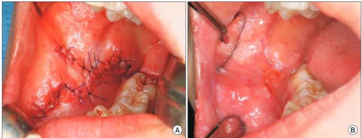
dilated cystic lesions containing erythrocytes under the epithelial tissue.(Fig. 2. A, 2. B) These characteristics were consistent with angiokeratoma. Under general anesthesia, the cheek lesion was completely excised, and the surgical defect was primarily closed with a cheek advancement flap.
(Fig. 3. A, 3. B) The patient was followed for more than three years without any evidence of recurrence.
To confirm the angiogenic characteristics of the tumor, the specimen was immunostained for VEGF and two of its receptors (VEGFR-1 and VEGFR-2). For immunohisto- chemical analysis, the tumor specimen was embedded in a paraffin block, cut into 4-μm sections, and mounted on glass slides. The sections were maintained at room temperature for 12 hours and deparaffinized. After hydration, immunohisto- chemical staining was performed using an automated immu- nostainer (BenchMark XT; Ventana Medical Systems Inc., Tucson, AZ, USA), and detection was performed using the UltraVIEW Universal DAB kit (Ventana Medical Systems Inc.) according to the manufacturer’s protocol. Briefly, sec- expression of vascular endothelial growth factor (VEGF) and
its two receptors, VEGFR-1 and VEGFR-2.
II. Case Report
An 18-year-old female patient was referred for an evalu- ation of a dark-colored, painless, elevated lesion in the oral cavity. Clinically, a 5×2-cm-sized, multi-lobular, irregular, raised lesion was observed on the right side of the buccal mucosa.(Fig. 1. A) The lesion was firm, did not bleed on ma- nipulation, and was not accompanied by mucosal ulceration.
The patient had no history of trauma. She had first recognized a small lesion on her cheek mucosa about three years prior, which had gradually increased in size. She had no history of systemic diseases or of skin or mucosal abnormalities.
Magnetic resonance imaging showed an ill-defined, heteroge- neously enhanced lesion on the right side of the buccal muco- sa.(Fig. 1. B, 1. C) A punch biopsy of the lesion showed epi- thelial hyperkeratosis, acanthosis, papillomatosis, and large
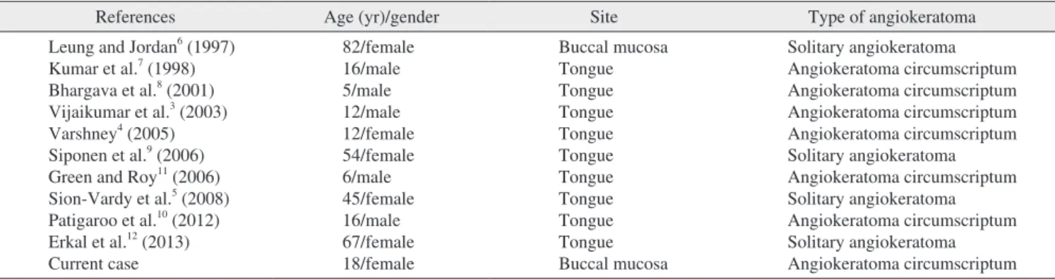
Table 1. Review of the literature on isolated angiokeratoma in the oral cavity
References Age (yr)/gender Site Type of angiokeratoma
Leung and Jordan6 (1997) Kumar et al.7 (1998) Bhargava et al.8 (2001) Vijaikumar et al.3 (2003) Varshney4 (2005) Siponen et al.9 (2006) Green and Roy11 (2006) Sion-Vardy et al.5 (2008) Patigaroo et al.10 (2012) Erkal et al.12 (2013) Current case
82/female 16/male 5/male 12/male 12/female 54/female 6/male 45/female 16/male 67/female 18/female
Buccal mucosa Tongue Tongue Tongue Tongue Tongue Tongue Tongue Tongue Tongue Buccal mucosa
Solitary angiokeratoma Angiokeratoma circumscriptum Angiokeratoma circumscriptum Angiokeratoma circumscriptum Angiokeratoma circumscriptum Solitary angiokeratoma Angiokeratoma circumscriptum Solitary angiokeratoma Angiokeratoma circumscriptum Solitary angiokeratoma Angiokeratoma circumscriptum Young-Hoon Kang et al: Angiokeratoma circumscriptum of the buccal mucosa: a case report and literature review. J Korean Assoc Oral Maxillofac Surg 2014
Fig. 1. Preoperative clinical and magnetic resonance imaging (MRI) findings. A. Primary lesions presented as multiple, small, irregular, raised lesions on the right side of the buccal mucosa. B, C. MRI shows an ill-defined heterogeneously enhanced lesion on the right side of the cheek (arrows).
Young-Hoon Kang et al: Angiokeratoma circumscriptum of the buccal mucosa: a case report and literature review. J Korean Assoc Oral Maxillofac Surg 2014
A B C
DAB+H2O2 substrate for 8 minutes, and nuclei were counter- stained in hematoxylin II and bluing reagent at 37oC. Immu- nostained sections were photographed under a Nikon Eclipse Ti-U microscope (Nikon Instruments Inc., Tokyo, Japan), and the proportion of positive staining was calculated for both endothelial and stromal cells in the lesion. At least three different specimens were used to calculate the proportion of positive cells. The primary antibodies used and the results of the immunohistochemical staining are summarized in Table 2.
Immunostaining revealed that the stromal cells in the lesion tions were deparaffinized using an EZ Prep solution (Ventana
Medical Systems Inc.) and then treated with CC1 standard (ethylenediaminetetraacetic acid [EDTA], pH 8.4) for 60 minutes at 100oC for antigen retrieval. Next, the slides were treated at 37oC for 4 minutes with UltraVIEW Universal DAB Inhibitor (3% H2O2) to block endogenous peroxidase activity. Slides were then incubated at 37oC first with each primary antibody for 32 minutes and then with the second- ary antibody (Universal HRP Multimer; Ventana Medical Systems Inc.) for 8 minutes. Finally slides were treated with
Fig. 3. Intraoperative and postoperative views. A. The cheek lesion was excised, and the surgical defect was primarily closed with an ad- vancement flap. B. Two months after the operation, the surgical site was healed uneventfully.
Young-Hoon Kang et al: Angiokeratoma circumscriptum of the buccal mucosa: a case report and literature review. J Korean Assoc Oral Maxillofac Surg 2014
A B
Fig. 2. H&E staining showing the histopathological features of the angiokeratoma circumscriptum lesion on the buccal mucosa (scale bars=200 μm). A. Low-power magnification shows epithelial hyperkeratosis, acanthosis, and large cystic lesions. B. High-power magnifi- cation shows erythrocytes filling the dilated cystic lesion. These pathological features are consistent with those of angiokeratoma circum- scriptum.
Young-Hoon Kang et al: Angiokeratoma circumscriptum of the buccal mucosa: a case report and literature review. J Korean Assoc Oral Maxillofac Surg 2014
B A
VEGFR-2 are shown in Table 2.
III. Discussion
The first case of angiokeratoma was reported by Mibelli in 1890, and the manifestation of the disorder found on fingers and toes is now known as angiokeratoma of Mibelli13. An- giokeratoma circumscriptum was first described in 1915 by Fabry as a localized lesion on a lower extremity13,14. It typi- and endothelial cells lining the pathologically dilated ves-
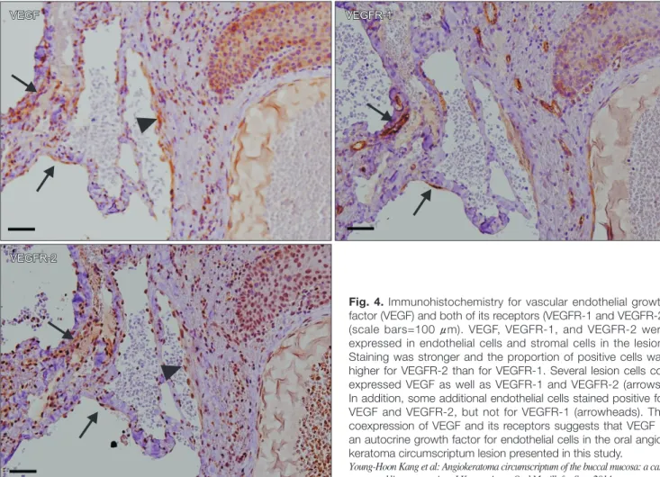
sels expressed VEGF and its two receptors (VEGFR-1 and VEGFR-2).(Fig. 4) Among both stromal and endothelial cells in the lesion, the percentage of positive-staining cells was higher for VEGFR-2 than that for VEGFR-1. Interestingly, some endothelial cells showed co-expression of VEGF and both of its receptors (arrows), whereas others co-expressed only VEGF and VEGF-2 (arrowhead). Proportions of the cells staining positive for each of VEGF, VEGFR-1, and
VEGF VEGFR-1
VEGFR-2
VEGFR-1
VEGFR-2 VEGF
Fig. 4. Immunohistochemistry for vascular endothelial growth factor (VEGF) and both of its receptors (VEGFR-1 and VEGFR-2) (scale bars=100 μm). VEGF, VEGFR-1, and VEGFR-2 were expressed in endothelial cells and stromal cells in the lesion.
Staining was stronger and the proportion of positive cells was higher for VEGFR-2 than for VEGFR-1. Several lesion cells co- expressed VEGF as well as VEGFR-1 and VEGFR-2 (arrows).
In addition, some additional endothelial cells stained positive for VEGF and VEGFR-2, but not for VEGFR-1 (arrowheads). The coexpression of VEGF and its receptors suggests that VEGF is an autocrine growth factor for endothelial cells in the oral angio- keratoma circumscriptum lesion presented in this study.
Young-Hoon Kang et al: Angiokeratoma circumscriptum of the buccal mucosa: a case report and literature review. J Korean Assoc Oral Maxillofac Surg 2014
Table 2. Primary antibodies, dilutions used, and the proportion of cells staining positive for each protein
Antibody Dilution Source Lot No. Positive-cell ratio (%)
Endothelial cells Stromal cells VEGF
VEGFR-1 VEGFR-2
1 : 400 1 : 400 1 : 400
Santa Cruz1 Abcam2 Abcam2
sc-152 ab9540 ab71772
68.1±6.0 22.8±3.8 81.8±2.9
37.3±3.1 11.1±1.9 72.8±1.7
(VEGF: vascular endothelial cell growth factor, VEGFR-1: vascular endothelial cell growth factor receptor-1, VEGFR-2: vascular endothelial cell growth factor receptor-2)
1Santa Cruz Biotechnology Inc., Dallas, TX, USA. 2Abcam, Cambridge, UK.
Values are presented as mean±standard deviation.
Young-Hoon Kang et al: Angiokeratoma circumscriptum of the buccal mucosa: a case report and literature review. J Korean Assoc Oral Maxillofac Surg 2014
the specimen analyzed in the present study, immunostaining intensity was stronger and the proportion of positive cells was higher for VEGFR-2 than for VEGFR-1, indicating en- hanced cell proliferation of endothelial cells. Additionally, the coexpression of VEGF and both of its receptors or only VEGF-2 was detected in some of the endothelial cells lining the pathologically dilated vessels. The coexpression of VEGF and VEGFRs by the endothelial cells in the lesion indicates that VEGF is an autocrine growth factor for these cells17. These results suggest that endothelial cells in the pathologi- cally dilated vessels possess VEGF autocrine growth activity involved in vasculogenesis and maintenance in the angio- keratoma lesion.
Conflict of Interest
No potential conflict of interest relevant to this article was reported.
References
1. Schiller PI, Itin PH. Angiokeratomas: an update. Dermatology 1996;193:275-82.
2. Ozdemir R, Karaaslan O, Tiftikcioglu YO, Kocer U. Angiokera- toma circumscriptum. Dermatol Surg 2004;30:1364-6.
3. Vijaikumar M, Thappa DM, Karthikeyan K, Jayanthi S. An- giokeratoma circumscriptum of the tongue. Pediatr Dermatol 2003;20:180-2.
4. Varshney S. Angiokeratoma circumscriptum of the tongue. Int J Dermatol 2005;44:886-8.
5. Sion-Vardy N, Manor E, Puterman M, Bodner L. Solitary an- giokeratoma of the tongue. Med Oral Patol Oral Cir Bucal 2008;13:E12-4.
6. Leung CS, Jordan RC. Solitary angiokeratoma of the oral cavity.
Oral Surg Oral Med Oral Pathol Oral Radiol Endod 1997;84:51-3.
7. Kumar MV, Thappa DM, Shanmugam S, Ratnakar C. Angio- keratoma circumscriptum of the oral cavity. Acta Derm Venereol 1998;78:472.
8. Bhargava P, Bhargava S, Mathur D, Agarwal US, Bhargava R.
Angiokeratoma of tongue. Indian J Dermatol Venereol Leprol 2001;67:270.
9. Siponen M, Penna T, Apaja-Sarkkinen M, Palatsi R, Salo T. Solitary angiokeratoma of the tongue. J Oral Pathol Med 2006;35:252-3.
10. Patigaroo S, Khan N, Manzoor S, Gupta N, Jain P, Shakeel M. Iso- lated multiple angiokeratoma of tongue--a case report and review of literature. Int J Pediatr Otorhinolaryngol Extra 2012;7:126-8.
11. Green JB, Roy S. Angiokeratoma circumscriptum of the dorsal tongue in a child. Int J Pediatr Otorhinolaryngol Extra 2006;1:107-9.
12. Erkal EY, Karabey MS, Vural Ç, Mutlu F, Aksu G, Sarper B, et al. Solitary angiokeratoma of the tongue in an adult patient treated with intensity modulated radiation therapy. Am J Otolaryngol 2013;34:582-5.
13. Mittal R, Aggarwal A, Srivastava G. Angiokeratoma circumscrip- tum: a case report and review of the literature. Int J Dermatol 2005;
44:1031-4.
14. Fabry J. Uber einen fall von angiokeratoma circumscriptum am linken oberschenkel. Derm Z 1915;22:1-4.
15. Kobayashi T, Sakuraoka K. A case of angiokeratoma circumscrip-
cally occurs unilaterally on the lower legs or feet, but it has also been observed on the thighs, buttocks, or elsewhere. The lesions are deep red to blue-black in color and are usually elevated, warty, purplish and compressible papules13. In most cases, angiokeratoma circumscriptum is present at birth, but in some cases, it develops during childhood or adulthood4,7.
Oral mucosal involvement is most commonly detected as a component of angiokeratoma corporis diffusum (Fabry disease and fucosidosis). However, the oral mucosa is very seldom involved in other types of angiokeratoma3,4. In ad- dition to the present case, a review of the literature revealed ten cases of isolated angiokeratoma that occurred in the oral cavity.(Table 1) Of those, four cases were solitary angiokera- toma and six were angiokeratoma circumscriptum. Except for one case of solitary angiokeratoma that involved the buccal mucosa6, the remaining nine occurred on the tongue (patho- logically, six were angiokeratoma circumscriptum and three were solitary angiokeratoma)3-5,7-12. To our knowledge, the present report is the second case published on isolated oral angiokeratoma confined to the buccal mucosa and the first case report on angiokeratoma circumscriptum involving the buccal mucosa.
The histopathological features of angiokeratoma in the oral cavity are similar to those in skin lesions, which are hyper- keratosis, acanthosis, papillomatosis of the squamous epithe- lium, and dilated subepithelial blood vessels with thrombosis5. The pathogenesis of angiokeratoma is still unknown. It has been thought that altered hemodynamics, such as local injury to papillary capillaries, cause telangiectatic lesions in the papillary dermis with overlying reactive hyperkeratosis and acanthosis of the epidermis3,4. Interestingly, matrix metal- loproteinase-9 (MMP-9) has been identified in the epidermis, particularly in hyperkeratotic lesions of angiokeratoma cir- cumscriptum, by immunohistochemistry15. The expression of MMP-9 may be related to the hyperkeratotic changes in angiokeratoma3,15.
In the surgical specimen analyzed in the present study, VEGF and its two receptors (VEGFR-1 and VEGFR-2) were expressed in the endothelial cells of dilated vessels. VEGF has been characterized as a heparin-binding angiogenic growth factor that plays a critical role in angiogenesis and vasculogenesis during tumor growth and tissue regenera-
tion16,17. Of the several VEGF receptors, VEGFR-1 is pri-
marily involved in cell migration and vascular maintenance rather than cell proliferation, whereas VEGFR-2 participates directly in regulating cell mitosis and proliferation; however, their precise roles have not been completely elucidated16. In
17. Masood R, Cai J, Zheng T, Smith DL, Hinton DR, Gill PS. Vascu- lar endothelial growth factor (VEGF) is an autocrine growth factor for VEGF receptor-positive human tumors. Blood 2001;98:1904- 13.
tum: immunolocalization of matrix metalloproteinase (MMP)-9. J Dermatol 1998;25:391-4.
16. Neufeld G, Cohen T, Gengrinovitch S, Poltorak Z. Vascular endo- thelial growth factor (VEGF) and its receptors. FASEB J 1999;13:
9-22.