171
브루너샘 샘종은 소장에 발생하는 드문 양성 종양으로상부위장관 내시경 검사에서 발견되는 십이지장 양성 종 양의 약 10%에서 관찰된다.1 대부분 크기는 1 ㎝를 넘지 않으며 용종형으로 다발성을 보이나 1 ㎝ 이상의 브루너샘 샘종은 대개 단일성이며 출혈과 폐쇄, 장중첩 등의 합병증 을 유발할 수 있는데 최근 악성화에 대해서도 보고되고 있다.2 현재까지 국내에서 8 ㎝ 이상의 브루너샘 샘종을 보고한 증례는 2 예가 있었으며 각각은 복부 불편감과 흑색변을 주소로 내원한 경우이나,3,4 본 증례와 같이 급성 출혈을 동반한 8cm 이상의 브루너샘 샘종은 보고된 바가 없다. 저자들은 만성적인 소화불량과 흑색변을 주소로 내
원한 환자에서, 급성 출혈을 동반한 약 8 cm 크기의 십이지 장의 브루너샘 샘종을 IT (insulated tip) 절개도를 이용하 여 내시경적으로 치료한 1 예를 경험하였기에 문헌 고찰과 함께 보고하는 바이다.
증례
환자: 46세 남자 박 O O 주소: 흑색변
현 병력: 환자는 수년 동안 악화와 호전을 반복하는 소화불량 및 조기포만감이 있었으며 특별한 치료 없이 지내던 중 내원 3일 전부터 발생한 흑색변을 주소로 내원
Kosin Medical Journal 2015;30:171-174.
http://dx.doi.org/10.7180/kmj.2015.30.2.171 KMJ
Case Report
A Case Of Huge Brunner’s Gland Adenoma With Acute Bleeding Treated By Endoscopic Resection
Pyung Kang Park
1, Woo-Cho Chung
1, Kyoung Yong Lee
1, Sung Hak Lee
1, Jae Jung Jang
2, Seungchul Suh
1Department of
1Internal Medicine and
2Pathology, Anyang SAM hospital, Anyang-si, Gyeonggi-do, Korea.
내시경적 절제술로 치료한 급성 출혈을 동반한 거대 브루너샘 샘종 1예
박평강1, 정우조1, 이경용1, 이성학1, 장재정2, 서승철1
안양 샘병원
1내과,
2병리과
Brunner’s gland adenoma is a rare benign small bowel neoplasm and it represents 10% of small bowel benign tumor. Most of adenoma manifest as polypoidal, multiple and size does not exceed 1 cm and mostly asymptomatic, but the lesion larger than 1 ㎝ is solitary and can cause bleeding, obstruction, intussusception and there are some reports of showing malignant transformation. Until the present, there are two cases of over 8㎝ huge Brunner’s gland adenoma in Korea and each of their chief complaint was abdominal discomfort and melena, but there is no case report of over 8 ㎝ Brunner’s gland adenoma accompanied with acute bleeding as seen in this case. We diagnosed an 8 ㎝ sized, huge duodenal Brunner’s gland adenoma which accompanied with acute bleeding and treated it by endoscopic resection using an IT-knife, successfully.
Key Words: adenoma, bleeding, Brunner’s gland, endoscopy
Corresponding Author: Seungchul Suh, M.D. Department of Internal medicine, Anyang SAM hospital, 9, Samdeok-ro, Manan-gu, Anyang-si, Gyeonggi-do 14030, Korea
Tel: +82-31-467-9110, Fax:+82-31-467-9198, E-mail: [email protected]
Received:
Revised:
Accepted:
Nov. 5, 2014
Jan. 30, 2015
Feb. 24, 2015
Kosin Medical Journal 2015;30:171-174.
172
하였다.과거력: 특이 소견 없음
사회력: 흡연력과 음주력은 없었다.
이학적 관찰: 내원 당시 혈압은 120/70 mmHg, 맥박 72회/분, 호흡 20회/분, 체온은 36.8℃였으며 전신쇠약감 을 호소하였다. 결막은 창백하지 않았고 공막에 황달 소견 보이지 않았으며 혀의 탈수 소견은 없었다. 흉부청진에서 폐음 및 심음 모두 정상이었고 부잡음은 들리지 않았다.
복부 촉진 시 심와부에 경한 압통이 있었고 장음은 약간 항진되어 있었으나 만져지는 종괴는 없었다.
검사 소견: 말초혈액검사에서 혈색소 11.8 g/dL, 헤마 토크릿 28.2%, 백혈구 7,000 /uL, 혈소판 179,000 /uL였 고 C 반응단백 0.6 mg/L였다. 일반화학검사에서 BUN 15 mg/dL, 크레아티닌 1.0 mg/dL, AST/ALT 20/38 U/
L, 총 콜레스테롤 165 mg/dL, 총 단백 7.2 g/dL, 알부민 4.5 g/dL였다. 혈액응고검사에서 PT 10.8 sec, aPTT 27.2 sec였다.
영상학적 소견: 흉부 및 복부 X선 검사는 정상 소견이었 다.
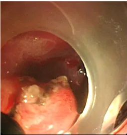
치료 및 경과: 상부 위장관 내시경 검사에서 십이지장의 구부에 기시부를 두고 제 2부위까지 걸쳐서 관찰되는 거대 용종이 신선혈과 함께 관찰되었고 이곳에서의 출혈이 확 인되었다(Fig. 1). 용종 경부의 하부 1/3 지점을 IT 절개도 를 이용하여 내시경적 점막 절제술을 시행하였고(Fig. 2)
Fig. 1. Endoscopic finding. It shows a huge pedunculated polyp with bleeding arising from the bulb.
Fig. 4. Histological finding. The polyp consists of glands, dilated cystic ducts, adipose tissue and lymphoid cells revealing as hamartoma.(H&E staining, x 100)
Fig. 2. Endoscopic finding. It shows a cross-section of the resected stalk using an IT knife
Fig. 3. Gross finding. The polyp is about 80mm long and
35mm wide.
huge Brunner’s gland adenoma
173
절단면의 삼출성 출혈에 대하여 전기적 소작술로 지혈하였다. 절제된 표본은 크기로 인해 유문륜 밖으로의 통과가 불가능하여 절개도를 이용하여 일부를 절제한 후에 올가 미로 포획하여 회수하였다. 종양의 크기는 약 8.0 x 3.5
㎝으로 측정되었고(Fig. 3) 병리조직 검사에서 도관, 지방 조직, 그리고 림프양 세포들이 혼합, 산재되어 있는 브루너 샘 과오종으로 진단되었다(Fig. 4). 시술 후 출혈 및 천공 등의 합병증 없이 퇴원하였다. 1년 후 외래에 방문 하였을 때 환자는 이전의 소화불량 및 조기포만감은 호전되었다 고 하였다.
고찰
브루너샘은 1688년 해부학자인 Brunner에 의하여 처음 지칭된 점막하 점액 분비샘으로 알칼리성 액체인 유로가스트 론 (urogastrone), 중탄산염, 창자가스트론 (enterogastrone) 등을 분비하여 위산으로부터 십이지장 점막을 보호한다.
5브루너샘 샘종은 이러한 브루너샘이 과증식으로 발생하는 십이지장 양성 종양 중의 하나로 19세기 말에 Cruveilhier가 처음으로 기술한 질환이다. 이 질환은 40-70대에 흔히 발견 되며 유병률에 있어 성별간 차이는 없다고 알려져 있다.
61934년 Feyrter는 2,800 예의 십이지장 해부연구에서 브루너 샘 과증식을 해부학적 형태에 따라서 분류하였는데 이는 미 세 다발성 결절로 십이지장 유두 상부에 국한되어 나타나는 미만성 결절성 증식형, 고립 결절 형태로 나타나는 국한성 결절성 증식, 크고 유경성인 선종성 과형성 세 가지로 분류된 다.
7조직학적으로는 과증식된 정상 브루너샘들이 소엽 형태 를 보이는 브루너샘 과증식과, 분비샘의 증식 외에도 도관, 지방조직, 근육조직 또는 간혹 이소성 췌장 조직까지도 포함 하는 브루너샘 과오종 두 가지로 나뉜다. 본 증례는 해부학적 으로는 유경성 선종성 과형성을 보이는 샘종이면서 조직학적 으로는 브루너샘 과오종으로 분류되는 경우로 볼 수 있다.
브루너샘 샘종의 병태생리에 대해서는 명확하게 알려져 있지 않으나 위산 과다증이 중요한 영향을 미치는 것으로 여겨지고 있으며, 만성 췌장염, 요독증, 소화성 궤양 및 헬리 코박터 감염증 등도 브루너샘 샘종의 유병률을 증가시키는
원인으로 알려져 있다.
8브루너샘 샘종은 십이지장 구부에 호발하며, 50%에서 임 상 증상을 야기하지만 대부분 비특이적 위장관 증세를 보이 며 드물게 출혈과 장폐쇄, 장중첩증과 같은 합병증을 유발한 다. 출혈은 종양 표면에 궤양, 미란이 있는 경우 증상을 보이 는 환자의 37%에서 나타날 수 있으며 종괴가 유경성인 경우 증상을 보이는 환자의 37%에서 장폐쇄을 유발하여 오심, 구토, 복부팽만감, 조기팽만감 등의 증상을 발생시킬 수 있 다.
6이번 증례는 미란을 동반한 유경성 거대 용종으로서 만성적인 소화 불량, 조기포만감과 함께 급성 위장관 출혈의 합병증을 유발한 경우라고 할 수 있다.
브루너샘 샘종은 내시경 검사를 통해 진단할 수 있으며 내시경 생검이 점막하층 까지 도달하지 못할 경우 위음성이 나타날 수 있으므로 고식적인 생검보다는 계제생검 (snare biopsy)이 요구된다. 내시경 초음파 검사가 도움이 되는데 경계가 비교적 명확한 종괴가 제3층인 점막하층에서 관찰되 며, 단발성 또는 다낭성 낭종 혹은 고형 형태의 종양 등 다양한 에코발생을 보이는 것이 특징이다.
9전산화단층촬영에서는 조영 증강을 보이는 저음영 병변으로 관찰될 수 있으나 진단 에 특이적이지 않다.
10브루너샘 샘종의 감별 질환으로는 지 방종, 평활근종, 신경종, 유암종, 이소성 췌장, 샘암 등이 있으 며 조직검사로서 확진할 수 있다.
11브루너샘 샘종의 치료는 증상을 유발하거나 합병증이 발 생한 경우에 제거하는 것이 원칙이나 최근 악성화에 대한 보고가 증가함에 따라 가능한 제거할 것을 권장하고 있다.
1,2Sakurai 등은 정확한 기전은 밝혀지지 않았으나 브루너샘 샘종의 크기의 증가가 점막 표면의 궤양형성을 유발하여 이 형성이나 악성변화를 나타낼 가능성이 비교적 높다고 하였고 이형성과 악성화는 위의 유두 소와세포 화생 (papillary foveolar metaplasia)과 관련이 있다고 하였는데,
12본 증례에 서는 크기는 8 ㎝ 이상이었으나 궤양을 동반하지 않았고, 이형성이나 악성변화는 관찰되지 않았다. 위산을 억제하는 내과적 치료는 브루너샘 과증식의 퇴화가 거의 없어 효과적 이지 않으며 용종의 크기 및 내시경적 접근성에 따라 내시경 적 절제술 또는 수술을 시행 할 수 있다.
국내에서 최근에 보고된 거대 브루너샘 종양 39 예의 임상
Kosin Medical Journal 2015;30:171-174.
174
및 고찰 연구에서 보면 대부분의 브루너샘 종양은 1-2 ㎝ 정도의 크기였다.
13이중 5 ㎝ 이상은 6 예에서 관찰되었는데 4 예에서 수술하였고 2 예에서 내시경으로 절제하였다.
13저자들은 급성 출혈의 합병증을 동반한 8 ㎝ 크기의 거대 브루너샘 샘종을 IT 절개도를 이용하여 성공적으로 제거하였 다. 본 증례와 같이 크기가 큰 경우에도 위치나 내시경적 접근의 유용성에 따라 수술이 아닌 내시경적 치료를 일차적 으로 고려해 볼 수 있을 것이다.
REFERENCES