281
Copyrights © 2013 The Korean Society of Radiology
INTRODUCTION
Acoustic schwannoma, the most common benign neoplasm of the internal auditory canal (IAC) and the cerebellopontine angle (CPA), arises from the perineural Schwann cells surrounding the vestibular and cochlear nerves. It most commonly arises near the fundus of the IAC at the Schwann cell-glial junction, but can be also found anywhere along the nerve from the IAC to the terminal ends of the vestibulocochlear nerve, within the vestibule, cochlea, and semicircular canals (1, 2). Intralabyrinthine schwannoma, an uncommon tumor with the likely underestimated incidence rate, develops from the Schwann cells of the intralabyrinthine branches of the vestibulocochlear nerve (1, 2). Recently, the imaging identifi- cation of intralabyrinthine schwannoma has increased along with
the use of the higher field magnets and the evolving magnetic resonance (MR) imaging sequences. As the rarest form of intral- abyrinthine schwannoma, transotic schwannoma is called when the neoplasm extends through the labyrinth into the IAC and the middle ear cavity (MEC). Transotic schwannoma is extremely rare. Only five cases have been reported in the English literature (1-5), while none has appeared in the Korean radiological litera- ture. We report a case of transotic schwannoma in a 38-year-old woman along with the CT, MRI, and pathologic findings.
CASE REPORT
A 38-year-old woman presented with an aggravated tinnitus and dizziness in the recent 2 days. She had experienced a pro-
Case Report
pISSN 1738-2637
J Korean Soc Radiol 2013;68(4):281-284 http://dx.doi.org/10.3348/jksr.2013.68.4.281
Received December 25, 2012; Accepted January 24, 2013 Corresponding author: Sang Kwon Lee, MD Department of Radiology, Dongsan Medical Center, Keimyung University School of Medicine, 56 Dalseong-ro, Jung-gu, Daegu 700-712, Korea.
Tel. 82-53-250-7735 Fax. 82-53-250-7766 E-mail: sklee@dsmc.or.kr
This is an Open Access article distributed under the terms of the Creative Commons Attribution Non-Commercial License (http://creativecommons.org/licenses/by-nc/3.0) which permits unrestricted non-commercial use, distri- bution, and reproduction in any medium, provided the original work is properly cited.
Transotic schwannoma is a condition extremely rare, and we herein report the CT, MRI, and pathologic findings of a transotic schwannoma case in a 38-year-old woman. The lesion was identified as an expansile mass in the internal auditory ca- nal (IAC) and a mass in the middle ear cavity (MEC) on the high-resolution, bone al- gorithm, temporal bone CT. It was shown as the following: the hypointense masses of the cerebellopontine angle (CPA)-IAC and MEC, and the hypointense replacement of the normal high-signal intensity fluid of the cochlea, vestibule, and semicircular canals on the 3-dimensional (3D) fast imaging, employing the steady-state acquisi- tion (FIESTA) sequence; and the intensely enhancing masses of the CPA-IAC and MEC, and the intense enhancement of the cochlea, vestibule, and semicircular ca- nals on the 3D gadolinium-enhanced spoiled gradient-recalled echo (SPGR) se- quence. The mass was removed via a transotic approach. The pathologic findings were consistent with schwannoma. The assessment of the extent of transotic schwannoma may be enhanced by the 3D FIESTA and the gadolinium-enhanced SPGR sequences.
Index terms
Transotic Schwannoma Tomography, X-Ray Computed Magnetic Resonance Imaging
The CT and Magnetic Resonance Imaging Features of Transotic Schwannoma: A Case Report
1경이 신경초종의 전산화단층촬영 및 자기공명영상 소견: 증례 보고1
Sang Kwon Lee, MD
1, Mi Sun Choe, MD
2Departments of 1Radiology, 2Pathology, Dongsan Medical Center, Keimyung University School of Medicine, Daegu, Korea
The CT and MR Imaging Features of Transotic Schwannoma
submit.radiology.or.kr
J Korean Soc Radiol 2013;68(4):281-284
282
kee, WI, USA). The axial 3-dimensional (3D) fast imaging em- ploying the steady-state acquisition (FIESTA) sequence [repeti- tion time (TR)/echo time (TE) = 4.6/1.4; flip angle = 45; matrix number = 384 × 256; number of excitation = 1; slice thickness = 1.0 mm] revealed an approximately 12 × 22 × 10-mm-sized, hy- pointense mass in the left CPA-IAC, a small hypointense mass in the MEC, and a hypointense replacement of the normal high- signal intensity fluid of the cochlea, vestibule, and semicircular canals (Fig. 1B). The gadolinium (Gd)-enhanced axial and coro- nal 3D spoiled gradient-recalled echo (SPGR) sequence (TR/TE
= 8.2/3.3; flip angle = 20; matrix number = 256 × 224, number of excitation = 2; slice thickness = 1.2 mm) demonstrated the heterogeneous, intensely enhancing masses in the CPA-IAC and gressive hearing loss of the left ear and dizziness for 7 years,
along with the intermittent tinnitus and otalgia of the left ear for 1 year. She denied otorrhea, vertigo, or headaches, and her facial expression was intact. The otoscopic examination revealed a mass behind the tympanic membrane. The axial and coronal, high-resolution, bone algorithm temporal bone CT, performed using the Sensation 16 scanner (Siemens Healthcare, Forch- heim, Germany) with the slice thickness of 0.8 mm, showed an expansile mass in the left IAC and a small mass in the MEC (Fig. 1A), while the cochlea, vestibule, and semicircular canals appeared to be grossly intact (Fig. 1A). A temporal bone MRI was performed for a better assessment of the extent of the lesion by using a 3.0-T unit (Signa Excite; GE Medical System, Milwau-
F
B C
G
D
E A
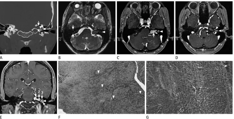
Fig. 1. CT, MRI, and pathologic features of transotic schwannoma in a 38-year-old woman.
A. A coronal high-resolution temporal bone CT image shows an expansile mass in the left internal auditory canal (IAC) (white arrow), and a mass in the left middle ear cavity (MEC) (black arrow) extending from the inner ear through the oval window (arrowhead).
B. An axial 3-dimensional (3D) fast imaging employing the steady-state acquisition (FIESTA) image reveals a hypointense mass in the left cerebel- lopontine angle (CPA)-IAC (black arrow), and hypointense replacement of normal high signal intensity fluid of the left lateral semicircular canal (arrowhead). Note normal high signal intensity of the right superior and posterior semicircular canals (white arrows).
C. A gadolinium (Gd)-enhanced axial 3D spoiled gradient-recalled echo (SPGR) image demonstrates heterogeneous, intensely enhancing mass (arrow) in the left CPA-IAC, and intense enhancement of the left lateral semicircular canal (arrowhead).
D. A Gd-enhanced axial 3D SPGR image shows an enhancing mass in the left MEC (white arrow) extending from the inner ear through the round window (black arrow). Also noted is intense enhancement of the left cochlea (arrowhead).
E. A Gd-enhanced coronal 3D SPGR image shows the components of transotic schwannoma consisting of a dumbbell-shaped mass of the left CPA (arrow 1)-IAC (arrow 2), intense enhancement of membranous labyrinth (arrow 3), and a mass in the MEC (arrow 4) extending from the in- ner ear through the oval window (arrowhead).
F, G. A photomicrograph of histological examination (F) of the specimen obtained by surgical resection demonstrates proliferation of the spindle cells in short bundles and nuclear palisading (arrowheads) (hematoxylin-eosin, × 200), and a photomicrograph of immunohistochemical staining (G) reveals strong positivity for S-100 protein in the cytoplasm and nuclei of tumor cells (original magnification, × 200), which are consistent with schwannoma.
123
4
Sang Kwon Lee, et al
submit.radiology.or.kr J Korean Soc Radiol 2013;68(4):281-284
283
and MEC in all patients referred for SNHL. Salzman et al. (2) have revealed intralabyrinthine schwannoma by using the high- resolution, thin-section fast spin-echo (FSE) T2-weighted imag- ing and the Gd-enhanced spin-echo (SE) T1-weighted imaging.
The lesions of intralabyrinthine schwannoma were identified as the filling defects with a replacement of the normal high-signal intensity fluid of the membranous labyrinth on the high-resolu- tion, thin-section FSE T2-weighted images, and the focal homo- geneously enhancing mass on the Gd-enhanced SE T1-weighted images (2). With the development of the high field magnets and the refined MR imaging sequences, more cases of intralabyrin- thine schwannoma may be detected with the MRI. The FIESTA sequence can provide a strong T2 contrast, which emphasizes the cerebrospinal fluid signals. In addition, this sequence has a high signal-to-noise ratio and the inherent flow compensation, and is suitable for the direct 3D imaging (6). Therefore, this se- quence can depict small structures surrounded by the cerebro- spinal fluid (7) and are thus used to evaluate the posterior fossa lesions, such as acoustic schwannomas (8). We have used the 3D thin-slice, high-resolution FIESTA and the Gd-enhanced SPGR sequences, which may have enhanced the delineation of the ex- tent of transotic schwannoma. In our case, transotic schwanno- ma was excellently demonstrated as a mass in the CPA-IAC and MEC and a hypointense replacement of the normal high-signal intensity fluid within the membranous labyrinth. These findings corresponded to the intensely enhancing masses in the CPA- IAC and MEC, and an intense enhancement of the membra- nous labyrinth. Notably, the extension of the schwannoma from the inner ear to the MEC occurred via the round and oval win- dows, which was excellently demonstrated by the high-resolu- tion, bone algorithm temporal bone CT and the Gd-enhanced 3D SPGR sequence, and was confirmed by the surgical findings.
Intralabyrinthine schwannoma is usually managed by obser- vation with the serial MR imaging. Surgery is indicated for in- tractable vertigo, extension of tumor to the CPA, or evidence of the tumor growth in patients who are medically fit for surgery.
In our case, the surgical resection was performed, because of the extension of the tumor into the IAC, CPA, and MEC. In sum- mary, transotic schwannoma, an extremely rare entity, repre- sents a tumor within the labyrinth with the extension into the IAC and MEC. Extension of the schwannoma from the inner ear into MEC may occur via the round and oval windows. The MEC, and an intense enhancement of the cochlea, vestibule,
and semicircular canals (Fig. 1C-E). The coronal Gd-enhanced 3D SPGR sequence revealed a dumbbell-shaped mass involving the CPA and IAC (Fig. 1E). The extension of the mass from the labyrinth to the MEC was thought to occur through the round and oval windows, which was excellently demonstrated on the high-resolution, bone algorithm temporal bone CT (Fig. 1A) and the Gd-enhanced 3D SPGR sequence (Fig. 1D, E). Under the impression of transotic schwannoma, she underwent a sur- gical resection of the mass via the transotic approach, and was found to have a tumor involving the CPA, IAC, whole membra- nous labyrinth, and MEC. The mass extended from the laby- rinth through the round and oval windows to the MEC. The histological and immunohistochemical findings of the resected specimen from the CPA, IAC, membranous labyrinth, and MEC were consistent with schwannoma (Fig. 1F, G). The post- operative course was unremarkable with intact facial nerve functions. She was followed-up clinically and with MR imaging without any evidence of recurrence until 22 months after the operation.
DISCUSSION
The management of acoustic schwannoma involves a surgical resection with a goal of preserving the hearing and facial nerve functions. When the schwannoma involves the inner ear, a hearing-preservation surgery is not an option, as removing the tumor from the labyrinth is expected to result in a profound sensorineural hearing loss (SNHL) (1). As such, the accurate preoperative delineation of the tumor extent is prerequisite.
Kennedy et al. (1) and Salzman et al. (2) have classified intralab- yrinthine schwannoma into six categories: intracochlear (a tu- mor confined to the cochlea), intravestibular (a tumor centered in the vestibule with or without extension into the semicircular canals), vestibulocochlear (a tumor within the vestibule and co- chlea), transmodiolar (a tumor centered in the cochlea with ex- tension through the modiolus into the IAC), transmacular (a tumor centered in the vestibule with extension into the IAC via the macula cribrosa), and transotic (a tumor within the laby- rinth with extension into the IAC and MEC). To make the accu- rate diagnosis of transotic schwannoma, the radiologist needs to carefully interrogate not just the CPA and IAC, but the labyrinth
The CT and MR Imaging Features of Transotic Schwannoma
submit.radiology.or.kr
J Korean Soc Radiol 2013;68(4):281-284
284
4. Amoils CP, Lanser MJ, Jackler RK. Acoustic neuroma pre- senting as a middle ear mass. Otolaryngol Head Neck Surg 1992;107:478-482
5. Stoney PJ, Rutka J, Dolan E, Hawke M. Acoustic neuroma presenting as a middle ear mass. J Otolaryngol 1991;20:
141-143
6. Tsuchiya K, Aoki C, Hachiya J. Evaluation of MR cisternog- raphy of the cerebellopontine angle using a balanced fast-field-echo sequence: preliminary findings. Eur Radiol 2004;14:239-242
7. Mikami T, Minamida Y, Yamaki T, Koyanagi I, Nonaka T, Houkin K. Cranial nerve assessment in posterior fossa tu- mors with fast imaging employing steady-state acquisi- tion (FIESTA). Neurosurg Rev 2005;28:261-266
8. Stuckey SL, Harris AJ, Mannolini SM. Detection of acoustic schwannoma: use of constructive interference in the steady state three-dimensional MR. AJNR Am J Neurora- diol 1996;17:1219-1225
thin-section, high-resolution, 3D FIESTA and the Gd-enhanced SPGR sequences under a 3.0-T unit may enhance the assess- ment of the extent of the transotic schwannoma.
REFERENCES
1. Kennedy RJ, Shelton C, Salzman KL, Davidson HC, Harns- berger HR. Intralabyrinthine schwannomas: diagnosis, man- agement, and a new classification system. Otol Neurotol 2004;25:160-167
2. Salzman KL, Childs AM, Davidson HC, Kennedy RJ, Shelton C, Harnsberger HR. Intralabyrinthine schwannomas: imag- ing diagnosis and classification. AJNR Am J Neuroradiol 2012;33:104-109
3. Tran Ba Huy P, Hassan JM, Wassef M, Mikol J, Thurel C.
Acoustic schwannoma presenting as a tumor of the exter- nal auditory canal. Case report. Ann Otol Rhinol Laryngol 1987;96:415-418
경이 신경초종의 전산화단층촬영 및 자기공명영상 소견: 증례 보고1
이상권
1· 최미선
2경이 신경초종은 극히 드물다. 저자들은 38세 여자에서 발생한 경이 신경초종 1예의 CT, MRI 및 병리 소견을 보고하고자 한다. 병변은 고해상도 골 알고리즘 측두골 CT에서 내이도의 팽창성의 종괴와 중이의 종괴로 관찰되었고, 3차원 fast imaging employing the steady-state acquisition (FIESTA) 영상에서 소뇌교각-내이도 및 중이의 저신호강도의 종괴와 달 팽이관, 전정기관, 세반고리관의 정상적인 물에 의한 고신호강도가 저신호강도로 대치되는 소견으로 나타났으며, 3차원 조영증강 후 spoiled gradient-recalled echo (SPGR) 영상에서 소뇌교각-내이도 및 중이에 강한 조영증강을 보이는 종괴 와 달팽이관, 전정기관, 세반고리관의 강한 조영증강으로 관찰되었다. 종괴는 경이적 접근에 의해 제거되었으며, 병리 소 견은 신경초종에 합당하였다. 경이 신경초종의 범위를 평가하는데 있어서 3차원 FIESTA 영상과 조영증강 후 SPGR 영상 은 그 정확도를 높여줄 것으로 생각된다.
계명대학교 의과대학 동산의료원 1영상의학과, 2병리과