Langerhans Cell Histiocytosis of the Clavicle in a 50-Year-Old Male: A Case Report
50세 남자에게서 발견된 쇄골의 랑게르한스 세포 조직구증:
증례 보고
Changhyun Park, MD1 , Yong Hoon Kim, MD1* , Soon Joo Cha, MD1 , Ji-Ye Kim, MD2
Departments of 1Radiology and 2Pathology, Ilsan Paik Hospital, Inje University, Goyang, Korea
Langerhans cell histiocytosis (LCH) is a rare condition that usually occurs in children and com- monly affects the skeletal system. It is extremely rare in adults, especially in the clavicles. In this report, we describe a pathologically confirmed case of LCH in the clavicle of a 50-year-old male.
We report various radiological findings, such as plain radiography, CT, MR, and PET-CT, along with a review of the literature.
Index terms Langerhans Cell Histiocytosis; Clavicle; Middle Age; Adult
INTRODUCTION
Langerhans cell histiocytosis (LCH) is a rare disease characterized by the abnormal proliferation of tissue macrophages called Langerhans cells. LCH predominantly oc- curs in children and approximately 80% of the cases occur in individuals under 15 years of age, with a predilection for bones within the skull, spinal column, ribcage, and
Received June 5, 2020 Revised September 23, 2020 Accepted October 15, 2020
*Corresponding author Yong Hoon Kim, MD Department of Radiology, Ilsan Paik Hospital, Inje University,
170 Juhwa-ro, Ilsanseo-gu, Goyang 10380, Korea.
Tel 82-31-910-7395 Fax 82-31-910-7366 E-mail [email protected] This is an Open Access article distributed under the terms of the Creative Commons Attribu- tion Non-Commercial License (https://creativecommons.org/
licenses/by-nc/4.0) which permits unrestricted non-commercial use, distribution, and reproduc- tion in any medium, provided the original work is properly cited.
ORCID iDs Changhyun Park https://
orcid.org/0000-0001-5558-3096 Yong Hoon Kim
https://
orcid.org/0000-0002-6276-9515 Soon Joo Cha
https://
orcid.org/0000-0002-3254-0189 Ji-Ye Kim
https://
orcid.org/0000-0003-4291-2967
CASE REPORT
A 50-year-old male visited our hospital with complaints of pain and discomfort in the mid- portion of his right clavicle. He reported that the symptoms started 3 months previously after this region was compressed by someone and had persisted since then. There was no history of trauma, fever, chills, night sweats, or weight loss.
Physical examination showed local tenderness in the mid-portion of the right clavicle.
There was no swelling or erythema of the skin in this region, and other findings of the physi- cal examination were unremarkable.
Laboratory examination showed mildly elevated levels of serum cholesterol. The results of other blood tests and urinalysis were within normal limits.
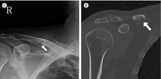
Plain radiography of the right clavicle showed a small osteolytic lesion with a narrow tran- sitional zone located in the inferior portion of the mid-body of the clavicle, with a definite cortical disruption. No sclerotic margins were observed (Fig. 1A).
CT scan revealed a focal osteolytic lesion with cortical disruption and an inferiorly protrud- ing soft tissue mass along the inferior margin of the mid-portion of the right clavicle (Fig. 1B).
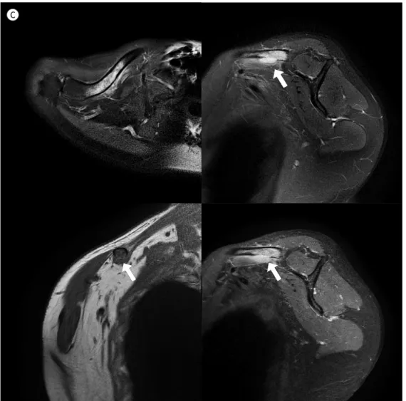
Subsequently, an MRI scan demonstrated a focal mass of a high signal intensity on a fat- saturated T2-weighted image and a low signal intensity on a T1-weighted image with an ill- defined margin in the mid-portion of the right clavicle near the inferior margin. Adjacent bone marrow showed diffuse high signal intensity on fat-saturated T2-weighted images. Cor- tical breakdown of the adjacent inferior margin of the right clavicle with nodular soft tissue extension was also observed on MRI. T1-weighted, fat-suppressed, contrast-enhanced scans showed diffuse homogeneous enhancement of the mass lesion and adjacent soft tissue in the right mid-shaft of the clavicle. Adjacent bone marrow also showed mild diffuse enhance-
Fig. 1. A 51-year-old male with Langerhans cell histiocytosis in the mid-portion of right clavicle.
A. Right clavicular antero-posterior plain radiography showing a small osteolytic lesion (arrow) with no scle- rotic margin and definite cortical disruption in the inferior margin of the mid-portion of the bone.
B. A coronal, reformatted CT image showing a focal osteolytic lesion (arrow) with cortical destruction and inferiorly protruding soft tissue mass along the inferior margin of the mid-portion of the right mid-shaft clavicle.
A B
Fig. 1. A 51-year-old male with Langerhans cell histiocytosis in the mid-portion of right clavicle.
C. Fat-saturated T2-weighted axial (upper left) and sagittal (upper right) MR images show ill-defined focal high signal intensity (arrow) with diffusely increased T2-weighted high signal intensity in the adjacent bone marrow. The adjacent soft tissue also shows diffuse, infiltrative, edematous, increased signal intensity. T1- weighted coronal image (lower left) shows a focal low signal intensity (arrow) in the right mid-shaft clavicle.
A T1-weighted fat-suppressed contrast-enhanced image (lower right) shows diffuse, homogenous enhance- ment of the mass lesion (arrow) and adjacent soft tissue in the right mid-shaft clavicle. Adjacent bone mar- row shows mild diffuse enhancement.
C
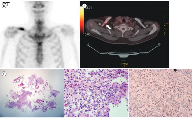
chemical staining revealed that the lesion was positive for S100 protein, which confirmed the diagnosis of LCH (Fig. 1F).
Four months after surgery, follow-up MRI showed an infiltrative pattern of high signal in- tensity on T2-weighted images of the soft tissue at the operation site on the right clavicle, which was thought to be a post-operative change. Nevertheless, the recurrence of LCH can- not be excluded with MRI alone. Therefore, an additional open curettage was performed, and the pathologic finding revealed post-operative changes with no evidence of recurrence.
The study protocol conformed to the ethical guidelines of the 1975 Declaration of Helsinki.
DISCUSSION
The most common causes of non-traumatic clavicular disease are neoplasms, infections, auto-immune disorders, and developmental abnormalities (3).
Although clavicular tumors are rare, a majority of them are malignant (4, 5).
Fig. 1. A 51-year-old male with Langerhans cell histiocytosis in the mid-portion of right clavicle.
D. Technetium 99m-methyl diphosphonate bone scan shows an intense focus of radioisotope accumulation within the mid-portion of the right clavicle.
E. PET/CT shows a maximum standardized uptake value of 3.6 in the mid-portion of the right clavicle (arrow).
F. The photomicrography (left, scan view) of H&E-stained tissue shows multiple bone and fibrovascular tissue fragments. Scanning photomi- crography (middle, × 40, H&E stain) shows aggregations of oval-shaped cells with complex folded nuclei, finely dispersed nuclear chromatin, and abundant cytoplasm (arrowheads). Eosinophils are abundantly present, producing eosinophilic microabscesses. Immunohistochemical staining (right, × 40) is positive for S100 protein.
H&E = hematoxylin and eosin D
F
E
The most frequent lesions in adult clavicles are metastatic lesions of carcinoma originating elsewhere. However, radiographic differentiation between metastasis, osteomyelitis, and LCH is often difficult (2).
LCH was formerly known as histiocytosis X, which includes eosinophilic granuloma, Hand–Schüller–Christian disease, and Letterer–Siwe disease (2).
The skeletal system is the most commonly involved organ of the single-system LCH (6), and the majority are solitary lesions (79%) (7). The most frequent site is the skull, followed by the femur, jaw, pelvis, ribs, spine, scapula, humerus, and clavicle in a decreasing order of fre- quency (7). Wester et al. (8) reported 61 cases of LCH occurring in adults, with only 6% in- volving the clavicle. A solitary lesion from an LCH in the clavicle is rare. In 2015, Udaka et al.
(2) reported a case of clavicular LCH which mentioned 12 published case reports of clavicu- lar LCH, and till date, there have been no additional reports on clavicular LCH.
The radiographic appearance of typical solitary LCH within a long bone is a lytic, medul- lary-based metaphyseal or diaphyseal lesion, with or without periosteal reaction. The perios- teal reaction depends on cortical erosion (7, 9). Medullary-based LCH may spread and erode the endosteal cortex, causing endosteal scalloping. Increased uptake of radiopharmaceuti- cals such as Tc99m-MDP is seen in bone scans, which are often performed to detect other sites of disease. CT and MRI are useful modalities for evaluating soft tissue involvement (9).
These lytic, expansive imaging features of osseous LCH may mimic fibrous dysplasia, uni- cameral or aneurysmal bone cysts, chondromyxoid fibromas, plasmacytomas, or metastatic disease. Moreover, if bone lesions with these imaging features are observed in adults, especial- ly in cases of multiple lesions, there is a tendency to focus on the possibility of malignant dis- ease, such as metastasis and multiple myeloma, due to the rarity of LCH beyond childhood (9).
Song at el. (9) analyzed the radiologic features of several adult patients with LCH and re- vealed some distinguishing features that may help differentiate LCH from metastasis and multiple myeloma. For example, multiple myeloma commonly presents imaging findings of punched-out lytic lesions, fractures, and rarely sclerosis, in contrast to LCH. Similar to adult LCH, metastatic lesions often present with cortical destruction and bony remodeling; how- ever, most metastatic lesions present with a more permeative appearance as opposed to ex- pansile bone destruction (9, 10).
Our case shows a medullary-based osteolytic lesion with adjacent cortical disruption in the right clavicle of a middle-aged male (50-years-old), and a pattern of the mass extending into
We also presented radiologic findings using various imaging modalities and pathologic test results that confirmed the diagnosis of LCH.
In conclusion, as demonstrated in this case report, it is important to consider the possibili- ty of LCH as a differential diagnosis when presented with an osteolytic lesion accompanied by bony disruption in the adult clavicle, although it is rare.
Author Contributions
Conceptualization, P.C., K.Y.H.; data curation, all authors; formal analysis, all authors; investiga- tion, all authors; methodology, P.C., K.Y.H.; project administration, P.C., K.Y.H.; resources, all authors;
software, all authors; supervision, K.Y.H.; validation, K.Y.H.; visualization, all authors; writing—origi- nal draft, P.C.; and writing—review & editing, all authors.
Conflicts of Interest
The authors have no potential conflicts of interest to disclose.
Funding None
REFERENCES
1. Weitzman S, Jaffe R. Uncommon histiocytic disorders: the non-Langerhans cell histiocytoses. Pediatr Blood Cancer 2005;45:256-264
2. Udaka T, Susa M, Kikuta K, Nishimoto K, Horiuchi K, Sasaki A, et al. Langerhans cell histiocytosis of the clavicle in an adult: a case report and review of the literature. Case Rep Oncol 2015;8:426-431
3. Franklin JL, Parker JC, King HA. Nontraumatic clavicle lesions in children. J Pediatr Orthop 1987;7:575-578 4. Kapoor S, Tiwari A, Kapoor S. Primary tumours and tumorous lesions of clavicle. Int Orthop 2008;32:829-
834
5. Parikh SN, Desai VR, Gupta A, Anton CG. Langerhans cell histiocytosis of the clavicle in a 13-year-old boy.
Case Rep Orthop 2014;2014:510287
6. Aricò M, Girschikofsky M, Généreau T, Klersy C, McClain K, Grois N, et al. Langerhans cell histiocytosis in adults. Report from the International Registry of the Histiocyte Society. Eur J Cancer 2003;39:2341-2348 7. Tsuchie H, Okada K, Nagasawa H, Yano M, Nanjyo H, Shimada Y. Langerhans cell histiocytosis of the ster-
num. Ups J Med Sci 2009;114:121-125
8. Wester SM, Beabout JW, Unni KK, Dahlin DC. Langerhans’ cell granulomatosis (histiocytosis X) of bone in adults. Am J Surg Pathol 1982;6:413-426
9. Song YS, Lee IS, Yi JH, Cho KH, Kim DK, Song JW. Radiologic findings of adult pelvis and appendicular skel- etal Langerhans cell histiocytosis in nine patients. Skeletal Radiol 2011;40:1421-1426
10. Christopher Z, Binitie O, Henderson-Jackson E, Perno J, Makanji RJ. Langerhans cell histiocytosis of bone in an adult: a case report. Radiol Case Rep 2018;13:310-314
50세 남자에게서 발견된 쇄골의 랑게르한스 세포 조직구증:
증례 보고
박창현1 · 김용훈1* · 차순주1 · 김지예2
랑게르한스 세포 조직구증은 희귀한 질환으로 일반적으로 근골격계를 침범하며, 대부분 소 아에게서 발생하고 성인의 쇄골에서 발병하는 경우는 극히 드물다. 이 증례 보고는 50세 남 자에게서 영상학적으로 발견되어 병리학적으로 확진된 쇄골의 랑게르한스 세포 조직구증 의 사례이다. 또한 저자들은 문헌고찰과 함께 단순촬영, 컴퓨터단층촬영, 자기공명영상법, 그리고 양전자방출단층촬영-컴퓨터단층촬영 영상 등의 다양한 영상검사 소견을 보고하고 자 한다.
인제대학교 일산백병원 1영상의학과, 2병리과