Epidemiological Aspects of Pertussis among Adults and Adolescents in a Korean Outpatient Setting: A Multicenter, PCR-Based Study
Epidemiological data of Bordetella pertussis infection among adolescents and adults are limited in Korea. Patients ( ≥ 11 yr of age) with a bothersome cough for less than 30 days were enrolled during a 1-yr period at 22 hospitals in Korea. Nasopharyngeal swabs were collected for polymerase chain reaction (PCR) and for bacteriologic culture. In total, 490 patients were finally enrolled, and 34 (6.9%) patients tested positive for B. pertussis;
cough duration (14.0 days [7.0-21.0 days]) and age distribution were diverse. The incidence was the highest in secondary referral hospitals, compared to primary care clinics or tertiary referral hospitals (24/226 [10.6%] vs. 3/88 [3.4%] vs. 7/176 [4.0%], P = 0.012), and the peak incidence was observed in February and August (15.8% and 15.9%), with no confirmed cases between March and June. In the multivariate analysis, post-tussive vomiting was significantly associated with pertussis (odds ratio, 2.508; 95% confidence interval, 1.146-5.486) and secondary referral hospital showed a borderline significance. In conclusion, using a PCR-based method, 6.9% of adolescent and adult patients with an acute cough illness had pertussis infection in an outpatient setting. However, hospital levels and seasonal trends must be taken into account to develop a better strategy for controlling pertussis.
Keywords: Pertussis; Adult; Hospitals; Incidence; Seasons Sunghoon Park,1* Sun Hwa Lee,2*
Ki-Hyun Seo,3 Kyeong-Cheol Shin,4 Yong Bum Park,5 Myung Goo Lee,6 Kwang Ha Yoo,7 Hui Jung Kim,8 Jae Seuk Park,9 Jae Hwa Cho,10 Yongchun Ko,11 Soo-Keol Lee,12 Ki Tae Cheon,13 Do Il Kim,14 Jun Wook Ha,15 Jae-Myung Lee,16 Ji-Won Suhr,17 Eui Hun Jeong,18 and Ki-Suck Jung1
1Division of Pulmonary, Allergy and Critical Care Medicine, Hallym University Sacred Heart Hospital, Anyang; 2Seegene Medical Foundation, Seoul;
3Division of Pulmonary and Critical Care Medicine, Soonchunhyang University Hospital, Cheonan;
4Division of Pulmonary, Allergy and Critical Care Medicine, Yeungnam University Medical Center, Daegu; 5Division of Pulmonary, Allergy and Critical Care Medicine, Kangdong Sacred Heart Hospital, Seoul; 6Division of Pulmonary, Allergy and Critical Care Medicine, Chuncheon Sacred Heart Hospital, Chuncheon; 7Division of Pulmonary, Allergy and Critical Care Medicine, Konkuk University Hospital, Seoul; 8Division of Pulmonary, Allergy and Critical Care Medicine, Wonkwnag University Sanbon Hospital, Gunpo; 9Division of Pulmonary, Allergy and Critical Care Medicine, Dankook University Hospital, Cheonan; 10Division of Pulmonary, Allergy and Critical Care Medicine, Inha University Hospital, Incheon; 11Division of Pulmonary Medicine, Gwangju Christian Hospital, Gwangju; 12Department of Allergy, Dong-A University Hospital, Busan; 13Onnuri Clinic of Internal Medicine, Jeonju; 14Rapha Clinic of Otolaryngology, Anyang; 15Cheongchun Clinic of Internal Medicine, Daegu; 16Leejaemyeong Clinic of Internal Medicine, Anyang; 17Hanaro Clinic of Internal Medicine, Daejeon; 18Hallym Clinic of Internal Medicine, Hongcheon, Korea
*Sunghoon Park and Sun Hwa Lee contributed equally to the work.
Received: 29 April 2014 Accepted: 12 June 2014 Address for Correspondence:
Ki-Suck Jung, MD
Division of Pulmonary, Allergy and Critical Care Medicine, Department of Internal Medicine, Hallym University Sacred Heart Hospital, 22 Gwanpyeong-ro, Anyang 431-070, Korea Tel: +82.31-380-3715, Fax: +82.31-380-3973 E-mail: [email protected]
Funding: This study was supported by the Korea Centers of Disease Control and Prevention (KCDC; 2011 Academic Research Fund, 2011-E46003-00).
http://dx.doi.org/10.3346/jkms.2014.29.9.1232 • J Korean Med Sci 2014; 29: 1232-1239
INTRODUCTION
Pertussis is a highly infectious disease and can cause significant morbidity in infants and the elderly (1, 2). However, the incidence of this vaccine-preventable disease has increased substantially in adolescents and adults since 1980, and pertussis has resurg- ed in many countries (3, 4). A large-scaled European study demonstrated an increased incidence of pertussis infection in adults rather than children, and a large outbreak oc- curred in the United States in 2012 (3, 5).
Studies on the epidemiology of pertussis demonstrated that 7%-17% of cases of pro- longed cough were attributable to Bordetella pertussis infection in adolescents and adults, and suggested that the disease is endemic in the present vaccine era (7-9). However, the diagnosis of pertussis is challenging in adult patients, and the waning immunity following vaccination can contribute to the persistence of pertussis; household contacts can be the main source of pertussis transmission (4, 6). Therefore, the early detection and prevention of adolescent and adult pertussis is of great importance (1). Although several Korean studies indicate a relatively low prevalence among adults (10, 11), re- cent outbreaks in other countries have alerted us to the dangers of pertussis (3, 5).
Up to date, many studies have been conducted about the epidemiological and clini- cal characteristics of adult pertussis. However, data are very limited among adult pa- tients in Korea. In addition, we hypothesized that their detection rates would be differ- ent by hospital levels (i.e., levels of healthcare system) and season in ordinary non-epi- demic settings. Serologic tests are important in epidemiological studies but they are Respiratory Diseases
not always readily available and are time-consuming in real- world clinical settings. Therefore, we conducted this multicenter study on patients with an acute cough illness (≤ 30 days) using a PCR-based test. We collaborated with a professional laborato- ry and the Korean Centers for Disease Control and Prevention (KCDC) to conduct the study.
MATERIALS AND METHODS Sites and subjects
This study was conducted from July 2011 to June 2012 at 22 hos- pitals (7 primary care clinics, 7 secondary referral hospitals, and 8 tertiary referral hospitals) in six major provinces in Korea; among them, 11 hospitals (or clinics) were selected in Seoul and Gyeong- gi-do because about a half of the nation’s population reside around the Seoul metropolitan area (Fig. 1). No standardized definitions of the level of the healthcare system exist, and the range of ser- vices provided by each level of system depends on the available resources. In Korea, secondary referral hospitals provide spe- cialized services at the regional level and usually have 30 to 500 beds (community or university hospitals). Tertiary referral hos- pitals are large-sized, university-affiliated, provide specialized services, train physicians and conduct research.
Outpatients (adolescents or adults aged ≥ 11 yr) who present- ed with a bothersome cough of a duration ≤ 30 days were en- rolled in this study. Exclusion criteria included a history of anti-
biotic treatment within 7 days, active lesion on the chest or pa- ranasal sinus radiographs (when available), a fever of > 38.0°C, immunocompromised status (e.g., AIDS, leukemia, aplastic ane- mia, organ transplant, autoimmune diseases, or chemotherapy), or a confirmed alternative cause for current illness (e.g., drugs [angiotensin-converting enzyme inhibitors], pneumonia, aller- gic rhinitis, sinusitis, or gastro-esophageal reflux).
Clinical data and specimen collection
Clinical information was collected by the participating investi- gators at outpatient departments. Data on age, sex, chronic re- spiratory diseases, comorbidities, tobacco usage, cough dura- tion, classical pertussis symptoms, other respiratory symptoms and vaccination history (e.g., diphtheria, tetanus, and pertussis [DTP] or diphtheria, tetanus, and acellular pertussis [DTaP]) were collected for all enrolled patients. All specimens were col- lected via nasopharyngeal swab (NPS; liquid Amies medium on flocked swabs; Copan Diagnostic, Inc., Murrieta, CA, USA) and transferred at room temperature to the central laboratory (i.e., Seegene Medical Foundation) and plated on culture me- dia within 24 hr. To ensure consistent specimen quality, inde- pendent personnel, usually trained registered nurses, performed the sampling procedure in each hospital; the KCDC educated all the participating hospitals, and all microbiological testing was performed in the central laboratory.
Case definition
To meet the clinical criteria for pertussis from the Centers for Disease Control and Prevention (CDC), patients must have a cough illness of ≥ 2 weeks duration and one of the following classical symptoms: 1) paroxysmal coughing, 2) inspiratory whooping, or 3) post-tussive vomiting (12). The laboratory cri- teria for diagnosis are isolation of B. pertussis or a positive poly- merase-chain-reaction (PCR) assay. A ‘confirmed case’ is diag- nosed when a patient satisfied both the clinical and laboratory criteria (16). In the present study, however, we define a confirm- ed case as a patient with a positive PCR or culture and a cough illness. We do not consider the duration of the cough illness in the diagnosis of pertussis due to the atypical characteristics of pertussis in adolescents and adults.
Specimens and microbiological tests
PCR and the bacteriologic culture were performed on all speci- mens. Regan and Lowe agar media (charcoal agar supplement- ed with 10% horse blood) with 40 mg/mL cephalexin were used for culture (13). After inoculation, the plates were incubated for at least 7 days under humid conditions (35-36°C). Identification was based on both biological characteristics and PCR (14).
For PCR, a portion of each specimen was boiled for 5 min.
After centrifugation, 1-2 μL of the supernatant was used as a PCR template. Although no standardized PCR method exists Fig. 1. Six provinces and numbers of participating hospitals. In total, 22 hospitals (or
clinics) participated in this study; numbers in parenthesis refer to those of participat- ing primary, secondary, and tertiary hospitals, respectively.
Gyeonggi-do (3, 3, 2)
Gangwon-do (2, 1, 0) Seoul (0, 2, 1)
Chungcheong-do (0, 1, 1)
Gyeongsang-do (1, 0, 2)
Jeolla-do (1, 0, 2)
for pertussis diagnosis, the “repeated-insertion sequence” and the “pertussis toxin (PT) promoter region” are used most com- monly as target regions (15, 16). In the present study, we used the repeated-insertion sequence: primers BP1 (5´-GATTCAAT- A GGTTGTATGCATGGTT-3´) and BP2 (5´-TTCAGGCACACA- AACTTGATGGGCG-3´). PCR was performed using a commer- cially available pre-mixed Taq polymerase (AccuPower PRC Pre- Mix; BIONEER, Daejeon, Korea).
PCR conditions were as follows: 95°C for 5 min (pre-denatur- ation), followed by 40 cycles of 95°C for 5 sec (denaturation) and 55°C for 10 sec (annealing). PCR products were resolved by elec- trophoresis on 2% agarose gels, and identification of a 180-bp band indicated a positive result.
Data analyses
The primary outcomes were the incidence rate of B. pertussis infection among adolescents and adults and its relationships with hospital levels and season. Secondary outcomes were com- parisons of incidence rates across age groups and six provinces.
Clinical characteristics of confirmed cases were also compared to cases of non-confirmed cough illness. Data are expressed as means ± standard deviations (or medians and interquartile ran- ges [IQRs]) for continuous variables and as percentages for cat- egorical variables, unless otherwise indicated. Student’s t-test was performed for continuous data, whereas chi-square or Fish- er’s exact tests were used for categorical data. A multivariate analysis by logistic regression (with a backward likelihood ratio method) was performed with covariates significant in univari- ate analysis to investigate risk factors for pertussis. All reported P values were two-sided, and P < 0.05 indicated statistical sig- nificance. All analyses were conducted using the SPSS statisti- cal software (IBM SPSS Statistics 21, Standard for Medical Net- work).
Ethics statement
All patients provided written informed consent, and the proto- col of this study was approved by the institutional review board of Hallym University Sacred Heart Hospital (IRB No. 2011-I050) and each participating hospital. The authors assert that all pro- cedures contributing to this work comply with the Helsinki De-
claration of 1975 and its later amendments.
RESULTS
Demographics and clinical symptoms
Among 504 patients initially enrolled, 14 were excluded due to their incomplete clinical data or inappropriate specimens (Fig.
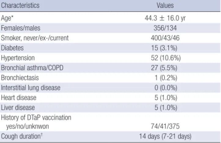
2). Therefore, a total of 490 patients (88 from primary care clin- ics, 226 from secondary referral hospitals and 176 from tertiary referral hospitals) were finally included in the study. The mean age of the patients was 44.3 ± 16.0 yr, and 72.7% were females (Table 1). Median cough duration was 14 days (7-21 days); and 72.4% of the patients reported a paroxysmal cough, while 13.1%
and 17.8% reported whooping cough and post-tussive vomit- ing, respectively. Hypertension was the most common co-mor- bidity reported, and 5.7% had a chronic airway disease. Among clinical symptoms, sputum (65.1%) and rhinorrhea (37.8%) were the most frequent symptoms, when cough was excluded. Only 15.1% of patients could recall if they had received the DTP (or DTaP) vaccination.
Microbiologic data
We collected 490 nasopharyngeal swab samples from 22 hospi- tals during the 1-yr period. In total, 34 (6.9%) patients were PCR positive (i.e., confirmed case), and of these, 10 (2.0%) were cul- ture positive (B. pertussis) concomitantly. We investigated fur- ther 20 close contacts including family members of the 11 con- firmed cases. Of 20 contacted cases, five (25.0%) were PCR pos- itive.
Incidence by hospital levels
When stratified by the hospital levels, the highest incidence was in secondary referral hospitals (10.6% [24/226] vs. 3.4% [3/88]
in primary care clinics vs. 4.0% [7/176] in tertiary referral hospi- tals; P = 0.012; Fig. 3). In terms of age, sex, and cough duration,
504 initially enrolled
490 finally analyzed PCR, 490 Culture, 490
14 excluded Incomplete data, 10 Inappropriate specimens, 4
Fig. 2. Flow diagram of enrolled patients. PCR, polymerase chain reaction.
Table 1. Baseline characteristics of the participants (n = 490)
Characteristics Values
Age* 44.3 ± 16.0 yr
Females/males 356/134
Smoker, never/ex-/current 400/43/46
Diabetes 15 (3.1%)
Hypertension 52 (10.6%)
Bronchial asthma/COPD 27 (5.5%)
Bronchiectasis 1 (0.2%)
Interstitial lung disease 0 (0.0%)
Heart disease 5 (1.0%)
Liver disease 5 (1.0%)
History of DTaP vaccination
yes/no/unknwon 74/41/375
Cough duration† 14 days (7-21 days)
*Mean ± SD; †Median (Interquartile range). COPD, chronic obstructive pulmonary disease; DTaP, diphtheria, tetanus, acellular pertussis.
there were no significant differences among the three hospital groups (data not shown).
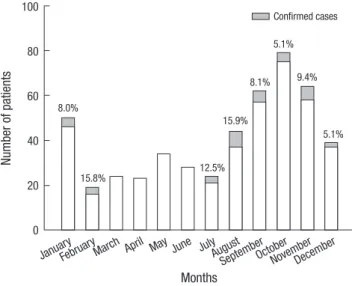
Incidence by season and province
As shown in Fig. 4, peak incidence was in February and August (15.8% [3/19] and 15.9% [7/44], respectively). However, there were no pertussis cases between March and June. In terms of regional distribution (Fig. 5), we identified confirmed cases from all the six provinces, and the incidence rates ranged from 3.8%
to 8.0%, with the lowest rates in the two southern provinces.
Clinical characteristics and risk factors
The age distribution of the confirmed cases was diverse, with the highest incidence rate among patients aged of 10 to < 20 yr (Fig. 6). The median cough duration was 14.0 days (7.0-21.0 days);
and 16 patients (45.7%) had a cough of duration < 14 days. Among
the confirmed cases, 27 patients (79.4%) complained of parox- ysmal cough, but only 11.8% and 33.4% had inspiratory whoop- ing and post-tussive vomiting, respectively. In particular, chills, crackles and wheezing were absent among the confirmed cas- es, and none of them had a history of chronic lung disease, ei- ther (Table 2).
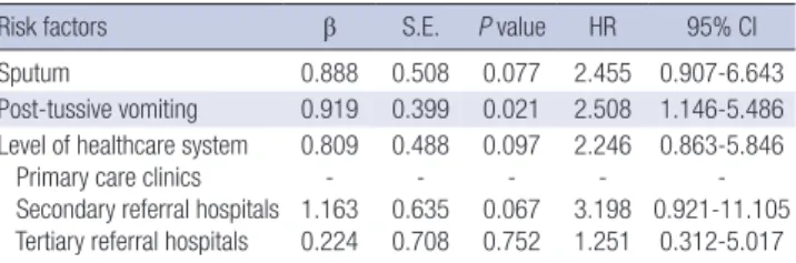
The clinical characteristics of the confirmed cases are shown in Table 2. There were no significant differences in co-morbid illnesses between the two groups. However, sputum, rhonchi, and post-tussive vomiting were more frequent in the confirmed cases when compared to the other cases of cough illness (P = 0.010, P = 0.011, and P = 0.021, respectively). In the multivari- ate analysis, where age, sputum, rhonchi, post-tussive vomiting, Fig. 3. Incidence of pertussis by hospital levels. The incidence was the highest in se-
condary referral hospitals compared to tertiary or primary care hospitals (P = 0.012).
Number of patients
Primary clinics
Secondary hospitalsTertiary hospitals 250
200
150
100
50
0
3.4%
10.6%
4.0%
Confirmed cases
Fig. 4. Seasonal trends of the incidence of pertussis. Peak incidences were observed in February and August; no pertussis cases were identified between March and June.
Number of patients
January Februar
yMarch April May June JulyAugust
SeptemberOctoberNovemberDecember 100
80
60
40
20
0 8.0%
15.8% 12.5%
15.9%
8.1%
5.1%
9.4%
5.1%
Confirmed cases
Months
Fig. 5. Incidence of pertussis by provinces. The incidence rates range from 3.8% to 8.0% among the six provinces and the two southern provinces (Gyeongsang-do and Jeolla-do) show the lowest rates, with no statistical significance.
Number of patients
Seoul
Gyeonggi-doGangwon-do
Chungcheong-doGyeongsang-do Jeolla-do 180
160 140 120 100 80 60 40 20 0
7.7%
8.0%
7.1% 7.5%
3.8%
4.8%
Confirmed cases
Provinces
Fig. 6. Incidence of pertussis by age group. The age distribution of the confirmed cases was diverse (P > 0.05 by chi-square test).
Number of patients
10 to <
20 20 to <
30 30 to <
40 40 to <
50 50 to <
60 60 to <
70 70 to <
80 ≥80
140 120 100 80 60 40 20 0
19.0%
9.6%
6.1%
7.9%
2.4%
4.5%
13.6%
Confirmed cases
Age groups (yr)
and hospital levels were included, post-tussive vomiting was significantly associated with pertussis (odds ratio [OR], 2.508;
95% confidence interval [CI], 1.146-5.486), and secondary re- ferral hospitals showed a borderline significance (OR, 3.198;
95% CI, 0.921-11.105; Table 3).
DISCUSSION
In the present study, we found several interesting results. First, the incidence of pertussis infection was 6.9% in a non-epidemic outpatient setting. Second, more than two-thirds of confirmed cases (70.6%) were diagnosed at secondary referral hospitals.
Third, the peak incidence of pertussis was in February and Au- gust, with no confirmed case during March-June. Fourth, post- tussive vomiting was significantly associated with PCR positivi- ty for B. pertussis.
Despite the introduction of a vaccine, a worldwide resurgence of pertussis infection has been reported in recent years, and the incidence in adolescents and adults has increased dramatically (1, 3). In particular, the US experienced a large outbreak in 2012, in which some US states experienced an incidence similar to those in the 1940s and 1950s (5). This worldwide increase in the incidence of pertussis may be explained by multiple mechani-
sms: 1) development of more sensitive diagnostic methods, 2) changes in nationwide surveillance systems for communicable diseases, 3) waning immunity among adolescents and adults, and 4) vaccine failure (or genetic changes) (1, 17). Although per- tussis are preventable by vaccination in childhood, the infec- tion in adolescents and adults remains common and seems to be an endemic feature in the present vaccine era (8).
In Korea, the surveillance of pertussis relies mostly on clinical notification systems, and most reported cases are in children (18). Studies of adult pertussis are rare. Park et al. (10) reported that the incidence of pertussis was 2.9% among adult patients;
in our previous study, it was 0.5% (11). These low positivity rates may be partly due to methodological problems such as the in- clusion of patients with viral upper respiratory infection, vari- able specimen quality, and the absence of serological tests. In the present study, we enrolled only patients who visited clinics or hospitals on weekdays to insure specimen transfer within 24 hr, and one professional laboratory performed all microbiologi- cal tests. We did not perform serological tests, which have prov- en useful for diagnosis of the infection after 3-4 weeks (19-21), because these tests are not readily available in routine clinical practice. Therefore, we decided to use both PCR and culture for sample testing, and only enrolled patients with a cough dura- tion of ≤ 30 days. However, the actual incidence of B. pertussis infection may have been underestimated. Additionally, PCR test- ing has also a risk of false positives (22, 23).
However, the incidence rate can vary by the length of cough illness used as an inclusion criterion. In several Western studies (7, 24), the incidence rate was reported to be 26%-31% among adults with subacute or chronic cough illnesses (a duration of
≥ 3 weeks), but a study from Israel reported a rate of 7% among adults with acute cough illnesses (25). According to the clinical definition of the CDC for B. pertussis infection, a cough illness of ≥ 2 weeks duration is required. In particular, previous stud- ies showed that a mean duration of cough illnesses was 36-48 days among adult pertussis (24). However, we only focused on patients with acute cough illnesses in the present study; the me- dian duration of cough illnesses was 14 days among the confirm- ed cases. Although the inclusion criteria were different from pre- vious studies, we think that our results can provide an evidence of B. pertussis infection among adolescents and adults with acute Table 2. Clinical characteristics between confirmed cases and other patients
Parameters Confirmed cases
(n = 34) Other patients
(n = 456) P value
Age (yr) 39.5 ± 16.9 44.6 ± 15.9 0.094
Females/males 27/7 329/127 0.359
Diabetes 2 (5.9%) 13 (2.9%) 0.279
Hypertension 3 (8.8%) 49 (10.7%) 1.000
BA/COPD 0 (0.0%) 27 (5.9%) 0.244
Bronchiectasis 0 (0.0%) 1 (0.2%) 1.000
Interstitial lung disease 0 (0.0%) 0 (0.0%) -
Heart disease 1 (2.9%) 4 (0.9%) 0.303
Liver disease 0 (0.0%) 5 (1.1%) 1.000
Sputum 29 (85.3%) 290 (63.6%) 0.010
Rhinorrhea 12 (35.3%) 173 (37.9%) 0.759
Sore throat 8 (23.5%) 123 (27.0%) 0.662
Chest pain 6 (17.6%) 93 (20.4%) 0.700
Headache 3 (8.8%) 68 (14.9%) 0.452
Dyspnea 4 (11.8%) 50 (11.0%) 0.780
Myalgia 2 (5.9%) 28 (6.1%) 1.000
Chilliness 0 (0.0%) 26 (5.7%) 0.243
Febrile sensation 2 (5.9%) 21 (4.6%) 0.669
Malaise 2 (5.9%) 14 (3.1%) 0.306
Hemoptysis 1 (2.9%) 8 (1.8%) 0.479
Rhonchi 7 (20.6%) 31 (6.8%) 0.011
Wheezing 0 (0.0%) 30 (6.6%) 0.253
Crackle 0 (0.0%) 23 (5.0%) 0.392
Stridor 1 (2.9%) 4 (0.9%) 0.303
Cough duration, days 14.0 (7.0-21.0) 14.0 (7.0-21.0) 0.760
Paroxysmal cough 27 (79.4%) 328 (71.9%) 0.346
Inspiratory whooping 4 (11.8%) 60 (13.2%) 1.000 Post-tussive vomiting 11 (32.4%) 76 (16.7%) 0.021 BA, bronchial asthma; COPD, chronic obstructive pulmonary disease.
Table 3. Multivariate analysis for risk factors for predicting pertussis infection*
Risk factors β S.E. P value HR 95% CI
Sputum 0.888 0.508 0.077 2.455 0.907-6.643
Post-tussive vomiting 0.919 0.399 0.021 2.508 1.146-5.486 Level of healthcare system
Primary care clinics Secondary referral hospitals Tertiary referral hospitals
0.809 - 1.163 0.224
0.488 - 0.635 0.708
0.097 - 0.067 0.752
2.246 - 3.198 1.251
0.863-5.846 - 0.921-11.105
0.312-5.017
*Hosmer-Lemeshow test, chi-square = 1.502 and P = 0.959. CI, confidence interval;
HR, hazard ratio; S.E., standard error.
cough illnesses.
One of the interesting findings in the present study was that confirmed cases were the most frequent in patients who visited secondary referral hospitals, which are usually community hos- pitals or medium-sized university hospitals. Only 3 of 34 con- firmed cases were diagnosed at primary care clinics. Although the statistical analysis showed a borderline significance, this re- sult can imply an important point that has been previously over- looked. That is, the patients with severe cough are less likely to attend primary care clinics. These patients can go directly to a secondary referral hospital in the Korean healthcare system.
Otherwise, it is possible that B. pertussis infection is rarely sus- pected or diagnostic tests are unlikely to be performed by pri- mary physicians. This can be due to the symptoms of B. pertus
sis infection that are not easily differentiated from those of the common cold in adults or adolescents. However, based on the results of our study, we can suggest that in countries such as Korea, we need to pay more attention to patients visiting sec- ondary referral hospitals, and the different incidence by level of healthcare system should be taken into account in a future strat- egy for controlling pertussis. In addition, a continued education of healthcare providers will also be critical to detect adult (or adolescent) pertussis cases in the early period of their illness, when they usually visit primary care clinics.
With regard to the seasonal trend, experts reported no defi- nite seasonal pattern of pertussis. However, they acknowledged that the incidence may increase in summer and fall (26). How- ever, a Danish study reported that the annual peak incidence was in August (27). The authors also found that monthly trends in adults showed a high correlation with trends in children but there was no evidence of a relationship between the increase in incidence and the opening of school. However, in a study by Rendi-Wagner et al. (28), there was no seasonal occurrence. In Korea, there seems to be no definite seasonal pattern when data from the national clinical reporting systems are analyzed (18).
However, we identified a peak incidence in February and Au- gust but no confirmed cases in March-June in the present study.
Although we evaluated the incidence of infection during an only 1-yr period and our study was not population-based, these findings are interesting. Therefore, the role of a seasonal effect should be further examined in the future. In addition, a study of the safety of concurrent vaccination for seasonal influenza and pertussis has been reported (29). Considering the similarity of the symptoms of the two illnesses such as cough, studies of the epidemiological relationship and a vaccination strategy to cov- er both diseases in adults would be intriguing.
In the present study, 72.4% of all patients reported a paroxys- mal cough, which was similar to our previous study. However, only 7.6% (n = 27) of patients reporting a paroxysmal cough were PCR positive. Additionally, only eight patients (1.6%) had all three classical pertussis symptoms, only two of which were PCR
positive. Although the diagnostic accuracy of the three classical symptoms remains questionable (11, 30, 31), we found that post- tussive vomiting was significantly associated with pertussis in the present study. Miyashita et al. (32) also demonstrated the importance of post-tussive vomiting in the clinical diagnosis of pertussis infection. Therefore, physicians should be alert to the presence of post-tussive vomiting among adolescent and adult patients with a cough illness. In addition, we plotted the receiv- er operating characteristic (ROC) curves for sputum, rhonchi, and the three classical pertussis symptoms for predicting B. per
tussis infection. However, none of these symptoms alone (or in combination) showed a significant area under the curve value (data not shown).
This study had several limitations. First, as mentioned above, because no serologic tests were performed, we may have under- estimated the true incidence of B. pertussis infection. Second, we only investigated 20 household contacts of confirmed cases.
Therefore, we were unable to calculate the secondary attack rate. Third, B. pertussis PCR testing is not standardized; therefore, the possibility of false positives should be considered. Fourth, the numbers of patients and participating hospitals were not distributed evenly across the six provinces. Therefore, we acknow- ledge that our results can be limited by several biases. However, this was a multi-center, prospective study, which included pri- mary care clinics as well as secondary and tertiary referral hos- pitals across the country. Our goal was to investigate several ep- idemiological characteristics of adult (and/or adolescent) per- tussis in a non-epidemic outpatient setting. Therefore, we be- lieve that our study has meaningful results for the future epide- miological strategy.
In conclusion, using a PCR-based method, 6.9% of adoles- cent and adult patients with a bothersome cough (≤ 30 days) were shown to have pertussis in a non-epidemic outpatient set- ting. The incidence was the highest in patients who visited sec- ondary referral hospitals and there were no confirmed cases in spring. Therefore, the different incidence by hospital levels and season must be taken into account to develop a better strategy for controlling B. pertussis infection.
ACKNOWLEDGMENTS
The authors would like to thank Seung Hun Jang, Yong Il Hwang, Joo-Hee Kim (Hallym University Sacred Heart Hospital), Kyeong Min Shon (Inseong Clinic, Chuncheon), Yong Cheol Lee (Chon- buk University Hospital), Yee Hyeong Kim (Kyung Hee Univer- sity Hospital) and A-Sook Shim (Seegen Medical Foundation) for their contribution to this multicenter study.
CONFLICTS OF INTEREST
The authors have no conflicts of interest to declare.
ORCID
Sunghoon Park http://orcid.org/0000000170046985 Sun Hwa Lee http://orcid.org/0000000220020282 Ki-Hyun Seo http://orcid.org/0000000206080676 Kyeong-Cheol Shin http://orcid.org/0000000319721847 Kwang Ha Yoo http://orcid.org/0000000199692657 Jae Seuk Park http://orcid.org/0000000193071155 Jae Hwa Cho http://orcid.org/0000000234323997 Yongchun Ko http://orcid.org/0000000156358579 Soo-Keol Lee http://orcid.org/0000000256089927 Do Il Kim http://orcid.org/0000000190463970 Jun Wook Ha http://orcid.org/0000000349672302 Jae-Myung Lee http://orcid.org/0000000262416349 Ji-Won Suhr http://orcid.org/0000000266499962 Ki-Suck Jung http://orcid.org/0000000268786543 REFERENCES
1. Hewlett EL, Edwards KM. Clinical practice: pertussis not just for kids.
N Engl J Med 2005; 352: 121522.
2. Stojanov S, Liese J, Belohradsky BH. Hospitalization and complications in children under 2 years of age with Bordetella pertussis infection. Infec
tion 2000; 28: 10610.
3. Celentano LP, Massari M, Paramatti D, Salmaso S, Tozzi AE; EUVAC- NET Group. Resurgence of pertussis in Europe. Pediatr Infect Dis J 2005;
24: 7615.
4. Mooi FR, Van Der Maas NA, De Melker HE. Pertussis resurgence: wan
ing immunity and pathogen adaptation two sides of the same coin. Epi
demiol Infect 2014; 142: 68594.
5. Cherry JD. Epidemic pertussis in 2012: the resurgence of a vaccinepre
ventable disease. N Engl J Med 2012; 367: 7857.
6. Kwon HJ, Yum SK, Choi UY, Lee SY, Kim JH, Kang JH. Infant pertussis and household transmission in Korea. J Korean Med Sci 2012; 27: 1547
51.
7. Cherry JD. The epidemiology of pertussis: a comparison of the epidemi
ology of the disease pertussis with the epidemiology of Bordetella pertus
sis infection. Pediatrics 2005; 115: 14227.
8. Cherry JD. Epidemiology of pertussis. Pediatr Infect Dis J 2006; 25: 3612.
9. Jackson LA, Cherry JD, Wang SP, Grayston JT. Frequency of serological evidence of Bordetella infections and mixed infections with other respi
ratory pathogens in university students with cough illnesses. Clin Infect Dis 2000; 31: 36.
10. Park S, Lee MG, Lee KH, Park YB, Yoo KH, Park JW, Kim C, Lee YC, Park JS, Kwon YS, et al. A Multicenter Study of Pertussis Infection in Adults with Coughing in Korea: PCRBased Study. Tuberc Respir Dis (Seoul) 2012;
73: 26672.
11. Park WB, Park SW, Kim HB, Kim EC, Oh M, Choe KW. Pertussis in adults with persistent cough in South Korea. Eur J Clin Microbiol Infect Dis 2005;
24: 1568.
12. Broder KR, Cortese MM, Iskander JK, Kretsinger K, Slade BA, Brown KH, Mijalski CM, Tiwari T, Weston EJ, Cohn AC, et al. Preventing teta
nus, diphtheria, and pertussis among adolescents: use of tetanus toxoid,
reduced diphtheria toxoid and acellular pertussis vaccines recommen
dations of the Advisory Committee on Immunization Practices (ACIP).
MMWR Recomm Rep 2006; 55: 134.
13. Regan J, Lowe F. Enrichment medium for the isolation of Bordetella. J Clin Microbiol 1977; 6: 3039.
14. Lautrop H. Laboratory diagnosis of whoopingcough or Bordetella in
fections. Bull World Health Organ 1960; 23: 1535.
15. Glare EM, Paton JC, Premier RR, Lawrence AJ, Nisbet IT. Analysis of a repetitive DNA sequence from Bordetella pertussis and its application to the diagnosis of pertussis using the polymerase chain reaction. J Clin Mi
crobiol 1990; 28: 19827.
16. Houard S, Hackel C, Herzog A, Bollen A. Specific identification of Bor
detella pertussis by the polymerase chain reaction. Res Microbiol 1989;
140: 47787.
17. Cherry JD. The science and fiction of the “resurgence” of pertussis. Pedi
atrics 2003; 112: 4056.
18. Korean Centers for Disease Control and Prevention. Sentinal surveil
lance systems for communicable infectious diseases. Available at http://
www.cdc.go.kr [accessed 30 December 2012].
19. Birkebaek NH, Kristiansen M, Seefeldt T, Degn J, Moller A, Heron I, An- dersen PL, Moller JK, Ostergård L. Bordetella pertussis and chronic cough in adults. Clin Infect Dis 1999; 29: 123942.
20. Cattaneo LA, Reed GW, Haase DH, Wills MJ, Edwards KM. The seroepi
demiology of Bordetella pertussis infections: a study of persons ages 165 years. J Infect Dis 1996; 173: 12569.
21. Miller E, Fleming DM, Ashworth LA, Mabbett DA, Vurdien JE, Elliott TS. Serological evidence of pertussis in patients presenting with cough in general practice in Birmingham. Commun Dis Public Health 2000; 3:
1324.
22. Loeffelholz MJ, Thompson CJ, Long KS, Gilchrist MJ. Comparison of PCR, culture, and direct fluorescentantibody testing for detection of Bor
detella pertussis. J Clin Microbiol 1999; 37: 28726.
23. Zouari A, Smaoui H, Kechrid A. The diagnosis of pertussis: which meth
od to choose? Crit Rev Microbiol 2012; 38: 11121.
24. Von König CH, Halperin S, Riffelmann M, Guiso N. Pertussis of adults and infants. Lancet Infect Dis 2002; 2: 74450.
25. Lieberman D, Shvartzman P, Lieberman D, Ben-Yaakov M, Lazarovich Z, Hoffman S, Mosckovitz R, Ohana B, Leinonen M, Luffy D, et al. Etiol
ogy of respiratory tract infection in adults in a general practice setting.
Eur J Clin Microbiol Infect Dis 1998; 17: 6859.
26. Centers for Disease Control and Prevention. Epidemiology and Preven
tion of VaccinePreventable Diseases. 12th ed. Atlanta: Pinkbook, 2012.
27. De Greeff SC, Dekkers AL, Teunis P, Rahamat-Langendoen JC, Mooi FR, De Melker HE. Seasonal patterns in time series of pertussis. Epide
miol Infect 2009; 137: 138895.
28. Rendi-Wagner P, Paulke-Korinek M, Stanek G, Khanakah G, Kollaritsch H. Impact of a pertussis booster vaccination program in adolescents and adults on the epidemiology of pertussis in Austria. Pediatr Infect Dis J 2007; 26: 80610.
29. Weston WM, Friedland LR, Wu X, Howe B. Vaccination of adults 65 years of age and older with tetanus toxoid, reduced diphtheria toxoid and acel
lular pertussis vaccine (Boostrix®): results of two randomized trials. Vac
cine 2012; 30: 17218.
30. Strebel P, Nordin J, Edwards K, Hunt J, Besser J, Burns S, Amundson G, Baughman A, Wattigney W. Populationbased incidence of pertussis
among adolescents and adults, Minnesota, 19951996. J Infect Dis 2001;
183: 13539.
31. Yaari E, Yafe-Zimerman Y, Schwartz SB, Slater PE, Shvartzman P, An- doren N, Branski D, Kerem E. Clinical manifestations of Bordetella per
tussis infection in immunized children and young adults. Chest 1999; 115:
12548.
32. Miyashita N, Akaike H, Teranishi H, Kawai Y, Ouchi K, Kato T, Hayashi T, Okimoto N. Diagnostic value of symptoms and laboratory data for per
tussis in adolescent and adult patients. BMC Infect Dis 2013; 13: 129.