A Nationwide Survey of Lymphangioleiomyomatosis in Korea:
Recent Increase in Newly Diagnosed Patients
In 2007, the Korean Interstitial Lung Disease Society had collected clinical data of patients who have diagnosed as Lymphangioleiomyomatosis (LAM) since 1990 through nationwide survey, which showed that LAM patients had increased sharply after 2004.
The present study was performed to show the clinical features of Korean patients with LAM, and to establish the reason for the recent increase in the diagnosis. All 63 patients were women and the mean age at diagnosis was 36 yr. The most common presenting symptom was dyspnea and 8 patients had tuberous sclerosis complex. The survival rate at 5 yr after diagnosis was 84%. Compared with patients diagnosed after 2004 (n=34), the patients diagnosed before 2004 (n=29) complained with dyspnea more (P=0.016) and had lower FEV1% predicted (P=0.003), and DLco% predicted (P=0.042). The higher proportion of patients diagnosed after 2004 showed the normal chest radiography, and they were detected by routine chest CT screening (P=0.016). This study showed that clinical features of Korean patients with LAM were not different from those reported elsewhere. It is concluded that the reason for the increase of newly diagnosed patients is the result of increase in detection of the early stage LAM by the widespread use of chest CT screening.
Key Words: Lymphangioleiomyomatosis; Korea; Registries; Respiratory Function Tests Hye Yun Park1,*, Hae-Seong Nam2,*,
Man Pyo Chung1, Sung Hwan Jeong3, Yu Jin Kim3, Seung-Ick Cha4, Young Whan Kim5, Jong Sun Park5, Soo-Taek Uh6, Choon-Sik Park7, Moo Suk Park8, Ji Ae Moon8, Kyung Soo Jung8, Yang Jin Jegal9, Dong Soon Kim10, Jin Woo Song10, Ho-Kee Yum11, and Young Bum Park12 Division of Pulmonary and Critical Care Medicine1, Department of Medicine, Samsung Medical Center, Sungkyunkwan University School of Medicine, Seoul;
Division of Pulmonary and Critical Care Medicine2, Department of Internal Medicine, Inha University Hospital, Inha University School of Medicine, Incheon;
Division of Pulmonary Medicine3, Department of Internal Medicine, Gachon Medical School Gil Medical Center, Incheon; Department of Internal Medicine4, Kyungpook National University Hospital, Daegu; Division of Pulmonary and Critical Care Medicine5, Department of Internal Medicine, Seoul National University College of Medicine, and Lung Institute, Seoul National University Hospital, Seoul; Division of Respiration and Allergy Medicine6, Soonchunhyang University Hospital, Seoul;
Department of Internal Medicine7, Bucheon Hospital, Soonchunhyang University School of Medicine, Bucheon;
Department of Internal Medicine8, Yonsei University College of Medicine, Seoul; Department of Internal Medicine9, Ulsan University Hospital, University of Ulsan College of Medicine, Ulsan; Department of Pulmonary and Critical Care Medicine10, Asan Medical Center, University of Ulsan College of Medicine, Seoul;
Department of Internal Medicine11, Seoul Paik Hospital, Inje University, Seoul; Department of Internal Medicine12, Hallym University College of Medicine, Kangdong Sacred Heart Hospital, Seoul, Korea
*Hye Yun Park and Hae-Seong Nam contributed equally to this work.
Received: 29 October 2009 Accepted: 25 January 2010 Address for Correspondence:
Man Pyo Chung, M.D.
Division of Pulmonary and Critical Care Medicine, Department of Medicine, Samsung Medical Center, Sungkyunkwan University School of Medicine, 81 Irwon-ro, Gangnam-gu, Seoul 135-710, Korea Tel: +82.2-3410-3429, Fax: +82.2-3410-3849
E-mail: [email protected]
DOI: 10.3346/jkms.2010.25.8.1182 • J Korean Med Sci 2010; 25: 1182-1186
INTRODUCTION
Lymphangioleiomyomatosis (LAM) is a rare disease that occurs mainly in women of reproductive age. The clinical features, in-
cluding recurrent spontaneous pneumothorax, slow progres- sive dyspnea, hemoptysis, chylothorax, and chylous ascites, re- sult from progressive cystic lung destruction and accumulation of smooth muscle cells (LAM cells) in the lungs and axial lym-
phatics (1-4). The primary treatment for LAM consists of hor- monal therapy and supportive management, although the re- sponse to hormonal therapy has not yet been demonstrated conclusively (4-6).
In 2007, the Korean Interstitial Lung Disease Society had col- lected data for patients diagnosed with LAM, by a nationwide survey, to establish their demographic, clinical, and radiograph- ic features. This survey showed that the number of new patients with LAM had increased sharply after 2004 (Fig. 1). Therefore, the present study aimed to understand the initial clinical radio- logical findings, management, and clinical course of patients with LAM in Korea. We also aimed to establish the reason for the recent increase in newly diagnosed patients by comparing the clinical and radiographic features between patients diag- nosed with LAM before and after 2004.
MATERIALS AND METHODS Study population and data collection
We enrolled 63 patients diagnosed with LAM between 1990 and 2007 in Korea. The nationwide survey was performed by collect- ing the questionnaire-based data sheet between May and Sep- tember 2007. The data sheet included age at diagnosis, smoking history, previous history of pneumothorax or pleural effusion, presence of tuberous sclerosis complex (TSC) or angiomyoli- poma (AML), initial clinical and radiological presentation, co- morbidity, initial pulmonary function tests, method of diagnosis, treatment modalities, and survival, and we reviewed retrospec- tively all items on the data sheets. The diagnosis of LAM was made by lung pathology or typical chest high-resolution com- puted tomography (HRCT) findings, along with compatible clin- ical history. Pathologically, LAM was diagnosed in 39 patients (62%), which was based on the results of surgical lung biopsies (n=33), transbronchial lung biopsies (n=4), excisional biopsy of retroperitoneal mass (n=1), or inguinal lymph node biopsy (n=
1). In the remaining 24 patients (48%), the diagnosis of LAM was
established from typical HRCT findings, which are diffuse, thin- walled cysts scattered in an even distribution throughout the lung fields, with normal intervening lung parenchyma (1).
Survival status was obtained from the death registry of the Korea National Statistical Office as of December 2007. This study was approved by the Institutional Review Board of the Samsung Medical Center (IRB approval number: 2009-12-066).
Statistical analysis
Values are expressed as number (percentile), mean±standard deviation or median (range). Statistical analysis was performed using the unpaired Student’s t test for continuous variables and the chi-squared or Fisher’s exact test for categorical values, as appropriate. Survival was calculated from the time of diagnosis of LAM until the endpoint (December 2007). Survival probabil- ity according to disease duration was estimated by the Kaplan–
Meier method. All P values were two-sided, with P<0.05 con- sidered to be statistically significant. Statistical analysis was per- formed using PASW version 17.0 (SPSS Inc., Chicago, IL, USA).
RESULTS
Demographic and clinical characteristics
All 63 patients were women, and the mean age at diagnosis was 36 yr (range, 19–65). The age at diagnosis was >40 yr in 35% of the patients. Three patients were ex-smokers and one was a cur- rent smoker. The most common past or coexisting disease was pulmonary tuberculosis (11%). AML was found in 21 patients
No. of patients
Years at diagnosis 1990-1991
1994-1995
1998-1999
2002-2003 1992-1993
1996-1997
2000-2001
2004-2005 2006-2007 18
16 14 12 10 8 6 4 2 0
Fig. 1. Annual number of new patients diagnosed with LAM.
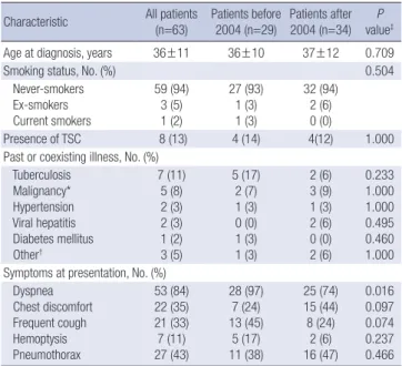
Table 1. Demographic and clinical features of 63 patients with LAM Characteristic All patients
(n=63) Patients before
2004 (n=29) Patients after 2004 (n=34) P
value‡
Age at diagnosis, years 36±11 36±10 37±12 0.709
Smoking status, No. (%) 0.504
Never-smokers Ex-smokers Current smokers
59 (94) 3 (5) 1 (2)
27 (93) 1 (3) 1 (3)
32 (94) 2 (6) 0 (0)
Presence of TSC 8 (13) 4 (14) 4(12) 1.000
Past or coexisting illness, No. (%) Tuberculosis
Malignancy*
Hypertension Viral hepatitis Diabetes mellitus Other†
7 (11) 5 (8) 2 (3) 2 (3) 1 (2) 3 (5)
5 (17) 2 (7) 1 (3) 0 (0) 1 (3) 1 (3)
2 (6) 3 (9) 1 (3) 2 (6) 0 (0) 2 (6)
0.233 1.000 1.000 0.495 0.460 1.000 Symptoms at presentation, No. (%)
Dyspnea Chest discomfort Frequent cough Hemoptysis Pneumothorax
53 (84) 22 (35) 21 (33) 7 (11) 27 (43)
28 (97) 7 (24) 13 (45)
5 (17) 11 (38)
25 (74) 15 (44) 8 (24)
2 (6) 16 (47)
0.016 0.097 0.074 0.237 0.466
*Malignancy includes advanced gastric carcinoma, papillary thyroid carcinoma, breast cancer, cervical carcinoma, and multiple myeloma; †Other includes nephrotic synd- rome, atrial septal defect, and hypothyroidism; ‡P values are for comparison between the patients diagnosed before and after 2004.
LAM, lymphangioleiomyomatosis; TSC, tuberous sclerosis complex.
(33%), and eight (13%) had TSC. The main presenting symptom was dyspnea (84%). Pneumothorax was observed in 43% of the patients at diagnosis, and 63% had a previous history of pneu- mothorax. The median number of patients who experienced at least one episode of pneumothorax was three (range, 1–10).
Twenty-nine patients were diagnosed with LAM before 2004, and the remaining 34 patients were diagnosed after that date.
No significant difference existed between the groups for age, history of smoking, and presence of TSC. However, exertional dyspnea was more frequent in the patients diagnosed before than after 2004 (97% vs. 74%; P=0.016; Table 1).
Pulmonary function tests
Baseline pulmonary function tests were performed <6 months before or after diagnosis of LAM (Table 2). Spirometry results were normal in 19 of 44 patients whose data were available. The mean values for forced vital capacity (FVC) % predicted, forced expiratory volume in 1 second (FEV1) % predicted (both n=44), total lung capacity (TLC) % predicted (n=24), and diffusing ca- pacity of carbon monoxide (DLCO) % predicted (n=34) were 82.5, 75.8, 99.3, and 74.2, respectively (Table 2). When lung func- tion was compared between patients diagnosed before and after 2004, those diagnosed before 2004 had more severe impairment in FEV1 % predicted (60.3 vs. 85.5, P=0.003), FEV1/FVC ratio (60.5 vs. 83.8, P<0.001), and DLco % predicted (60.9 vs. 80.6, P=0.042;
Table 2).
Radiological findings at the time of diagnosis
The main chest radiography finding was the presence of pneu- mothorax (43%). The reticulonodular infiltration and presence of cysts or bulla upon chest radiography were more common in patients diagnosed before than after 2004 (48% vs. 15%, P=0.004 and 41% vs. 15%, P=0.017), while normal chest radiographic appearance was more common in patients diagnosed after 2004 than before (3% vs. 26%, P=0.017). All patients showed typical, thin-walled parenchymal cysts that were usually distributed homogeneously in both lung fields upon HRCT. Other abnor- malities seen with HRCT included nodular densities in 13%, ground-glass opacities in 11%, pleural effusion in 13%, and peri- cardial effusion in 3% (Table 3).
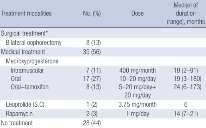
Treatment modalities and outcome
Several medical treatment modalities were tried in 35 patients.
Seven of these patients received intramuscular medroxyproges- terone (400 mg/month for a median duration of 19 months), 17 received oral progesterone monotherapy (10–20 mg/day for a median duration of 19 months), and eight received oral proges- terone with tamoxifen (median, 24 months). Among the remain- ing three patients, one was taking gonadotropin-releasing hor- mone agonist and two were taking rapamycin (Table 4).
Eight patients who received medical treatment underwent oophorectomy. Ten of 21 patients with AML were treated with surgical resection and four with arterial embolization. Of all pa- Table 2. Baseline pulmonary function testsw
Characteristics All patients Patients before 2004 Patients after 2004
P value†
No.* Mean±S.D. No.* Mean±S.D. No.* Mean±S.D.
FVC % predicted 44 82.5±23.3 17 76.8±23.8 27 86.0±22.8 0.203
FEV1 % predicted 44 75.8±28.5 17 60.3±28.7 27 85.5±24.1 0.003
FEV1/FVC ratio 44 74.8±19.3 17 60.5±14.6 27 83.8±16.3 <0.001
TLC % predicted 24 99.3±11.7 6 105.5±7.0 18 97.2±12.4 0.139
DLCO % predicted 34 74. 2±26.7 11 60.9±27.7 23 80.6±24.3 0.042
*Number of patients with information available; †P values are for comparison between the patients diagnosed before and after 2004.
FVC, forced vital capacity; FEV1, forced expiratory volume in one second; TLC, total lung capacity; DLCO, diffusing capacity of carbon monoxide.
Table 3. Radiographic finding at the time of diagnosis
Findings All patients Patients
before 2004 Patients after 2004 P
value*
Chest X-ray, No. (%) 63 29 34
Normal appearance Reticulonodular infiltrates Pneumothorax Cysts or bullae Pleural effusion
10 (16) 19 (30) 27 (43) 17 (27) 7 (11)
1 (3) 14 (48) 11 (38) 12 (41) 3 (10)
9 (27) 5 (15) 16 (47) 5 (15) 4 (12)
0.016 0.004 0.466 0.017 1.000
HRCT scan, No. (%) 63 29 34
Multiple cysts in both lung Nodular densities Ground-glass opacities Pleural effusion Pericardial effusion
63 (100) 8 (13) 7 (11) 8 (13) 2 (3)
29 (100) 2 (7) 6 (21) 3 (10) 1 (3)
34 (100) 5 (15)
1 (3) 5 (15)
1 (3) - 0.437 0.042 0.716 1.000
*P values are for comparison between patients diagnosed before and after 2004.
HRCT, high resolution computed tomography.
Table 4. Modality of hormonal therapy in 63 patients with LAM
Treatment modalities No. (%) Dose
Median of duration (range), months Surgical treatment*
Bilateral oophorectomy 8 (13) Medical treatment 35 (56) Medroxyprogesterone
Intramuscular Oral
Oral+tamoxifen
7 (11) 17 (27) 8 (13)
400 mg/month 10–20 mg/day 5–20 mg/day+
20 mg/day
19 (2–91) 19 (3–180) 24 (6–173)
Leuprolide (S.C) 1 (2) 3.75 mg/month 6
Rapamycin 2 (3) 1 mg/day 14 (7–21)
No treatment 28 (44)
*All subjects for surgical treatment received medical treatment.
S.C, subcutaneously.
tients, two patients underwent lung transplantation. One of these patients underwent left lung transplantation and died from chronic graft-versus-host disease at 38 months after trans- plantation. The other patient is still being followed-up, since April 2007.
Disease course and survival
During follow-up, 12 patients died within a median duration of 60 months (1–144 months). Cause of death was attributed to LAM in all patients, directly from respiratory failure (n=11) and lung transplantation morbidity (n=1). The survival probability, evaluated by the Kaplan–Meier method, is shown in Fig. 2. The probability of being alive was 84% at 5 yr and 65% at 8.5 yr after diagnosis.
DISCUSSION
This report provided the clinical and radiological features, and therapeutic modalities of patients with LAM in Korea. The clini- cal features of Korean patients with LAM do not differ from those reported elsewhere and we also found that the patients recently diagnosed as LAM can be detected at the early stage with mild respiratory symptoms and lung impairment by frequent use of chest CT screening.
Recently, a large number of patients with LAM have been re- ported (7, 8). These reports have demonstrated that the age range of women with LAM was broader, and normal spirometry at di- agnosis was more common than previously reported (2, 9). In addition, the clinical course of these patients with LAM was better than that described earlier, when the majority of patients died from respiratory failure, usually within 10 yr after the ap- parent onset of the disease (2, 10, 11).
According to the present Korea nationwide survey conduct- ed for patients diagnosed as LAM between 1990 and 2007, the number of LAM patients showed a tendency to increase and it is noticeable that the number of patient newly diagnosed as LAM increased rapidly after 2004 (Fig. 1). In this nationwide survey, although the mean age at diagnosis was similar to that reported previously (2, 9, 12, 13), the age range (19–65 yr) was as broad as data shown in the National Heart, Lung and Blood Institute (NHLBI) registry of 2006. The most common presenting symp- toms were exertional dyspnea and pneumothorax, which was consistent with other studies (8, 12). The prevalence of chylo- thorax was 20–30% in previous studies (4, 8, 9), but in our study, chylothorax was seen in only one patient at initial diagnosis and in two more during the course of the disease.
Nineteen patients were not available for pulmonary function tests because of dyspnea or pneumothorax at initial diagnosis, Fig. 2. Kaplan–Meier survival analysis of mortality of 63 patients with LAM. The
probability of being alive was 84% at 5 yr and 65% at 8.5 yr after diagnosis.
Survival probability
1.0
0.8
0.6
0.4
0.2
0.0
Years from diagnosis
0.00 5.00 10.00 15.00 20.00
Fig. 3. Radiographic features of a 46-yr old woman with LAM. (A) Chest radiography of 46-yr old woman with LAM. Chest radiography discloses no clue to the cystic lung disease. (B) High resolution CT scan of 46-yr old woman with LAM. High resolution CT scan at the level of lower lobar bronchus shows innumerable ovoid or round cystic lesions (arrows) in both lungs. The walls of these cysts are thin and even in thickness, which were considered as typical morphologic features of LAM.
A B
but the overall pulmonary function was better when compared to that in previous studies (2, 9, 13). In patients newly diagnosed with LAM after 2004, FEV1 predicted, FEV1/FVC, and DLco were significantly higher than those in patients diagnosed before 2004.
Declines in FEV1 and the FEV1/FVC ratio, which represent pro- gressive obstructive defects, and decreased DLco are sensitive factors for disease progression in LAM (8). Therefore, we sug- gest that patients diagnosed after 2004 were detected with mild lung impairment, which may mean that it was at an earlier stage compared to those diagnosed before 2004. This was consistent with the finding that clinical symptoms were significantly better, and normal radiographic findings were more frequent in recent- ly diagnosed patients. This earlier detection of LAM in patients with mild symptoms and normal chest radiography might be explained by the widespread and liberal use of CT scanning, which is superior to chest radiography for the assessment of patients with LAM (14). In our study, among 10 patients with normal chest radiographic findings, seven were detected by CT screening (Fig. 3) and three without respiratory symptoms were diagnosed coincidentally for LAM, after undergoing CT scan- ning for breast cancer, thyroid cancer, or abdominal pain.
In our nationwide survey, 56% of patients received medical therapy and the remainder did not receive it due to rejection of therapy for pregnancy or postmenopausal status, or they had no subjective symptoms. Although the beneficial effect of hormon- al manipulation is inconclusive (2, 4, 8), it is the only therapeu- tic option for LAM. However, a recent nonrandomized, open- label trial showed that treatment with sirolimus was associated with improvement of lung function in sporadic LAM (15). Addi- tional trials for the relative risks and benefits are needed; how- ever, the use of sirolimus is expected to be promising in the treat- ment of LAM. Thus, it might be helpful for the treatment of early- detected patients with LAM who show mild symptoms and signs.
In the present study, Kaplan–Meier plots showed survival probabilities of 84% after 5 yr and 65% after 8.5 yr from the time of diagnosis. Previous studies have reported a 3-yr time differ- ence between the time of onset of symptoms and diagnosis, and our result was therefore similar to that of Taylor et al. (4) and Urban et al. (8) who reported 78% and 79% probability of 8.5 yr survival after disease onset.
Our study had several limitations. First, because it was a mul- ticenter retrospective study performed on patients who were diagnosed with LAM over a 18-yr period, treatment protocols were very diverse. And the schedules for following lung func- tion tests were not consistent among hospitals. Thus, the effect of therapeutic interventions on lung function could not be eval- uated. In addition, because the follow-up period in patients di- agnosed with LAM since 2004 was short, we cannot compare and discuss the natural course of the disease.
In conclusion, our study showed that the clinical features of Korean patients with LAM do not differ from those reported
elsewhere. Moreover, we demonstrated that the increment of newly diagnosed patients is the result of increase in detection of the early stage LAM with mild respiratory symptoms and lung impairment by the widespread use of chest CT screening.
ACKNOWLEDGMENTS
We are especially grateful to the staff of all hospitals who con- tributed to this survey.
REFERENCES
1. Johnson SR. Lymphangioleiomyomatosis. Eur Respir J 2006; 27: 1056-65.
2. Kitaichi M, Nishimura K, Itoh H, Izumi T. Pulmonary lymphangioleio- myomatosis: a report of 46 patients including a clinicopathologic study of prognostic factors. Am J Respir Crit Care Med 1995; 151: 527-33.
3. McCormack FX. Lymphangioleiomyomatosis: a clinical update. Chest 2008; 133: 507-16.
4. Taylor JR, Ryu J, Colby TV, Raffin TA. Lymphangioleiomyomatosis. Clin- ical course in 32 patients. N Engl J Med 1990; 323: 1254-60.
5. Carrington CB, Cugell DW, Gaensler EA, Marks A, Redding RA, Schaaf JT, Tomasian A. Lymphangioleiomyomatosis. Physiologic-pathologic- radiologic correlations. Am Rev Respir Dis 1977; 116: 977-95.
6. Eliasson AH, Phillips YY, Tenholder MF. Treatment of lymphangioleio- myomatosis. A meta-analysis. Chest 1989; 96: 1352-5.
7. Ryu JH, Moss J, Beck GJ, Lee JC, Brown KK, Chapman JT, Finlay GA, Ol- son EJ, Ruoss SJ, Maurer JR, Raffin TA, Peavy HH, McCarthy K, Taveira- Dasilva A, McCormack FX, Avila NA, Decastro RM, Jacobs SS, Stylianou M, Fanburg BL. The NHLBI lymphangioleiomyomatosis registry: char- acteristics of 230 patients at enrollment. Am J Respir Crit Care Med 2006;
173: 105-11.
8. Urban T, Lazor R, Lacronique J, Murris M, Labrune S, Valeyre D, Cordier JF. Pulmonary lymphangioleiomyomatosis. A study of 69 patients. Groupe d’Etudes et de Recherche sur les Maladies “Orphelines” Pulmonaires (GERM”O”P). Medicine (Baltimore) 1999; 78: 321-37.
9. Chu SC, Horiba K, Usuki J, Avila NA, Chen CC, Travis WD, Ferrans VJ, Moss J. Comprehensive evaluation of 35 patients with lymphangioleio- myomatosis. Chest 1999; 115: 1041-52.
10. Kalassian KG, Doyle R, Kao P, Ruoss S, Raffin TA. Lymphangioleiomyo- matosis: new insights. Am J Respir Crit Care Med 1997; 155: 1183-6.
11. Silverstein EF, Ellis K, Wolff M, Jaretzki A 3rd. Pulmonary lymphangio- myomatosis. Am J Roentgenol Radium Ther Nucl Med 1974; 120: 832-50.
12. Johnson SR, Tattersfield AE. Clinical experience of lymphangioleiomyo- matosis in the UK. Thorax 2000; 55: 1052-7.
13. Oh YM, Mo EK, Jang SH, Yoo CG, Kim YW, Seo JW, Han SK, Im JG, Shim YS. Pulmonary lymphangioleiomyomatosis in Korea. Thorax 1999; 54:
618-21.
14. Muller NL, Chiles C, Kullnig P. Pulmonary lymphangiomyomatosis:
correlation of CT with radiographic and functional findings. Radiology 1990; 175: 335-9.
15. Bissler JJ, McCormack FX, Young LR, Elwing JM, Chuck G, Leonard JM, Schmithorst VJ, Laor T, Brody AS, Bean J, Salisbury S, Franz DN. Siroli- mus for angiomyolipoma in tuberous sclerosis complex or lymphangi- oleiomyomatosis. N Engl J Med 2008; 358: 140-51.