https://doi.org/10.12750/JET.2017.32.3.131
돼지 난자의 체외성숙에서 Caffeine 처리가 난자 성숙과 체세포 핵이식 배아의 체외발육에 미치는 영향
이주형1,a, 유진영2,a, 이한나2, 신혜지2, 이근식2, 이승태3, 이은송1,2†
1강원대학교 동물의학종합연구소, 2강원대학교 수의과대학, 3강원대학교 동물생명과학대학
Caffeine treatment during in vitro maturation improves developmental competence of morphologically poor oocytes after somatic cell nuclear transfer in pigs
Joohyeong Lee1,a, Jinyoung You2,a, Hanna Lee2, Hyeji Shin2, Geun-Shik Lee2, Seung Tae Lee3and Eunsong Lee1,2†
1Institute of Veterinary Science, Kangwon National University, Chuncheon 24341, Korea
2College of Veterinary Medicine, Kangwon National University, Chuncheon 24341, Korea
3Division of Applied Animal Science, College of Animal Life Science, Kangwon National University, Chuncheon 24341, Korea
ABSTRACT
In most mammals, metaphase II (MII) oocytes having high maturation promoting factor (MPF) activity have been considered as good oocytes and then used for assisted reproductive technologies including somatic cell nuclear transfer (SCNT). Caffeine increases MPF activity in mammalian oocytes by inhibiting p34cdc2 phosphorylation. The objective of this study was to investigate the effects of caffeine treatment during in vitro maturation (IVM) on oocyte maturation and embryonic development after SCNT in pigs. To this end, morphologically good (MGCOCs) and poor oocytes (MPCOCs) based on the thickness of cumulus cell layer were untreated or treated with 2.5 mM caffeine during 22-42, 34-42, or 38-42 h of IVM according to the experimental design. Caffeine treatment for 20 h during 22-42 h of IVM significantly inhibited nuclear maturation compared to no treatment. Blastocyst formation of SCNT embryos was not influenced by the caffeine treatment during 38-42 h of IVM in MGCOCs (41.1-42.1%) but was significantly improved in MPCOCs compared to no treatment (43.4 vs. 30.1%, P<0.05). No significant effects of caffeine treatment was observed in embryo cleavage (78.7-88.0%) and mean cell number in blastocyst (38.7-43.5 cells). The MPF activity of MII oocytes in terms of p34cdc2 kinase activity was not influenced by the caffeine treatment in MGCOCs (160.4 vs. 194.3 pg/ml) but significantly increased in MPCOCs (133.9 vs. 204.8 pg/ml). Our results demonstrate that caffeine treatment during 38-42 h of IVM improves developmental competence of SCNT embryos derived from MPCOCs by influencing cytoplasmic maturation including increased MPF activity in IVM oocytes in pigs.
(Key words: caffeine, oocyte maturation, maturation promoting factor, somatic cell nuclear transfer, pig)
†Correspondence: Eunsong Lee
Phone: 82-33-250-8670; Fax: 82-33-259-5625 E-mail: [email protected]
a These two authors contributed equally to this work.
서 론
가축에서 인공번식 기술의 발달과 함께 체세포 복제 동물 또는 유전자 도입을 통한 형질전환 동물의 생산 효율을 증가 시키기 위한 연구가 활발히 진행되고 있다. 인공번식 기술의 성공 여부는 체외에서 생산된 난자 또는 배아의 품질, 대리모 의 발정주기와 배아 발육단계와의 일치 여부 및 이식된 배아 의 수 등 다양한 요인에 의해 결정된다(Polge와 Day, 1977;
Pope 등, 1986; Hyun 등, 2006). 현재까지 인공 번식기술은 자 연번식에 비해 낮은 동물생산 효율을 보이고 있는데 이러한
낮은 효율을 극복하기 위하여 체외생산 난자 및 배아의 발육 능을 개선하기 위한 다양한 연구가 수행되어 왔다(Hyun 등, 2003; Im 등, 2004; Lee 등 2017). 체세포 핵이식을 통해 생산 된 배아를 체내에 이식하여 정상적인 산자를 생산하기 위해 서는 많은 수의 양질의 체외성숙 난자를 안정적으로 생산할 수 있는 배양체계의 확립이 필요하다. 특히, 돼지의 경우 복제 돼지를 생산하기 위해 대리모에 이식되는 배아의 수는 100~300개에 달한다(Bang 등, 2013; Li 등, 2013). 돼지에서 다수의 배아를 생산하기 위해서는 도축된 난소로부터 회수된 난자를 체외에서 성숙을 유도한 후 보조생식술에 이용하는
것이 일반적이며, 이 경우 체외수정 또는 체세포 핵이식 후 약 20~50%의 난자가 배반포 단계까지 발육되고 있는데 이는 체내 수정 배아의 발육능에 비해 현저히 낮은 수준으로, 여전 히 난자의 체외성숙 및 배아의 체외생산 체계에는 개선되어 야 할 부분이 남아 있다.
체내에서 자연적으로 일어나는 난자의 성숙 과정과 달리 미성숙 난자의 체외성숙에서는 여러 가지 요인에 의해 난자 의 성숙 여부가 결정된다. 본 연구에서는 체세포 핵이식 배아 의 생산 효율성을 개선하기 위하여 우선적으로 수핵 난자의 품질 향상에 초점을 두고, 다수의 고품질의 체외성숙 난자를 생산하는 것을 목표로 연구를 수행하였다. 일반적으로 돼지 난소에서 채취된 미성숙 난자는 난포의 크기에 따라 난자의 발육 정도가 다르게 분포한다. 체외 성숙에 사용되는 돼지 미 성숙 난자를 선발할 때는 현미경을 이용하여 난자의 크기 (Kim 등, 2010), 난자 세포질의 균일 정도, 위란강의 크기(Lee 등, 2013), 난구세포의 부착 정도(Gordon, 2003) 등 형태학적 특징을 기준으로 선발하게 된다. 선행 연구결과에 따르면 난 구세포가 치밀하지 못하고 세포질이 균일하지 못한 형태의 난자를 이용하였을 때 단위발생 및 체세포 핵이식 이후 낮은 배반포 발육률을 보이는 것으로 조사되었다(Lee 등, 2015). 따 라서 이러한 저품질 미성숙 난자의 품질을 개선할 수 있는 체 외배양 시스템이 확보되고 개선된 품질의 체외성숙 난자를 보조생식술에 이용할 수 있다면 궁극적으로는 우수한 유전자 원의 확보 및 인공번식 기술의 효율을 향상시킬 수 있는 중요 한 연구가 될 수 있을 것으로 생각된다.
난자가 성숙과정을 시작하게 되면 핵막 붕괴(germinal vesicle break down; GVBD)를 시작으로 난자성숙이 진행되며 이와 동시 에 염색체들이 정렬하여 제1 감수분열 중기(Metaphase I; MI) 단계를 거쳐 수정이 가능한 제2 감수분열 중기(Metaphase II;
MII) 단계에서 정지하게 된다. 난자의 감수분열이 재개되는 현상 은 maturation promoting factor (MPF)의 활성에 의하여 조절된다 (Villa-Diaz 등, 2004). Caffeine은 난자의 세포질 내 MPF의 높은 활성을 유지하고 돼지 난자의 노화를 억제 하는 것으로 알려져 있다(Kikuchi 등, 2000). 뿐만 아니라 체세포 핵이식 배아의 발달 과정에서 MPF 및 mitogen-activated protein kinase (MAPK)를 인위적으로 증가시키기 위하여 널리 이용되고 있다(Lee와 Campbell, 2006; Kwon 등, 2008). 본 연구에서는 돼지 미성숙 난자의 체외성숙 과정에서 caffeine 처리가 난세포질 내 MPF의 활성화를 인위적으로 유도함으로써 난자의 품질을 개선할 수 있으며 이로 인해 체세포 핵이식 배아의 발육능을 증가시킬 수 있다는 가설을 설정하였다. 이 가설 검증을 위하여 체외성숙 배양 동안 난자를 caffeine으로 처리한 후 핵 성숙, 난자 내 glutathione (GSH) 함량과 MPF 활성도를 측정하였으며, 단위발 생 및 핵이식 후 배 발육에 미치는 영향을 검토하였다.
재료 및 방법
1. 배양액 및 시약
본 연구에 사용한 모든 시약은 특별한 설명이 없는 한 Sigma-Aldrich (St. Louis, MO, USA)의 제품을 사용하였다. 미성 숙난자의 체외성숙에 사용된 기본 배양액으로는 TCM-199 (Invitrogen, Grand Island, NY, USA)에 10% (v/v) 돼지 난포액, 0.6 mM cysteine, 0.91 mM pyruvate, 10 ng/ml 상피세포 성장인자, 75 μg/ml kanamycin과 1 μg/ml insulin을 첨가하여 사용하였다.
체외배양에는 0.4% (w/v) 소 혈청알부민(bovine serum albumin;
BSA)이 포함된 porcine zygote medium-3 (PZM)-3 배양액에 2.77 mM myo-inositol, 10 μM β-mercaptoethanol, 0.34 mM trisodium citrate를 첨가하여 사용하였다(Lee 등, 2017).
2. 난자의 채취 및 체외성숙
도축장에서 도축된 미경산 돼지의 난소를 채취한 후 35-3 8℃의 멸균 생리식염수에 넣어 실험실로 운반하였다. 일회용 주사기(10 ml 용량)에 18G의 주사침을 부착하여 3-8 mm 직 경의 난포에서 난포 내용물을 흡인하였다. 난포 내용물을 15 ml 원심관에 담아 침전물이 가라앉도록 5분간 정치 하였다.
상층액을 제거한 후 난자가 포함된 침전물을 0.05% (w/v) polyvinyl alcohol (PVA)과 HEPES buffer가 포함된 Tyrode’s medium (TLH-PVA)(Bavister 등, 1983)에 옮긴 후 실체현미경 하에서 난자를 회수하였다. 난자는 Lee 등(2015)의 연구에서 기술된 바와 같이 실험설계에 따라 형태학적으로 세 층 이상 의 치밀한 난구세포로 둘러싸인 난자(morphologically good;
MGCOCs) 및 난구세포가 적고 세포질이 균일하지 못한 난자 (morphologically poor; MPCOCs)를 구별하여 채취한 후 체외 성숙에 이용하였다. COCs는 3회 이상 TLH-PVA에서 세정한 후 체외성숙 배양액으로 1회 세정하였다. 그 후 80 μg/ml follicle stimulating hormone (Antrin R-10; Kyoritsu Seiyaku, Tokyo, Japan)과 10 IU/ml human chorionic gonadotropin (Intervet International BV, Boxmeer, Holland)가 포함된 500μl의 체외성숙 배양액이 들어 있는 4-well multi-dish (Nunc, Roskilde, Denmark)의 각 well 에 40-80개의 COCs를 넣어 39°C, 5% CO2, 95% 공기 조건의 인큐베이터에서 배양하였 다. 체외배양 22시간 후에 COCs를 호르몬이 포함되지 않은 체외성숙 배양액으로 3회 세정한 후 호르몬이 첨가되지 않은 체외성숙 배양액에서 20-22시간 추가 배양하여 체외성숙을 유도하였다.
3. 난자 세포질 내 glutathione 함량 분석
세포질 내 GSH 함량 분석은 이전 연구에서 사용된 방법을 토대로 진행하였다(King 등, 2004; Sakatani 등, 2007). 세포질 내 GSH 농도를 검출하기 위하여 CellTracker Blue CMF2HC (4-chloromethyl-6.8-difluoro-7-hydroxycoumarin; Invitrogen)로 난자를 염색하여 형광 현미경하에서 형광 밝기를 관찰하여 분 석하였다. 실험 설계에 따라 성숙된 난자들 중 제1극체가 방출 된 난자만을 선별한 후 11-12개의 난자를 10 μM CellTracker 가 첨가된 TLH-PVA 배양액에서 30분간 배양하였다. 이 후 PZM-3 배양액으로 3회 세척 후 30 분간 추가 배양하였다. 추 가배양이 끝난 난자는 0.1% (w/v) PVA가 첨가된 Dulbecco's phosphate-buffered saline (D-PBS; Invitrogen, Grand Island, NY)으로 3회 세척 후 2 μl의 미소적으로 옮겨 UV filter (370 nm)가 부착된 형광현미경(TE-300; Nikon, Tokyo, Japan) 하에 서 관찰하였고 이미지 촬영 후 ImageJ software (version 1.46r;
National Institutes of Health, Bethesda, MD, USA)를 이용하여 형광의 강도를 분석하였다.
4. 난 세포질 내 maturation promoting factor 활성의 분석 성숙 난자의 세포질 내 MPF 활성 측정을 위해 MPF의 정량분 석을 위해 고안된 시판되는 ELISA kit (Porcine Maturation promoting factor ELISA Kit, My BioSource, Southern California, USA)를 이용하여 제조사에서 제공한 방법에 따라 분석을 진행 하였다. 각 50개의 난자를 0.1% PVA 함유 DPBS가 들어 있는 microtube에 넣어 -80℃에서 측정 전까지 보관하였다. 처리군 당 200개의 난자를 수집하여 측정에 이용하였다. Buffer와 혼합 된 시료는 코팅된 플레이트에서 MPF-HRP 접합체와 함께 1시간 동안 배양하였다. 배양 후 각 well의 내용물을 제거한 후 5회 세척하였다. 마지막으로 stop solution을 첨가하여 반응을 정지시 킨 후 450 nm filter가 장착된 microplate reader를 이용하여 색도 를 측정하였다. 각 시료의 MPF 농도는 표준곡선과의 비교를 통하여 산출하였다.
5. 공여핵 세포의 준비
미니돼지 신생자돈의 귀 조직에서 채취한 섬유아세포를 15% (v/v) 소 태아혈청이 포함된 DMEM/F12 (Invitrogen, Grand Island, NY) 배양액에서 단일층 (monolayer)이 형성될 때까지 5-7일간 배양하였다. 공여핵 세포는 72-96 시간 동안 접촉저지에 의해 세포주기가 G0 / G1 단계에서 동기화되도록 유도하였다. 본 실험에서는 3-8 passage의 세포를 핵이식에 공 여하였다. 핵이식 당일 trypsin 처리로 세포 부유액을 제조한 후 이를 공여핵 세포로 사용하였다.
6. 체세포 핵이식, 단위발생 및 난자 활성화
체외성숙 38-42시간 후 난자를 0.1% (w/v) hyaluronidase가 첨가된 체외성숙 배양액 내에서 부드럽게 피펫팅 함으로써 난자에 부착되어 있는 난구세포를 제거하였다. 체외성숙 난자 를 5 μg/ml cytochalasin B (CB) 및 5 μg/ml bis benzimide가 들어있는 미세조작 배양액에서 15분간 정치시킨 후 미세조작 용 배양액의 미소적으로 옮겨 탈핵용 피펫(내경 16-17 μm)을 이용하여 제1극체 및 중기 염색체(metaphase chromosome)를 흡인, 제거하였다. 이 과정에서 난자를 자외선에 순간적으로 노출시켜 염색체의 위치 및 탈핵 여부를 확인하였다. 탈핵이 끝난 난자는 새로운 미세조작용 배양액으로 옮겨 공여핵 세 포 주입 전까지 39℃에서 정치하였다. 난자 미세조작용 배양 액으로는 Hepes-buffered Tyrode's medium (Bavister 등, 1983) 에 0.4%(w/v) BSA 및 0.6 mM cysteine을 첨가하여 사용하였 다. 탈핵 후 세포주입용 피펫(내경 16-17 μm) 내부로 20-30개 의 체세포를 흡인한 후 탈핵 난자의 위란강(perivitelline space)에 1개씩의 세포를 주입하였다. 공여핵 세포가 주입된 난자를 융합용 배지에 3분간 정치시킨 후 융합배지가 도포된 1 mm 간격의 두 개의 전극 사이에 난자를 위치시켰다. 직류 전압 1.4~1.8 kV/cm로 20-40 μsec동안 2회 통전하여 난자와 공여핵 세포의 융합을 유도하였다. 전기자극 후 핵이식란을 미세조작용 배양액으로 3회 세정 후 체외배양액으로 옮겨 활 성화 처리 전까지 1시간 정치하였다. 세포융합 배양액으로는 0.001 mM CaCl2와 0.05 mM MgCl2가 첨가된 0.28 M mannitol 액을 사용하였으며, 미세조작을 포함한 모든 난자의 조작은 39℃에서 수행하였다. 체세포 핵이식란의 활성화 및 단위발생을 유도하기 위하여 체외성숙 후 제1극체를 방출한 MII기의 성숙난자를 선별하였다. MII기 난자를 0.01 mM CaCl2와 0.05 mM MgCl2가 포함된 280 mM mannitol 용액에 넣어 1분간 평형시킨 후 120 V/cm의 직류 전압으로 60 μsec 동안 2회 통전하여 난자의 활성화를 유도하였다.
7. 후활성화 처리 및 배아의 체외 배양
활성화 후 단위발생 난자는 7.5 μg/ml CB가 포함된 배양액에 서, 그리고 체세포 핵이식 난자는 1.9 mM 6-dimethylaminopurine 과 0.4 μg/ml demecolcine이 포함된 배양액에서 4시간 동안 배양 함으로써 후활성화 처치를 하였다. 그 다음 단위발생 및 체세포 핵이식 배아를 신선한 배양액으로 세정한 후 미네랄오일이 도포 된 30 μl 체외배양액 미소적으로 옮겨 39°C, 5% CO2, 5% O2
및 90% N2의 기상조건 하에서 7일간 배양하였다. 체세포핵이식 또는 단위발생의 날을 0일로 하여 각각 체외배양 2일과 7일에 분할 및 배반포 형성을 관찰하였다. 배반포 배아의 평균 세포수를 산정하기 위하여 배반포를 Hoechst 33342로 염색한 후 형광현미 경 하에서 염색된 핵을 관찰하여 세포수를 산정하였다.
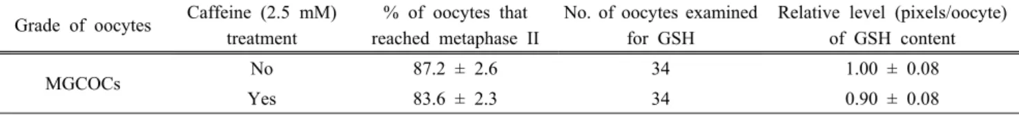
Table 1. Effect of caffeine treatment during the late stage (38-42 h) of in vitro maturation on oocyte maturation and intra-oocyte glutathione (GSH) content of morphologically good oocytes (MGCOCs)
Grade of oocytes Caffeine (2.5 mM) treatment
% of oocytes that reached metaphase II
No. of oocytes examined for GSH
Relative level (pixels/oocyte) of GSH content
MGCOCs No 87.2 ± 2.6 34 1.00 ± 0.08
Yes 83.6 ± 2.3 34 0.90 ± 0.08
Five replicates.
8. 실험 설계
실험 1에서는 체외성숙 배양액 내에 2.5 mM caffeine 첨가 가 MGCOCs 유래 돼지 난자의 체외성숙 및 세포질 내 GSH 함량에 미치는 영향을 조사하였고, 실험 2에서는 난자의 단위 발생 후 배아의 체외 발육능에 미치는 영향을 조사하였다. 실 험 3과 4에서는 각각 체외성숙배양액 내 2.5 mM caffeine 첨 가 시기(22-42, 34-42, 38-42시간)에 따른 MGCOCs 및 MPCOCs 유래 돼지 난자의 체외성숙 및 체세포 핵이식 후 배 아의 체외 발육능에 미치는 영향을 조사하였고, 실험5에서는 체외성숙배양액 내 caffeine 첨가가 돼지 난자의 MPF에 미치 는 영향을 조사하였다.
9. 통계 분석
실험 결과는 Statistical Analysis System(SAS, version 9.4;
Statistical Analysis System Institute, Cary, NC, USA)을 이용 한 일반 선형모델(general linear model)로 분산분석을 실시하 였다. 처리 평균간의 차이는 least significant difference (LSD) test를 이용하여 p<0.05 수준에서 통계학적 유의성을 검정하 였다. 실험 결과는 평균±표준오차(standard error of the mean;
SEM)로 표기하였다.
결 과
1. Caffeine 처리가 MGCOCs의 체외성숙 및 세포질 내 glutathione 함량에 미치는 영향
MGCOCs의 체외배양에서 배양액에 2.5 mM의 caffeine을 첨가하여 체외성숙 후기 38-42시간 동안 배양하였을 때 난자 의 핵 성숙률은 83.6%로 대조군의 87.2%와 유의적 차이가 없 었다. 난자 내 GSH 함량은 caffeine 처리군(0.90 pixels/oocyte) 과 대조군(1.00 pixels/oocyte) 사이에 유의적인 차이는 관찰되 지 않았다(Table 1).
2. Caffeine 처리가 MGCOCs의 단위발생 후 배 발육에 미치는 영향 MGCOCs를 체외성숙 38-42시간에 2.5 mM caffeine으로 처리하여 체외성숙을 유도한 후 단위발생을 유도하였다. 단위
발생 난자의 배 발육을 관찰한 결과 caffeine 처리군에서 분할 률, 배반포 형성률 및 배반포 세포수는 각각 89.8%, 37.4% 및 41.1개로 무처리 대조군의 92.0%, 34.2% 및 42.1개와 비교했 을 때 유의적인 차이는 관찰되지 않았다(Table 2).
3. Caffeine 처리 시간에 따른 MGCOCs의 체외성숙 및 체세포 핵이식 후 배 발육능
체외성숙 배양액 내에 2.5 mM caffeine을 첨가하여 체외성 숙 후기 22-42, 34-42, 및 38-42시간 동안 MGCOCs를 배양하였 다. 체외성숙 후기 20시간 동안 caffeine을 처리한 군의 핵 성숙 률은 82.0%로 대조군의 89.8%와 8시간 및 4시간 처치군의 89.8%, 93.2%에 비해 유의적으로(P<0.05) 낮았다. 체세포 핵이 식 후 세포융합률은 4시간 처치군이 군이 66.4%로 대조군의 77.3%에 비해 유의적으로(P<0.05) 낮은 결과를 보였다. 그러나 분할률, 배반포 형성률 및 배반포 세포수에 대해서는 caffeine 처리에 의한 유의적인 효과가 관찰되지 않았다(Table 3).
4. Caffeine 처리가 MPCOCs의 체외성숙 및 체세포 핵이식 후 배 발육에 미치는 영향
MPCOCs의 체외성숙을 위하여 체외성숙 후기 22-42, 34-42, 및 38-42시간에 2.5 mM caffeine이 첨가된 배양액으로 난자를 배양하였다. 체외성숙 22-42시간 동안 caffeine으로 처리한 난 자(76.4%)는 대조군(88.6%) 및 4-8시간 처치군(84.4-92.6%)에 비해 유의적으로 낮은 핵 성숙률을 보였다. 핵이식 후 배 발육 능을 조사한 결과 체외성숙 후기 4시간 동안 caffeine 처리군 (43.4%)이 대조군(30.1%), 22시간(22.2%) 및 8시간 처리군에 비해 유의적으로(P<0.05) 높은 배반포 발육률을 보였다. 세포 융합률, 분할률 및 배반포 세포수에는 caffeine 첨가에 따른 유 의적인 차이가 관찰되지 않았다(Table 4).
5. 체외성숙배양액 내 caffeine 첨가가 체외성숙 난자의 MPF 함량에 미치는 영향
체외성숙 과정에서 caffeine 처리가 난자 내 MPF 활성에 미치는 영향을 검토하였다. 체외성숙 후기 38-42시간에 난자 를 2.5 mM caffeine으로 처리했을 때 MGCOCs 유래 성숙난 자의 MPF 활성은 194.3 ± 7.7 pg/ml로 무처리 대조군의 160.4
Table 2. Effect of caffeine treatment during the late stage (38-42 h) of in vitro maturation on embryonic development after parthenogenesis of morphologically good oocytes (MGCOCs)
Grade of oocytes
Caffeine (2.5 mM) treatment
No. of embryos cultured
% of embryos developed to No. of cells in blastocyst
≥ 2-cells Blastocyst
MGCOCs No 146 92.0 ± 1.1 34.2 ± 2.3 42.1 ± 2.6
Yes 136 89.8 ± 2.6 37.4 ± 8.2 41.1 ± 2.3
Five replicates.
Table 3. Effects of caffeine treatment during various stage of in vitro maturation medium on oocyte maturation and development after somatic cell nuclear transfer of morphologically good oocytes (MGCOCs)
Grade of oocytes
Caffeine treatment
% of oocytes that reached MII
% of fused oocytes
No. of SCNT oocytes cultured
% of embryos developed to No. of cells in blastocyst
≥ 2-cell Blastocyst
MGCOCs
No 89.8 ± 2.3a 77.3 ± 3.6a 121 81.8 ± 2.1 24.0 ± 3.7 37.9 ± 2.8 22-42h 82.0 ± 2.3b 72.8 ± 4.7ab 94 76.5 ± 2.8 19.8 ± 6.3 45.3 ± 4.0 34-42h 89.8 ± 2.6a 70.2 ± 2.5ab 96 85.9 ± 3.7 30.8 ± 5.6 41.1 ± 3.5 38-42h 93.2 ± 1.4a 66.4 ± 2.5b 100 87.0 ± 4.7 29.6 ± 6.8 46.8 ± 3.9 Four replicates.
abValues in the same column with different superscript letters are different (P<0.05).
Table 4. Effects of caffeine treatment during various stage of in vitro maturation medium on oocyte maturation and development after somatic cell nuclear transfer of morphologically poor oocytes (MPCOCs)
Grade of oocytes
Caffeine treatment during IVM
% of oocytes that reached MII
Fusion rate (%)
No. of SCNT oocytes cultured
% of embryos developed to No. of cells in blastocyst
≥ 2-cell Blastocyst
MGCOCs No 94.8 ± 1.0a 68.9 ± 4.8 131 86.1 ± 3.9 36.8 ± 3.7ab 43.3 ± 2.8
MPCOCs
No 88.6 ± 3.0ab 66.9 ± 4.6 123 78.7 ± 8.3 30.1 ± 4.2bc 42.5 ± 2.9 22-42h 76.4 ± 3.9c 70.2 ± 6.4 109 81.7 ± 6.1 22.2 ± 3.3c 42.1 ± 3.5 34-42h 84.4 ± 4.0bc 74.8 ± 5.6 113 88.0 ± 2.3 26.2 ± 4.6bc 38.7 ± 2.5 38-42h 92.6 ± 1.9a 71.7 ± 6.7 130 80.7 ± 2.8 43.4 ± 4.0a 43.5 ± 2.8 Five replicates.
abcValues in the same column with different superscript letters are different (P<0.05).
Table 5. Effect of caffeine treatment during 38-42 h of in vitro maturation on maturation promoting factor activity in morphologically good (MGCOCs) and poor oocytes (MPCOCs)
Grade of oocytes Caffeine treatment during 38-42 h of IVM Time of MPF assay after IVM MPF activity (pg/ml)
MGCOCs
- 38 h 210.6 ± 16.0a
No 42 h 160.4 ± 7.7ab
Yes 42 h 194.3 ± 31.9a
MPCOCs
- 38 h 161.3 ± 15.8ab
No 42 h 133.9 ± 24.2b
Yes 42 h 204.8 ± 21.1a
abValues in the same column with different superscript letters are different (P<0.05).
± 7.7 pg/ml과 유의적인 차이가 없었다. 그러나 MPCOCs 유 래 체외성숙 난자의 MPF 활성은 204.8 ± 21.1 pg/ml로 대조
군의 133.9 ± 24.2 pg/ml에 비해 caffeine 처리에 의해 유의적 으로(P<0.05) 증가하였다(Table 5).
고 찰
체세포 핵이식 기술은 인체 유용단백질의 대량생산, 인공 장기, 질환모델동물과 같은 형질전환동물을 생산함에 있어 유 용한 방법으로 활용되고 있다. 그럼에도 불구하고 낮은 복제 효율성이 큰 문제점으로 남아있다. 돼지 체세포 핵이식의 효 율을 높이기 위해서는 다수의 고품질 성숙난자 확보가 뒷받 침 되어야 한다. 돼지의 경우 체세포 핵이식에 이용되는 난자 는 대부분 도축된 미경산돈의 난소로부터 채취된 미성숙 난 자를 체외에서 배양하여 생산하고 있다. 그러나 체외에서 성 숙된 난자는 자연적으로 체내에서 성숙되어 배란된 난자에 비해 생존성 또는 발생능이 떨어지는 것으로 보고되고 있다 (Blanco 등, 2011). 따라서 인공번식기술의 효율을 향상시키 기 위해서는 고품질의 난자를 다수 확보하기 위한 새로운 체 외성숙 배양법 개발에 관한 연구가 필요하다.
세포 주기를 조절하는 인자인 MPF는 난자의 유사분열을 조 절하는 중요한 요소 중의 하나로서 Cdc2(CDK1이라고도 함) 와 cyclin B1의 복합체로 구성되어 있다(Labbe 등, 1989). 난자 의 감수분열 과정에서 난자 내 MPF 활성은 성숙단계에 따라 증가 또는 감소하는 현상을 보이며, 난세포질 내에서 생성되는 소량의 MPF가 증폭되어 일정 농도가 되면 난의 성숙을 유발 한다. 본 연구에서 우리는 인위적으로 난자의 MPF 활성을 높 여주기 위해 체외성숙 배양액 내에 2.5 mM의 caffeine을 첨가 하여 체외성숙에 이용하였다. 기존 연구 결과에서 난자 배양액 에 첨가되는 caffeine 농도의 허용범위는 2.5-5.0 mM 로 보고 되어 있다(Kawahara 등, 2005; Kren 등, 2004; Abeydeera 등, 1997). 돼지에서는 5 mM 카페인이 체세포 핵이식 과정에서 전기적 자극 후 분열 속도를 감소시키는 효과를 보였으며, 노 화난자에 caffeine을 주입하였을 경우 핵 리모델링을 촉진 시 켰다는 보고도 있다(Iwamoto 등 2005). 뿐만 아니라 카페인이 세포 중심체의 완전성을 회복하고 노화된 돼지 난자에서 방추 사를 유지할 수 있음도 발견되었다(Miao 등, 2009). 본 연구에 서는 난자의 체외성숙 과정에서 caffeine 처리 시간이 난자성 숙에 미치는 영향에 대해 조사하였다. 그 결과 체외성숙 후기 22시간 동안 caffeine 처리는 무처리 및 4시간 처리군에 비해 유의적으로 낮은 성숙률을 보였다. 기존 연구에 따르면 caffeine 은 난자의 성숙과정에서 cyclic adenosine monophosphate (cAMP) 수준을 유의적으로 하게 증가시켰으며 cdc2 kinase와 MAP kinase 활성화를 억제시켰다고 보고되었다(Kren 등, 2004; Bernal-Ulloa 등, 2016). 본 연구 결과에서도 22시간 동안 의 caffeine 처리는 위와 같은 이유로 인해 난자의 핵 성숙률을 감소시킨 것으로 추정해 볼 수 있다. 그러나 체외성숙 후기 4 시간 동안의 caffeine 처리는 난자의 성숙률 감소 없이 체세포 핵이식 후 증가된 배반포 발달률을 보였다. 그 이유는 대부분
의 난자가 제1극체가 방출된 시점인 체외성숙 38시간 이후에 caffeine으로 처리 함으로서 핵 성숙에 영향을 주지 않으면서 MPF 활성을 증가시킴으로써 체세포 핵이식 후 배 발육을 개 선한 것으로 판단된다. 또한 본 연구에서 체외성숙 후기 4시간 동안 난자에서 카페인 첨가 효과를 검토한 결과, MGCOCs 유 래 난자와 달리 MPCOCs 유래 난자에서는 caffeine의 첨가가 체세포 핵이식 이후에 배반포 발달률을 증가시키는 것으로 조 사되었다. 이전 연구결과에 따르면 원시난포의 성장이 진행되 면 난자는 난포성장과 함께 발육, 성숙하면서 충분한 양의 cdc2와 cyclin B1 단백질이 모두 만들어져야만 난자성숙이 완 성될 수 있다고 보고되었다(Kanatsu-Shinohara 등; 2000; Fulka 등, 1986). 그러나 원시난포로부터 회수된 난자나 혹은 미성숙 개체로부터 채취된 난자의 경우 체외배양을 해도 난자성숙이 원활히 유도되지 않는다. 그 원인으로는 cdc2나 cyclin B1 단 백질의 양이 불충분 하거나 혹은 두 단백질 간의 상호작용이 충분히 이뤄지지 않기 때문인 것으로 기존 연구들에서 보고되 고 있다(Kanatsu-Shinohara M등, 2000; Mitra 등, 1996). 상대 적으로 cdc2 나 Cyclin B1 단백질의 양이 불충분한 MPCOCs 유래 난자의 경우 caffeine 처리에 의한 인위적인 MPF 활성 증가가 MGCOCs에 비해 더 효과적으로 나타났는데 이것이 MPCOCs 유래 난자에서 핵이식 후 배 발육능이 유의적으로 개선된 원인인 것으로 판단된다. 실제로 체외성숙 후기 42시 간에 측정한 난자 세포질 내 MPF 활성을 측정한 결과 MGCOCs 군에서는 caffeine의 처리에 따른 MPF 활성에 유의 적인 변화를 확인할 수 없었으나 MPCOCs 유래 난자에서는 유의적으로 MPF가 증가하는 것을 확인할 수 있었다.
적 요
본 연구에서는 체외성숙 배양액 내 caffeine 첨가가 돼지 난 자의 성숙과 단위발생 및 체세포 핵이식 후 배 발육에 미치는 영향을 조사하였다. 난세포질 및 난구세포 부착정도의 형태학 적 특징에 따라 MGCOCs와 MPCOCs로 구분된 미성숙난자 를 각각 무처리군(대조군)과 2.5 mM caffeine이 첨가된 배양 액에서 체외성숙 22-42(20시간), 34-42(8시간), 38-42(4시간) 동안 처리하는 군으로 나누어 체외성숙을 유도하였다. 또한 체외성숙 난자를 단위발생 및 체세포 핵이식에 공여하여 배 아를 생산한 후 7일 동안 체외배양하여 체외성숙 동안 caffeine 처리가 분할률, 배반포 형성률 및 배반포의 세포수에 미치는 영향을 조사하였다. 연구 결과, 분할률 및 배반포 세포 수는 caffeine의 처리 시간에 따라 유의적인 영향을 받지 않았 다. 그러나 MPCOCs 유래 난자에서 체외성숙 후기 4시간 동 안 caffeine 처리는 체세포 핵이식 배아의 배반포 형성률을 유
의적으로 증가시켰다. 이 결과는 caffeine 처리가 체외성숙 동 안 난자의 MPF 수준의 감소를 억제시킴으로써 체세포 핵의 리모델링이나 리프로그래밍에 영향을 미쳐 핵이식 배아의 발 육능에 영향을 미친 것으로 사료된다.
ACKNOWLEDGMENTS
This research was supported by Basic Science Research Program through the National Research Foundation of Korea (NRF) funded by the Ministry of Science, ICT and Future Planning (Grant No. 2015R1A2A2A01005490) and 2016 Research Grant from Kangwon National University (No.
520150282).
REFERENCES
Abeydeera L, and Day B. 1997. In vitro penetration of pig oocytes in a modified Tris-buffered medium: effect of BSA, caffeine and calcium. Theriogenology 48:537-544.
Alvarez GM, Dalvit GC, Achi MV, Miguez MS, and Cetica PD. 2009. Immature oocyte quality and maturational competence of porcine cumulus-oocyte complexes subpopulations. Biocell 33:167-177.
Bang J, Yoo J, Park M, Shin T, Cho B, Lee H, Kim B, Kang T, Kong I, and Kim J. 2013. The effects of artificial activation timing on the development of SCNT-derived embryos and newborn piglets. Reproductive biology 13:127-132.
Bavister BD, Lorraine Leibfried M, and Lieberman G. 1983.
Development of preimplantation embryos of the golden hamster in a defined culture medium. Biol. Reprod.
28:235-247.
Bernal-Ulloa SM, Heinzmann J, Herrmann D, Hadeler K, Aldag P, Winkler S, Pache D, Baulain U, Lucas-Hahn A, and Niemann H. 2016. Cyclic AMP affects oocyte maturation and embryo development in prepubertal and adult cattle. PloS one 11:e0150264.
Demyda S, and Genero E. 2011. Developmental competence of in vivo and in vitro matured oocytes: a review.
Biotechnology and Molecular Biology Reviews 6:155-165.
Fulka Jr J, Motlík J, Fulka J, and Crozet N. 1986. Activity of maturation promoting factor in mammalian oocytes after its
dilution by single and multiple fusions. Dev. Biol.
118:176-181.
Hyun S, Lee G, Kim D, Kim H, Lee S, Kim S, Lee E, Lim J, Kang S, and Lee B. 2003. Effect of maturation media and oocytes derived from sows or gilts on the development of cloned pig embryos. Theriogenology 59:1641-1649.
Hyun S, Jeung Y, Lee E, Kim H, Kim G and Jeung E. 2006.
Selection of surrogates and analysis of its ovulation status for the production of somatic cell cloned piglets. J. Vet.
Clin. 23:123-128.
Im G, Lai L, Liu Z, Hao Y, Wax D, Bonk A, and Prather RS.
2004. In vitro development of preimplantation porcine nuclear transfer embryos cultured in different media and gas atmospheres. Theriogenology 61:1125-1135.
Iwamoto M, Onishi A, Fuchimoto D, Somfai T, Suzuki S, Yazaki S, Hashimoto M, Takeda K, Tagami T, and Hanada H. 2005. Effects of caffeine treatment on aged porcine oocytes: parthenogenetic activation ability, chromosome condensation and development to the blastocyst stage after somatic cell nuclear transfer. Zygote 13:335-345.
Kanatsu-Shinohara M, Schultz RM, and Kopf GS. 2000.
Acquisition of meiotic competence in mouse oocytes:
absolute amounts of p34cdc2, cyclin B1, cdc25C, and wee1 in meiotically incompetent and competent oocytes. Biol.
Reprod. 63:1610-1616.
Kawahara M, Wakai T, Yamanaka K, Kobayashi J, Sugimura S, Shimizu T, Matsumoto H, Kim JH, Sasada H, and Sato E. 2005. Caffeine promotes premature chromosome condensation formation and in vitro development in porcine reconstructed embryos via a high level of maturation promoting factor activity during nuclear transfer. Reproduction 130:351-357.
Kikuchi K, Naito K, Noguchi J, Shimada A, Kaneko H, Yamashita M, Aoki F, Tojo H, and Toyoda Y. 2000. Maturation/M-phase promoting factor: a regulator of aging in porcine oocytes.
Biol. Reprod. 63:715-722.
Kim J, You J, Hyun S, Lee G, Lim J, and Lee E. 2010.
Developmental competence of morphologically poor oocytes in relation to follicular size and oocyte diameter in the pig.
Mol. Reprod. Dev. 77:330-339.
King N, Korolchuk S, McGivan J, and Suleiman M. 2004. A new method of quantifying glutathione levels in freshly isolated single superfused rat cardiomyocytes. J. Pharmacol.
Toxicol. Methods 50:215-222.
Kren R, Ogushi S, and Miyano T. 2004. Effect of caffeine on meiotic maturation of porcine oocytes. Zygote 12:31-38.
Kwon DJ, Park CK, Yang BK, and Cheong HT. 2008. Control of nuclear remodelling and subsequent in vitro development and methylation status of porcine nuclear transfer embryos. Reproduction 135:649-656.
Labbe JC, Capony JP, Caput D, Cavadore JC, Derancourt J, Kaghad M, Lelias JM, Picard A, and Doree M. 1989. MPF from starfish oocytes at first meiotic metaphase is a heterodimer containing one molecule of cdc2 and one molecule of cyclin B. EMBO J. 8:3053-3058.
Lee J, Lee H, Lee Y, Park B, Elahi F, Lee ST, Park C, Hyun S, and Lee E. 2017. In vitro oocyte maturation in a medium containing reduced sodium chloride improves the developmental competence of pig oocytes after parthenogenesis and somatic cell nuclear transfer. Reproduction, Fertility and Development 29:1625-1634.
Lee J, Park J, Yun JI, Lee Y, Yong H, Lee ST, Park C, Hyun S, Lee G, and Lee E. 2015. Rapamycin treatment during in vitro maturation of oocytes improves embryonic development after parthenogenesis and somatic cell nuclear transfer in pigs. Journal of veterinary science 16:373-380.
Lee J, and Campbell KH. 2006. Effects of enucleation and caffeine on maturation-promoting factor (MPF) and mitogen-activated protein kinase (MAPK) activities in ovine oocytes used as recipient cytoplasts for nuclear transfer. Biol. Reprod.
74:691-698.
Li Z, Shi J, Liu D, Zhou R, Zeng H, Zhou X, Mai R, Zeng S, Luo L, and Yu W. 2013. Effects of donor fibroblast cell type and transferred cloned embryo number on the efficiency of pig cloning. Cellular Reprogramming (Formerly “Cloning and Stem Cells”) 15:35-42.
Miao Y, Kikuchi K, Sun Q, and Schatten H. 2009. Oocyte
aging: cellular and molecular changes, developmental potential and reversal possibility. Hum Reprod Update 15:573-585.
Mitra J, and Schultz RM. 1996. Regulation of the acquisition of meiotic competence in the mouse: changes in the subcellular localization of cdc2, cyclin B1, cdc25C and wee1, and in the concentration of these proteins and their transcripts. J.
Cell Sci. 109(Pt 9):2407-2415.
Polge C and Day BN. 1977. Transfer of preimplantation pig embryos following in vitro culture for 24 or 48 hours. J.
Anim. Sci. 44:1036-1040.
Pope WF, Lawyer MS, Nara BS and First NL. 1986. Effect of asynchronous superinduction on embryo survival and range of blastocyst development in swine. Biol. Reprod.
35:133-137.
Sakatani M, Suda I, Oki T, Kobayashi S, Kobayashi S, and Takahashi M. 2007. Effects of purple sweet potato anthocyanins on development and intracellular redox status of bovine preimplantation embryos exposed to heat shock.
Journal of Reproduction and Development 53:605-614.
Villa-Diaz LG, and Miyano T. 2004. Activation of p38 MAPK during porcine oocyte maturation. Biol. Reprod.
71:691-696.
You J, Lee J, Hyun S, and Lee E. 2012. L-carnitine treatment during oocyte maturation improves in vitro development of cloned pig embryos by influencing intracellular glutathione synthesis and embryonic gene expression. Theriogenology 78:235-243.
Received September 18 2017, Revised September 22, 2017, Accepted September 25, 2017