Antiadipogenic Activity of Solvent-partitioned Fractions from Limonium tetragonum in 3T3-L1 Preadipocytes
Myeong Sook Kwon1, Jung-Ae Kim1, Jung Hwan Oh1, Fatih Karadeniz2, Jung Im Lee2, Youngwan Seo3,4 and Chang-Suk Kong1,2*
1Department of Food and Nutrition, College of Medical and Life Sciences, Silla University, Busan 46958, Korea
2Marine Biotechnology Center for Pharmaceuticals and Foods, College of Medical and Life Sciences, Silla University, Busan 46958, Korea
3Division of Marine Bioscience, College of Ocean Science and Technology, Korea Maritime and Ocean University, Busan 49112, Korea
4Department of Convergence Study on the Ocean Science and Technology, Ocean Science and Technology School, Korea Maritime and Ocean University, Busan 49112, Korea
Received August 14, 2018 /Revised October 25, 2018 /Accepted November 14, 2018
Limonium tetragonum, an edible halophyte that grows on salt marshes in Korea, is thought to possess various health benefits (e.g., antioxidant, antitumor, and hepatoprotective). In the present study, differ- ent solvent partitioned subfractions, water (H2O), buthanol (n-BuOH), 85% aqueous methanol (85% aq.
MeOH), and hexane (n-hexane), from crude extract of L. tetragonum were tested for their ability to pre- vent adipogenesis in differentiating 3T3-L1 preadipocytes. The treatment of differentiating 3T3-L1 pre- adipocytes with L. tetragonum subfractions (LTFs) resulted in suppressed adipogenesis and reduced ex- pression of adipogenesis-related transcription factors such as peroxisome proliferator-activated re- ceptor gamma (PPARγ), CCAATT/enhancer-binding protein alpha (C/EBPα), and sterol regulatory el- ement-binding protein 1c (SREBP-1c) at both mRNA and protein levels. In addition, the LTF treatment notably decreased the levels of phosphorylated p38, extracellular signal-regulated kinase (ERK), and c-Jun N-terminal kinase (JNK) of the mitogen-activated protein kinase (MAPK) pathway in association with PPARγ-linked adipogenesis. Among all the tested LTFs, H2O and n-hexane were the most effec- tive in lowering lipid accumulation and regulating the adipocyte differentiation via PPARγ pathway.
Taken together, the results indicated that the H2O and n-hexane LTFs contain bioactive compounds that may exhibit significant antiadipogenesis activity by downregulation of the PPARγ pathway and inactivation of the MAPK signal pathway in 3T3-L1 preadipocytes.
Key words : 3T3-L1, Adipogenesis, Limonium tetragonum, MAPK, PPARγ
*Corresponding author
*Tel : +82-51-999-5429, Fax : +82-51-999-5457
*E-mail : [email protected]
This is an Open-Access article distributed under the terms of the Creative Commons Attribution Non-Commercial License (http://creativecommons.org/licenses/by-nc/3.0) which permits unrestricted non-commercial use, distribution, and reproduction in any medium, provided the original work is properly cited.
Journal of Life Science 2019 Vol. 29. No. 1. 60~68 DOI : https://doi.org/10.5352/JLS.2019.29.1.60
Introduction
Excessive fat accumulation in the body resulting in body mass index to exceed 30 is commonly regarded as a situation called obesity. Obesity is found to be one of the leading caus- es of many life-threatening conditions such as Type II dia- betes [14], heart diseases [12], hypertension [24] and cancer [1]. Molecular onset and progress of obesity is linked with both environmental and genetic factors which mostly ex- pressed as impaired adipocyte function [15, 17]. Triacylgly- cerols are highly efficient sources of energy in the body, and mammals have developed complicated pathways to store ex-
cess energy in the body through transforming triacylglycer- ols into fats to minimize the loss of energy in adipose tissue [22]. Adipose tissue is an important part of the body in con- nection with most fatal organs and formed by specialized cell types called adipocytes, which are responsible for stor- ing the excessive energy as fat and secreting cell-specific hor- mones that play pivotal roles in almost all metabolic path- ways [6]. Obesity progression starts with the rapid increase of adipocyte numbers and in turn expansion of the adipose tissue. Due to their significant role in the fat metabolism, adipocyte function is gaining interest as the main target for prevention and treatment of obesity along other metabolic complications which are linked to irregular adipocyte behav- ior [8]. Formation of adipocytes is carried out by the differ- entiation of pre-adipocytes through adipogenesis where the pre-adipocytes mature into adipocytes through specific path- ways in the presence of differentiation markers.
Adipogenesis is regulated by an intricate mechanism in- volving peroxisome proliferator-activated receptor γ (PPAR
γ) and mitogen activated protein kinase (MAPK) pathways as the main coordinators [7, 18]. PPARγ is a member of the nuclear-receptor superfamily and has been considered as main factor in adipogenesis as well as sequentially activated CCAAT-enhancer-binding protein α (C/EBPα). Activation of both proteins induces the expression of required proteins and enzymes which are responsible maintain and express adipocyte-specific function. Several on the market obesity drugs, acting as PPARγ ligands, target and inhibit PPARγ activation [4]. PPARγ and C/EBPα pathways are regulated by different metabolic pathways such as mitogen-activated protein kinase (MAPK) and 5' adenosine monophosphate- activated protein kinase (AMPK) that are required to main- tain energy metabolism-linked cellular functions as adipo- cytes [2].
Recently, attention from preventive treatment studies has been directed to development of active ingredients from nat- ural sources with minor side effects and high biocompatibi- lity for preventing and alleviating obesity. Limonium tetrago- num is a halophyte growing in salt marshes and rocky shores of Korea and known to possess health beneficial properties.
Common to halophytes L. tetragonum is reported to have an- tioxidant substances and in addition some potential bio- activities such as hepatoprotection and anti-cancer [13, 25].
Therefore, current study investigated the potential effect of L. tetragonum and its sub-fractions on adipocyte differ- entiation to provide data regarding its anti-obesity potential and possible bioactive constituents.
Materials and Methods
Chemicals and Materials
Chemicals and reagents for cell culture and differentiation were acquired from Gibco BRL (Grand Island, NY) unless otherwise stated. Primary and secondary antibodies for Western blot analysis were procured from Cell Signaling Technology (Danvers, MA, USA). Specific amplification pri- mers for reverse transcription polymerase chain reaction was purchased from Bio-RAD (Hercules, CA, USA). Remaining materials and chemicals were ordered from Sigma-Aldrich (St. Louis, MO, USA) unless noted.
Plant material
The sample (1 kg) of L. tetragonum was air dried and ground. Plant material was grounded using a cutter and ex- tracted subsequently with 3 l methylene chloride (CH2Cl2)
and 3 l MeOH for 24 hr each solvent at room temperature.
Each extract from two solvents were concentrated in vacuo with a rotary evaporator, and dried crude extracts were ob- tained and combined. The combined crude extracts (32.06 g) from CH2Cl2 and MeOH extraction were partitioned be- tween methylene chloride and water (H2O) following the preparation of a stock sample (1 mg/ml) in 10% dimethyl sulfoxide (DMSO) to use in experiments. Crude extracts were dissolved in a 2 l mix containing 1:1 (v/v) CH2Cl2 and H2O in a separating funnel. The mix was shaken vigorously for 5 min and the funnel was kept in room temperature for 4 hr afterwards. This procedure was repeated two times un- til the separation was concluded. The methylene chloride layer was transferred to another flask and dried under re- duced pressure using a rotary evaporator. The residue was dissolved and partitioned between 2 l mix (1:1, v/v) of n-Hexane and 85% aq. MeOH with same procedure above.
The aqueous layer was also transferred to another flask and concentrated to dryness. The residue was partitioned be- tween 2 l mix (1:1, v/v) of n-BuOH and H2O, with same procedure. Each layer of n-Hexane, 85% aq. MeOH, n-BuOH and H2O were concentrated using a rotary evaporator and solvent-partitioned fractions of L. tetragonum were obtained and labelled according to final separation solvents; n-Hexane (2.64 g), 85% aq. MeOH (1.42 g), n-BuOH (1.53 g) and H2O (26.47 g). Sub-fractions were dissolved in methanol and di- luted with DMEM to be used in experiments.
Cell culture and adipocyte differentiation
Mouse 3T3-L1 pre-adipocytes were cultured in DMEM supplemented with 10% fetal bovine serum (FBS) at 37℃
atmosphere supplemented with 5% CO2. At the second day after the cells reached confluence, adipogenic differentiation of the pre-adipocytes was induced with an adipogenesis cocktail containing insulin (5 μg/ml), dexamethasone (0.25 μM), and methylisobutylxanthine (0.5 mM) in DMEM. Two days after the differentiation inducement, the adipogenesis cocktail was replaced with DMEM with 10% FBS and insulin (5 μg/ml). Following a 48 hr incubation, after the con- firmation of adipogenesis progression through the observa- tion of cell morphology, cells were fed 10% FBS-supple- mented DMEM until the time of experiment. L. tetragonum samples were administered to the cell culture medium start- ing with adipogenesis cocktail and included in all new medium. Cytotoxicity levels of L. tetragonum solvent frac- tions (LTFs) to 3T3-L1 cells was evaluated by MTT assay
as previously described [10].
Oil-Red O staining of the lipid droplets
Cell culture medium was removed at the day 8 after the inducement of adipogenesis, and cells were washed twice with phosphate buffer saline (PBS). Cells were then fixed on wells with 3.7% fresh formaldehyde dissolved in PBS and left at room temperature for 1 hr. Removal of fixation sol- ution from wells were followed by the administration of fil- tered staining solution containing Oil-Red O (0.5% w/v) dis- solved in 60% isopropanol and 40% water. After 1 hr of in- cubation, staining solution was aspired from the plates.
Images of cells were taken with a Nikon Instruments micro- scope (Tokyo, Japan). Oil-Red O stain was eluted from wells with 100% isopropanol and quantified by its absorbance val- ue at 500 nm using a microplate reader (Tecan Austria GmbH, Austria).
Reverse transcription polymerase chain reaction analysis
Total RNA was isolated from differentiated adipocytes us- ing Trizol according to reagent’s manual (Invitrogen Co., CA, USA). Total RNA (2 μg) was added to RNase-free water containing oligo (dT) and denatured for 5 min at 70℃ for cDNA synthesis. Reverse transcription of the cDNA was car- ried out in a master mix (1X RT buffer, 1mM dNTPs, 500 ng oligo (dT), 140 U M-MLV reserve transcriptase and 40 U RNase inhibitor) using an automatic T100 Thermal Cycler (Bio-Rad, UK). The reverse transcription cycle was consisted of 42℃ for 60 min and 72℃ for 5 min. The amplification of the synthesized cDNA was performed using the following sense and antisense primers for specific target proteins: for- ward 5'-TTT-TCA-AGG-GTG-CCA-GTT-TC-3' and reverse 5'-AAT-CCT-TGG-CCC-TCT-GAG-AT-3' for PPARγ; for- ward 5‘-TGT-TGG-CAT-CCT-GCT-ATC-TG-3’ and reverse 5‘-AGG-GAA-AGC-TTT-GGG-GTC-TA-3‘ for SREBP-1c; for- ward 5’-TTA-CAA-CAG-GCC-AGG-TTT-CC-3‘ and reverse 5’-GGC-TGG-CGA-CAT-ACA-GTA-CA-3‘ for C/EBPα; for- ward 5'-CCA-CAG-CTG-AGA-GGG-AAA-TC-3' and reverse 5'-AAG-GAA-GGC-TGG-AAA-AGA-GC-3' for β-actin. The cDNA amplification on T100 Thermal Cycler (Bio-Rad, UK) was carried out by 30 cycles where each cycle included the steps of 95℃ for 45 sec, 60℃ for 1 min and 72℃ for 45 sec.
Amplified products were separated by gel electrophoresis on 1.5% agarose gel for 30 min at 100 V. Gels were stained with 1 mg/ml ethidium bromide and gel images were taken
under UV light using Davinch-Chemi imagerTM (CAS-400 SM, Seoul, Korea).
Western blot
Levels of target proteins were assessed with standard Western blotting procedures. Briefly, cells were lysed in RIPA lysis buffer (Sigma–Aldrich Corp., St. Louis, MO, USA) at 4℃ for 30 min. Cell lysates (25 μg) were used for Western blot analysis. Separation of proteins in total cell ly- sate were completed by sodium dodecyl sulphate-poly- acrylamide gel electrophoresis (SDS-PAGE) on 4% stacking and 10% separating gels. Proteins were then transferred to a polyvinylidene fluoride membrane (Amersham Pharmacia Biotech., England, UK) from gels, and the membrane was blocked with 5% skim milk powder in TBST buffer after transfer. The membrane was then incubated with primary antibodies (1:1,000 in 1X TBST with 5% bovine serum albu- min) at 4℃ overnight. Membranes were incubated with horseradish-peroxidase-conjugated secondary antibody at room temperature for 2 hr. Proteins bands were imaged by a luminol-based chemiluminescence assay kit (Amersham Pharmacia Biosciences, England, UK) according to the man- ufacturer's instructions. Images of the membranes with pro- tein bands were captured using a Davinch-Chemi imagerTM (CAS-400SM, Seoul, Korea).
Statistical analysis
The data was presented as mean of three independent experiments ± SD. Statistically significant differences be- tween the means of the individual groups were determined by one-way analysis of variance (ANOVA) followed by Duncan’s multiple range tests. SAS v9.1 software (SAS Institute, Cary, NC, USA) was used for statistical analysis and calculations. The significance of differences was defined at the p<0.05 level.
Results and Discussion
Effect of LTFs on lipid accumulation in differentiat- ing 3T3-L1 adipocytes
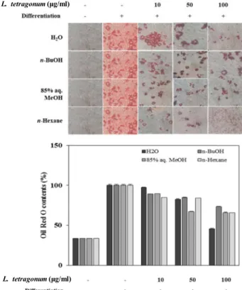
To evaluate the possible anti-adipogenic effect of LTFs, 3T3-L1 mouse pre-adipocyte fibroblasts were induced to dif- ferentiate into mature adipocytes in the presence of sol- vent-partitioned LTFs namely 85% aq. MeOH, n-Hexane, H2O and n-BuOH LTFs.
Prior to adipogenesis-linked examinations, the toxicity of
Concentration (μg/ml)
Fig. 1. Cytotoxic effect of LTFs on 3T3-L1 cells. The data repre- sent the mean ± SD of three separate experiments.
L. tetragonum (μg/ml)
L. tetragonum (μg/ml)
Fig. 2. Effect of LTFs on the lipid accumulation of differentiated 3T3-L1 adipocytes depicted by Oil red O staining and the quantification of the stain bound to lipid droplets.
The data represent the mean ± SD of three separate experiments.
LTFs was observed via their effect on the viability of 3T3-L1 cells by MTT assay. None of LTFs exerted any toxicity to cultured 3T3-L1 pre-adipocytes at varying concentrations of 10, 50 and 100 μg/ml (Fig. 1). Next, the inhibitory effect of LTFs on the adipogenic differentiation of 3T3-L1 cells was examined via lipid accumulation of the mature adipocytes.
Lower intracellular lipids were regarded as hindered adipo- genesis as fat accumulation is the key characteristic of a ma- ture adipocyte. The differentiating cells were shown to con- tain relatively higher amounts of lipid compared to non-dif- ferentiated blank group which was dose-dependently in- hibited by all tested LTFs (Fig. 2). Lipid levels of 3T3-L1 cells were lowered by 52% of the untreated fully differ- entiated control group after treatment with 100 μg/ml H2O LTF, followed by n-Hexane, MeOH and n-BuOH LTFs re- spective to their inhibitory percentage. These results sug- gested an adipogenesis inhibitory effect for LTFs on 3T3-L1 pre-adipocytes.
Effect of LTFs on PPARγ pathway during adipo- genesis of 3T3-L1 cells
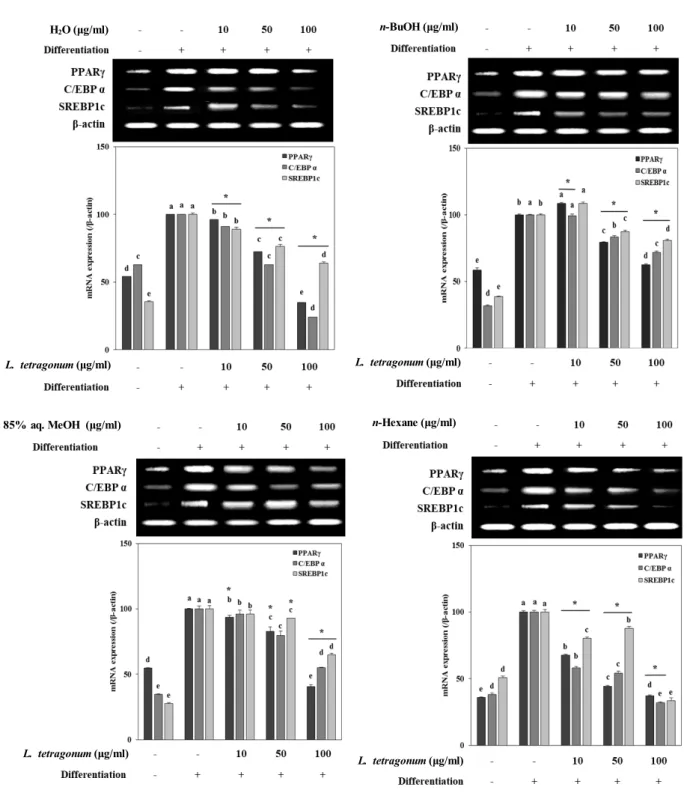
In order to provide insights towards the possible mecha- nism behind the adipogenesis inhibitory effect of LTFs, mRNA expression and protein levels of crucial adipogenesis markers (PPARγ, C/EBPα and SREBP1c) and linked tran- scription factors from MAPK pathways (p38, p-ERK and p-JNK) were observed. Studies suggested strong links be- tween obesity and irregularly elevated adipogenesis as the adipose tissue-dependent factors induce the PPARγ con- trolled differentiation [3]. Expectedly, mRNA expression and protein levels of all adipogenesis-related factors was notably
increased following the differentiation of 3T3-L1 cells into mature adipocytes (Fig. 3, Fig. 4). While all LTFs were able to decrease the mRNA expression levels of PPARγ, C/EBPα and SREBP1c to a content dose-dependently, H2O and n-Hexane LTFs were the most active samples among all (Fig.
3). Similar results were observed in protein levels of PPARγ, C/EBPα and SREBP1c which were lowered significantly by H2O and n-Hexane LTFs compared to untreated fully differ- entiated control cells (Fig. 4). Regulating the activation of PPARγ was suggested as a potential mode of action for LTFs following the decrease in both mRNA and protein levels af- ter the treatment.
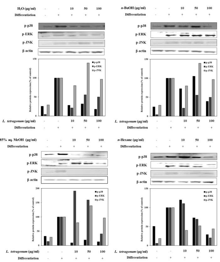
Effect of LTFs on MAPK pathway during adipo- genesis of 3T3-L1 cells
After the suggested inhibition of PPARγ pathway-linked adipogenesis in 3T3-L1s, levels of MAPK-related phosphory- lated proteins, namely p38, ERK and JNK, were also eval- uated (Fig. 5). It was shown that ERK and JNK were acti- vated sequentially during early adipogenesis by PPARγ signaling. In a MAPK-independent way, levels of phos-
L. tetragonum (μg/ml) H2O (μg/ml)
L. tetragonum (μg/ml)
L. tetragonum (μg/ml) L. tetragonum (μg/ml)
n-BuOH (μg/ml)
n-Hexane (μg/ml) 85% aq. MeOH (μg/ml)
Fig. 3. mRNA expression levels of key adipogenic factors from PPARγ in differentiated mature 3T3-L1 adipocytes following treatment of LTFs (H2O; n-BuOH; 85% aq. MeOH; n-Hexane) assayed by RT-PCR. β-actin was used as a housekeeping protein control.
The data represent the mean ± SD of three separate experiments. a-eMeans with the different letters are significantly different by Duncan’s multiple range test. (significant as compared to control. *p<0.05).
phorylated (p-) ERK, JNK and p38 proteins were also ob- served to be increased during elevated adipocyte differ- entiation [5]. In this regard, a possible intervention of the PPARγ pathway would also result in lowered activation of ERK, JNK and p38 which in turn hinder the adipogenesis
the transcription factor cascade. In a same dose-dependent manner, LTFs were able to lower the p-ERK, p-JNK, and p-p38 levels (Fig. 5) indicating a prevention of MAPK tran- scription factor relocation into nucleus which evidently lead to regulation of PPARγ-stimulated adipogenesis of 3T3-L1
H2O (μg/ml) n-BuOH (μg/ml)
L. tetragonum (μg/ml) L. tetragonum (μg/ml)
L. tetragonum (μg/ml) L. tetragonum (μg/ml)
85% aq. MeOH (μg/ml) n-Hexane (μg/ml)
Fig. 4. Effect of LTFs (H2O; n-BuOH; 85% aq. MeOH; n-Hexane) on the protein levels of the PPARγ pathway transcription actors analyzed by Western blotting. The protein levels were showed as protein bands and effects were observed as the band density. β-actin was used as a housekeeping protein control. Data presented as percentage of untreated differentiated control cell group normalized against β-actin.
H2O (μg/ml) n-BuOH (μg/ml)
L. tetragonum (μg/ml) L. tetragonum (μg/ml)
L. tetragonum (μg/ml) L. tetragonum (μg/ml)
85% aq. MeOH (μg/ml) n-Hexane (μg/ml)
Fig. 5. Effect of LTFs (H2O; n-BuOH; 85% aq. MeOH; n-Hexane) on the protein levels of the phosphorylated (p-) MAPK pathway proteins p38, ERK and JNK analyzed by Western blotting. The protein levels were showed as protein bands and effects were observed as the band density. β-actin was used as a housekeeping protein control. Data presented as percentage of untreated differentiated control cell group normalized against β-actin.
cells.
Adipogenesis of pre-adipocytes for all adipose tissue in the body was presented to share the same pathways of
PPARγ and MAPK which would potentially name these transcription factors as targets for alleviating irregular adi- pogenesis [20]. As irregular adipogenesis was credited to be
sone of the underlying mechanisms of obesity progression, inhibiting adipogenesis may serve as an effective way to pre- vent and treat obesity. Current results suggested the H2O and n-Hexane LTFs were effectively downregulated the PPARγ pathway with lowered activation of MAPKs indicat- ing a promising anti-obesity effect. Both fractions showed different efficiency in inhibiting adipogenesis via different steps. While H2O fraction was the most active sample in in- hibiting lipid accumulation during adipogenesis, Western blot and RT-PCR results showed n-Hexane as the most active fraction followed by H2O. Difference of anti-adipogenesis ef- fect level was suggested to be due to difference in chemical composition of the fractions. There are different bioactive molecules with various chemical structure bases that can act against adipogenesis. Studies showed that n-Hexane frac- tions yield mostly non-polar compounds and fatty acids. On the other hand, H2O fractions contained mostly phenolic based compounds which might inhibit adipogenesis via hin- dering lipid accumulation but lack the biocompatibility and efficiency on specific intracellular pathways due to non-spe- cific binding to the cellular membrane receptors. Several studies reported bioactive substances from H2O and n- Hexane solvent fractions of other species of plants. There- fore, according to literature and previous studies it was sug- gested that bioactive content composition of active LTFs were suggested to be phenolic-based constituents for H2O LTF [11, 23], and fatty acid-based compounds for n-Hexane LTF [16, 21]. These chemical compositions were also sug- gested to be the reason behind the different levels of an- ti-adipogenesis effects of the fractions on different steps of adipogenesis. Both chemical compositions were known to yield bioactive substances that can prevent adipogenesis [9, 19].
In conclusion, sub-fractions obtained from crude extract of L. tetragonum were able to prevent 3T3-L1 pre-adipocytes from adipogenic differentiation providing valuable insights on development of natural products against obesity and obe- sity-related complications by hindering the excess fat accumulation. Downregulation of PPARγ pathway with pre- vention of MAPK activation was suggested to be mode of action LTFs exerted their anti-adipogenic activity. Because of H2O and n-Hexane LTFs being the most active sub-frac- tions, L. tetragonum was suggested to express anti-obesity effect through compounds of phenol and fatty acid chemical backbone. In this context, L. tetragonum was suggested to be a potential source for active metabolites that could be
developed into efficient anti-adipogenic substances.
Acknowledgment
This work was supported by the National Research Foundation of Korea (NRF) grant funded by the Korea gov- ernment (MSIP) (NRF-2017R1A2B4009588).
References
1. Anderson, A. S. and Caswell, S. 2009. Obesity management - an opportunity for cancer prevention. Surgeon 7, 282-285.
2. Bost, F., Aouadi, M., Caron, L. and Binétruy, B. 2005. The role of MAPKs in adipocyte differentiation and obesity.
Biochimie 87, 51-56.
3. Burns, K. A. and Vanden Heuvel, J. P. 2007. Modulation of PPAR activity via phosphorylation. Biochim. Biophys. Acta 1771, 952-960.
4. Choi, J. H., Banks, A. S., Estall, J. L., Kajimura, S., Bostrom, P., Laznik, D., Ruas, J. L., Chalmers, M. J., Kamenecka, T.
M., Bluher, M., Griffin, P. R. and Spiegelman, B. M. 2010.
Obesity-linked phosphorylation of PPARγ by cdk5 is a di- rect target of the anti-diabetic PPARγ ligands. Nature 466, 451-456.
5. Dagon, Y., Avraham, Y. and Berry, E. 2006. M. AMPK acti- vation regulates apoptosis, adipogenesis, and lipolysis by eIF2α in adipocytes. Biochem. Biophys. Res. Commun. 340, 43- 47.
6. de Ferranti, S. and Mozaffarian, D. 2008. The perfect storm:
Obesity, adipocyte dysfunction, and metabolic consequences.
Clin. Chem. 54, 945-955.
7. Fajas, L., Fruchart, J. C. and Auwerx, J. 1998. Transcriptional control of adipogenesis. Curr. Opin. Cell Biol. 10, 165-173.
8. Hajer, G. R., van Haeften, T. W. and Visseren, F. L. J. 2008.
Adipose tissue dysfunction in obesity, diabetes, and vas- cular diseases. Eur. Heart J. 29, 2959-2971.
9. Hsu, C. L. and Yen, G. C. 2007. Effects of flavonoids and phenolic acids on the inhibition of adipogenesis in 3T3-L1 adipocytes. J. Agric. Food Chem. 55, 8404-8410.
10. Jeong, H., Kim, H., Ju, E., Kong, C. S. and Seo, Y. 2016.
Antioxidant effect of the halophyte Atriplex gmelinii. KSBB J. 31, 200-207.
11. Kim, I. S., Yang, M., Lee, O. H. and Kang, S. N. 2011. The antioxidant activity and the bioactive compound content of Stevia rebaudiana water extracts. LWT Food Sci. Technol. 44, 1328-1332.
12. Lavie, C. J., Milani, R. V. and Ventura, H. O. 2009. Obesity and cardiovascular disease: Risk factor, paradox, and impact of weight loss. J. Am. College Cardiol. 53, 1925-1932.
13. Lee, J. I., Kong, C. S., Jung, M. E., Hong, J. W., Noh, I. and Seo, Y. 2011. Peroxynitrite-scavenging activity of the hal- ophyte Limonium tetragonum. Ocean Polar Res. 33, 185-191.
14. Malnick, S. D. and Knobler, H. 2006. The medical complica- tions of obesity. QJM. 99, 565-579.
초록:갯질경이 용매분획물의 3T3-L1전지방세포에서의 지방생성억제 효과
권명숙1․김정애1․오정환1․파티 카라데니즈2․이정임2․서영완3,4․공창숙1,2*
(1신라대학교 의생명과학대학 식품영양학과, 2신라대학교 해양식의약소재융합기술연구소, 3한국해양대학교 해양
과학기술대학 해양생명과학부, 4한국해양대학교 해양과학기술전문대학 해양과학기술융합학과)
갯질경이(Limonium tetragonum)는 질경이과에 속하는 여러해살이 풀로 습지에 자생하는 염생식물의 일종이며, 항산화, 항종양 및 간보호 효능이 있는 것으로 알려져 있다. 본 연구에서는 갯질경이 추출물로부터 용매 극성에 따라 분획한 분획물(H2O, n-BuOH, 85% aq. MeOH 및 n-Hexane)을 이용하여 지방세포내 중성지방 생성 및 지방 세포 분화조절 인자 발현에 미치는 영향을 검토하였다. 마우스 유래 지방전구세포3T3-L1을 지방세포로 분화하여 Oil Red O염색법으로 지방 세포 분화정도를 확인한 결과, 갯질경이 분획물에 의해 지방세포의 형성이 농도의존적 으로 억제되었다. 또한 지방생성조절에 관여하는 전사인자 PPARγ, C/EBPα 및 SREBP-1c의 발현을 mRNA와 단 백질 수준에서 확인한 결과 갯질경이 분획물 처리시 지방세포 분화 인자의 발현이 유의적으로 감소하였다. 지방 세포 분화에 관여하는 것으로 알려진 MAPK 신호 전달 경로를 확인한 결과 갯질경이 분획물 처리군에서 p38,
ERK 및 JNK의 인산화가 억제되었다. 용매 분획물중에서 H2O 및 n-Hexane 분획물이 가장 우수한 지방생성 억제
활성을 나타내었는데 이는, 분획물 중 페놀 또는 지방 유도체에 의한 것으로 사료된다. 본 연구 결과로부터 갯질 경이 분획물의 MAPK 신호전달 경로 억제를 통한 항비만 효과를 확인하였으며, 나아가 건강기능성 식품 소재로 서의 개발 가능성이 기대된다.
15. Marti, A., Martinez-Gonzalez, M. A. and Martinez, J. A.
2008. Interaction between genes and lifestyle factors on obesity. Proc. Nutr. Soc. 67, 1-8.
16 Musa, A. M., Ibrahim, M. A., Aliyu, A. B., Abdullahi, M.
S., Tajuddeen, N., Ibrahim, H. and Oyewale, A. O. 2015.
Chemical composition and antimicrobial activity of hexane leaf extract of Anisopus mannii (Asclepiadaceae). J. Intercult.
Ethnopharmacol. 4, 129-133.
17. Ordovas, J. M. and Shen, J. 2008. Gene–environment inter- actions and susceptibility to metabolic syndrome and other chronic diseases. J. Periodontol. 79, 1508-1513.
18. Otto, T. C. and Lane, M. D. 2005. Adipose development:
from stem cell to adipocyte. Crit. Rev. Biochem. Mol. Biol.
40, 229-242.
19. Pariza, M. W., Park, Y. and Cook, M. E. 2001. The bio- logically active isomers of conjugated linoleic acid. Prog.
Lipid Res. 40, 283-298.
20. Rosen, E. D. and MacDougald, O. A. 2006. Adipocyte differ- entiation from the inside out. Nat. Rev. Mol. Cell Biol. 7, 885-896.
21. Sasidharan, S., Chen, Y., Saravanan, D., Sundram, K. M. and Yoga Latha, L. 2011. Extraction, isolation and character- ization of bioactive compounds from plants’ extracts. Afr.
J. Tradit. Complement. Altern. Med. 8, 1-10.
22. Sun, K., Kusminski, C. M. and Scherer, P. E. 2011. Adipose tissue remodeling and obesity. J. Clin. Investig. 121, 2094- 2101.
23. Uribe, E., Delgadillo, A., Giovagnoli-Vicuña, C., Quispe- Fuentes, I. and Zura-Bravo, L. 2015. Extraction techniques for bioactive compounds and antioxidant capacity determi- nation of Chilean papaya (Vasconcellea pubescens) fruit. J.
Chem. 2015, 47532.
24. Wofford, M. R. and Hall, J. E. 2004. Pathophysiology and treatment of obesity hypertension. Curr. Pharm. Des. 10, 3621-3637.
25. Yang, M. H., Kim, N. H., Heo, J. D., Sung, S. H. and Jeong, E. J. 2014. Hepatoprotective effects of Limonium tetragonum, edible medicinal halophyte growing near seashores. Pharma- cogn. Mag. 10, 563-568.