Antiinflammatory Activity of Solvent-partitioned Fractions from Atriplex gmelinii C. A. Mey. in LPS-stimulated RAW264.7 Macrophages
Heejeong Jeong1, Hojun Kim2, Eunsin Ju2, Seul-Gi Lee3, Chang-Suk Kong3 and Youngwan Seo1,2*
1Department of Convergence Study on the Ocean Science and Technology, Ocean Science and Technology School, Korea Maritime and Ocean University, Busan 49112, Korea
2Division of Marine Bioscience, College of Ocean Science and Technology, Korea Maritime and Ocean University, Busan 49112, Korea
3Department of Food and Nutrition, College of Medical and Life Sciences, Silla University, Busan 46958, Korea
Received September 27, 2016 /Revised October 27, 2016 /Accepted November 2, 2016
As a part of ongoing research to elucidate and characterize antiinflammatory nutraceuticals, the crude extracts from Atriplex gmelinii C. A. Mey. and their solvent-partitioned fractions were tested for their antiinflammatory potential in lipopolysaccharide (LPS)-stimulated RAW 264.7 mouse macrophages.
The crude extracts of A. gmelinii C. A. Mey. were fractioned according to polarity with n-hexane, 85%
aqueous methanol (85% aq. MeOH), n-butanol, and H2O. Their antiinflammatory activities were inves- tigated in LPS-induced inflammation in mouse macrophages by measuring nitric oxide (NO) gen- eration and mRNA expression of inflammation mediators, namely, inducible nitric oxide synthase (iNOS), cyclooxygenase-2 (COX-2), interleukin-1β (IL-1β), and IL-6. As a result, we confirmed that the crude extracts of A. gmelinii C. A. Mey. inhibited LPS-stimulated NO production and mRNA ex- pression of iNOS and COX-2 as important inflammatory factors. The inhibition of NO production through the downregulation of important inflammatory factors such as iNOS, COX-2, IL-1β, and IL-6 was found by treatment with all solvent-partitioned fractions. Among all tested fractions, 85% aq.
MeOH showed the strongest antiinflammatory response. Based on the current results, A. gmelinii C.
A. Mey. was suggested to possess natural antiinflammatory components, indicating that it could be used as a valuable source of antiinflammatory substances.
Key words : Anti-inflammation, Atriplex gmelinii C.A.Mey., lipopolysaccharide, macrophages, nitric oxide
*Corresponding author
*Tel : +82-51-410-4328, Fax : +82-51-404-3538
*E-mail : [email protected]
This is an Open-Access article distributed under the terms of the Creative Commons Attribution Non-Commercial License (http://creativecommons.org/licenses/by-nc/3.0) which permits unrestricted non-commercial use, distribution, and reproduction in any medium, provided the original work is properly cited.
Journal of Life Science 2017 Vol. 27. No. 2. 187~193 DOI : https://doi.org/10.5352/JLS.2017.27.2.187
서 론
염증반응은 외부자극원에 의해 신체에서 일어나는 다양한 위해 상황에서 조직손상 및 감염 등에 대항하여 일어나는 방 어보호반응으로, 조직의 항상성을 유지하기 위해 일어나는 필 수적인 조절반응이다[10]. 하지만 염증반응이 지속적으로 진 행하게 되면 세포조직의 손상뿐만 아니라 cyclooxygenase-2 (COX-2), inducible nitric oxide synthase (iNOS), inter- leukin-6 (IL-6), IL-1β 등과 같은 염증반응 촉진 인자들의 생성 에 의해 천식, 뇌질환, 알츠하이머병, 동맥경화 및 암 등의 여 러 질병 유발 및 노화의 직·간접적 원인이 되는 것으로 알려져 있다[5, 12, 17, 18].
최근 해조류 등의 해양생물자원이 가지고 있는 다양한 기능
성 성분의 발굴 및 그 효능에 대한 연구가 활발히 진행되기 시작하면서 예로부터 건강기능성 소재로서 이용되어 왔거나 그 기능성이 아직 검증하지 못한 다양한 해양생물자원에 대한 연구가 주목받고 있는 실정이다[11-13]. 염생식물은 바닷가, 염분이 있는 호숫가, 암염이 있는 지대 등 염분기가 많은 토양 에서 자라는 식물로, 이러한 식물이 자라는 염생습지는 강한 바람, 해무, 뜨거운 햇빛, 염분, 침수 등이 섞인 지하수의 영향 을 받는 극한환경에 속한다. 염생식물은 이러한 극한환경에서 생존하기 위해 특이대사산물의 생성을 통해 생리적 및 생화학 적 대사 과정이 이루어지는 것으로 보고되고 있다[1, 6, 14, 15]. 염생식물 중 갯질경이, 함초, 광나무 등을 시작으로 염생 식물의 다양한 생리활성에 대한 연구가 이루어지고 있다[11, 12]. 가는갯능쟁이(Atriplex gmelinii C. A. Mey.)는 석죽목 명아 주과 갯능쟁이속에 속하는 염생식물로 가는명아주라고도 불 리는 한해살이풀이다. 주로 한국, 일본, 러시아, 북아메리카에 분포하며, 우리나라에서는 경기도, 충청남도, 남해안, 울릉도, 독도, 제주도 등 중부이남 해변에서도 폭넓게 자생하고 있으 며 나물, 쌈, 국 등의 형태로 식용으로 사용될 뿐만아니라 사료 로도 이용되고 있다[3, 7, 8]. 또한 예로부터 가는갯능쟁이는 백전풍, 충독, 한창, 개선 등의 약효가 있는 것으로 알려져 있 으나[7], 가는갯능쟁이의 생리활성 관련 과학적인 연구는 거의
보고 되지 않고 있다. 따라서 본 연구에서는 염생식물을 이용 한 생리활성 소재 개발 연구의 일환으로 가는갯능쟁이의 용매 추출물 및 분획물을 제조하여 LPS로 염증을 유도한 마우스 유래 macrophages에서의 NO 생성 및 염증관련인자의 발현 에 미치는 영향을 검토하여 가는갯능쟁이의 항염증 소재로서 의 이용 가능성을 알아보고자 하였다.
재료 및 방법
실험재료
실험에 사용된 가는갯능쟁이(Atriplex gmelinii C. A. Mey.) 는 2007년 전라남도 무안군 현경면 현화리에서 채집하여 사용 하였으며 응달에서 건조하여 잘게 잘라 밀봉한 후 추출하기 전까지 -25℃에서 냉동보관하였다.
시료추출 및 순차분획[11, 12]
냉동보관된 가는갯능쟁이는 해빙하여 추출에 적합한 크기 로 세절한 후, methylene chloride로 침지시켜 24시간 동안 방치한 후, 여과하여 용매 추출액을 얻었으며 이 과정을 2회 반복하였다. 여과하고 남은 잔사에 동량의 methanol로 동일한 과정을 반복하여 용매 추출액을 얻었다. 얻어진 두 종류의 용 매 추출액을 진공증발기(EYELA JAPAN, N-N series)로 농축 하여 각각 methylene chloride와 methanol 추출물을 얻었으 며, 두 추출물을 혼합하여 조추출물(crude extract, 48.3 mg)을 얻었다. 혼합한 조추출물은 용매극성에 따라 단계적으로 분획 하여 n-hexane 층(11.5 mg), 85% aqueous methanol 층(85%
aq. MeOH, 11.3 mg), n-butanol 층 (n-BuOH, 11.2 mg)과 wa- ter 층(12.3 mg)을 얻었다.
세포배양
RAW264.7 대식세포는 10% fetal bovin serum (FBS, Atlas Biologicals, Fort Collins, Colorado, USA)과 1% L-Glutamine penicillin streptomycin solution (Sigma-Aldrich Co., St.
Louis, MO, USA)이 함유된 Dulbecco’s modified Eagle’s me- dium (DMEM, Corning, Manassas, VA, USA)을 배양액으로 하여 37℃, 5% CO2조건에서 배양하였다.
세포독성
RAW264.7 대식세포에 대한 세포독성은 3-(4,5-dimethyl- thiazole-2-yl) -2,5-diphenyl-tetrazolium bromide (MTT) as- say를 이용하여 측정하였다[12]. 세포는 1×10³ cells/well가 되 도록 96-well plate에 분주하여 37℃, 5% CO2 incubator에서 24시간 동안 배양한 후, 농도별로 준비한 시료를 세포주에 처 리하여 동일한 조건에서 24시간 동안 배양하였다. 각 well에 배지를 제거한 후 100 μl의 MTT용액(finc. 1 mg/ml)을 첨가하 여 동일한 배양 조건에서 4시간 동안 더 배양하여 formazan
crystal의 생성을 유도하였다. 이때 생성되는 formazan을 100 μl의 dimethyl sulfoxide (Sigma-Aldrich Co.)용액을 가하여 환원반응으로 생성된 보라색의 formazan crystal을 완전히 용 해한 후 ELISA reader (Bio-Tek instruments, Winooski, VT)로 540 nm에서 흡광도를 측정하여 생존율(%)을 구하였다.
Cell viability (%)=
대조군의 흡광도 - 시료처리군의 흡광도
대조군의 흡광도 ×100
Cell viability (%) =×100
Nitric oxide (NO) 함량
대식세포인 RAW264.7 세포를 96-well plates에 1×10³ cells/well로 well당 100 μl씩 분주하여 37℃, 5% CO2 in- cubator에서 24시간 배양한 후, 실험에 사용하였다. 페놀레드 가 제거된 새로운 배지로 교체한 다음 농도별로 준비한 시료 를 세포주에 처리한 후 NO생성을 유도하기 위해 LPS를 1 μg/
ml 농도로 처리하여 37℃, 5% CO2 incubator에서 48시간동안 배양하였다. 세포배양액을 수거하여 Griess reagent (Sigma- Aldrich Co. St. Louis, MO, USA)와 1:1의 비율로 혼합하여 10분 간 실온에서 암반응시킨 후, ELISA reader (Bio-Tek in- struments, Winooski, VT)로 540 nm에서 흡광도를 측정하였 다[12]. 생성된 NO (μM)의 농도는 표준물질인 sodium nitrate (NaNO2)용액의 표준곡선을 기준으로 하여 계산하였다.
Reverse Transcription-Polymerase Chain Reaction (RT-PCR)
상기와 동일한 조건에서 배양한 RAW264.7 대식세포를 phosphate buffered saline (PBS)로 세척하고 Trizol reagent (ambion, life technologies™, USA)로 RNA를 추출한 후 chloroform을 가하여 phenol 성분을 제거하였다. 상등액과 isopropanol을 동량으로 넣어 RNA를 침전시키고 알코올을 가하여 세척 및 건조하였다. 건조한 RNA에 DEPC water 30 μl를 첨가한 후 spectrophotometer를 이용하여 260 nm/ 280 nm 에서 흡광도를 측정하여RNA농도를 정량하였다. 분리한 동량의 RNA (2 μg)로부터 components mixture (Invitrogen, life technologies™, USA)을 이용하여 cDNA를 합성하였다.
PCR PreMix에 합성된 cDNA와 iNOS, COX-2, interleukin-1β (IL-1β) 및 IL-6 primer를 반응시킨 다음(Table 1), T100 Thermal Cycler (Bio-Rad, CA, USA)을 이용하여 94℃에서 5 분간 pre-denature, 94℃에서 30초간 denature, 50~54℃에서 30초간 annealing cycle, 72℃에서 1분간 Extension과정을 35 번 반복함으로써 증폭시킨 후, 72℃에서 10분 동안 final ex- tension시키고 4℃에서 종결, 보관하였다. 증폭된 PCR 산물들 은 TAE buffer (Biosesang, Kunggido, Korea)를 전해질로 사 용한 1.5% agarose gel을 이용하여 100 V에서 30분간 전기영 동시킨 후 ethidium bromide 용액에서 30분 간 반응시킨 후
Table 1. Sequences of primes used for RT-PCR
Gene Direction Sequence
iNOS Forward
Reverse
5'-TTC-CAG-AAT-CCC-TGG-ACA-AG-3' 5'-TGG-TCA-AAC-TCT-TGG-GGT-TC-3'
COX-2 Forward
Reverse
5'-AGA-AGG-AAA-TGG-CTG-CAG-AA-3' 5'-GCT-CGG-CTT-CCA-GTA-TTG-AG-3'
IL-1β Forward
Reverse
5'-GGG-CCT-CAA-AGG-AAA-GAA-TC-3' 5'-TAC-CAG-TTG-GGG-AAC-TCT-GC-3'
IL-6 Forward
Reverse
5'-AGT-TGC-CTT-CTT-GGG-ACT-GA-3' 5'-CAG-AAT-TGC-CAT-TGC-ACA-AC-3'
β-Actin Forward
Reverse
5'-CCA-CAG-CTG-AGA-GGG-AAA-TC-3' 5'-AAG-GAA-GGC-TGG-AAA-AGA-GC-3' iNOSCOX-2IL-1βIL-6β-Actin
Fig. 1. Cytotoxicity of the crude extracts and their solvent-partitioned fractions from A. gmelinii C. A. Mey. in RAW264.7 cells.
UV (Davinch-Chemi imager™, CAS-400SM, Davinch-K, Seoul, Korea) 하에서 mRNA 발현 정도를 관찰하였다.
통계처리
실험결과는 평균±표준편차(Mean ± Standard deviation, SD)로 표시하였으며, SPSS+/WIN12.0 (Statistical Package for Social Science, version 12.0) 통계프로그램을 이용하여 통계적 유의성을 검토하였다. 집단간의 유의성을 검정하기 위해 일원 배치 분산분석(Oneway Analysis Of Variance: ANOVA)을 통 해 분석하였고, 사후검증은 Duncan's multiple range test를 실시하여 p<0.05 수준에서 검증하였다.
결과 및 고찰
세포독성에 미치는 효과
가는갯능쟁이의 조추출물 및 분획물의 RAW264.7 대식세 포에 대한 독성 여부는 MTT assay로 확인하였다(Fig. 1). 조추 출물 및 분획물을 각각 10, 50, 100, 200 μg/ml 농도로 처리하 여 세포생존율을 측정한 결과, 헥산층을 제외한 모든 시료에 서 RAW264.7 대식세포에 대하여 200 μg/ml의 처리 농도까지
90%이상의 생존율을 보여 세포에 대한 독성이 나타나지 않음 을 확인하였으며, 이 농도에서 추출물 및 분획물의 항염증 활 성을 확인하기 위한 실험을 진행하였다.
조추출물의 Nitric oxide (NO) 생성 및 염증 인자 발현에 미치는 효과
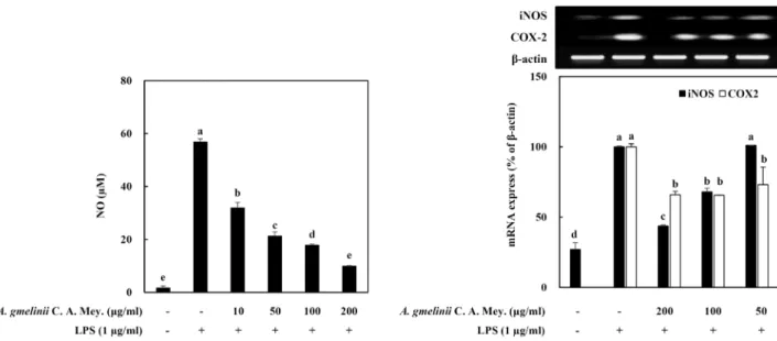
내독소(Lipopolysaccharide, LPS)는 그람음성균의 세포 외 막 구성성분으로, 단구세포와 대식세포 등의 여러 세포에 강 한 자극효과를 가지고 있다. 특히 대식세포는 LPS의 자극에 의해 활성화되면 전염증성 인자의 조절을 통해 NO와 같은 염증반응의 매개물질 등을 생산 및 방출하게 된다[9, 12]. 과도 하게 분비된 NO는 생체내의 O2, O2-와의 반응으로 반응성 산 화질소로 변화하여 산화스트레스 및 DNA손상 등을 유발시켜 각종 염증성질환의 발생에 관여하게 된다[9]. 이러한 사실을 근거로 LPS 자극에 의해 활성화된 RAW264.7 세포 내에서 생 성되는 NO에 대한 가는갯능쟁이 조추출물의 억제 효과를 측 정하였다(Fig. 2). LPS 자극에 의해 1.6±0.8 μM에서 56.8±1.1 μM로 NO의 생성이 현저히 증가하였으며, 이러한 LPS에 의한 NO의 증가는 조추출물의 처리에 의해 농도 의존적으로 유의 성있게 억제되었다. 즉 처리 농도 10, 50, 100, 200 μg/ml에서
Fig. 2. Effect of crude extracts from A. gmelinii C. A. Mey. on intracellular NO level (A) and iNOS and COX-2 gene expressions in LPS-stimulated RAW264.7 cells. The cells were pretreated with LPS (1 μg/ml) and followed by treating with the crude extracts for 24 hr. a-eMeans with the different letters are significantly different (p<0.05) by Duncan's multiple range test.
NO 생성 정도를 비교한 결과 각각 31.9±2.1 μM, 21.3±1.6 μM, 17.8±0.5 μM, 9.9±0.2 μM의 값을 나타내었다(Fig. 2A). 이들 결과로부터 NO 생성 및 염증과 관련 있는 iNOS와 COX2의 발현에 미치는 효과를 검토하였다. iNOS와 COX-2는 면역세 포의 활성을 유도하여 NO를 포함한 다량의 염증성 매개체를 생합성하는데 관여하는 것으로 알려져 있다[4, 16, 19]. COX-2 는 아라키돈산(arachidonic acid)를 이용하여 또다른 염증성 매개자로 염증반응에서 발열이나 통증 등에 영향을 끼치는 것으로 알려진 프로스타글란딘(prostaglandin)을 합성하는 효 소로 COX-2의 억제는 암 치료뿐만 아니라 염증의 억제에도 관련이 있는 것으로 알려져 있다[20, 21]. 산화질소는 산화질소 합성효소인 NOS에 의해 생성되며, NOS는 eNOS, nNOS와 iNOS의 3가지 형태가 존재한다. 이 중 eNOS와 nNOS는 세포 내에서 항시 발현되어 있는 효소로 체내에서 산화질소를 낮은 농도로 유지하고 역할을 하는 반면, iNOS는 외부 자극에 의해 생성되는 효소로 일단 LPS 등에 의해 유도가 진행되면 장기간 다량의 산화질소 농도를 생성하여 염증반응을 촉진시키는 것 으로 알려져 있다[20, 22]. 가는갯능쟁이 조추출물의 NO 생성 억제 효과가 이들 염증관련 유전자의 발현 조절과 연관성이 있는지를 확인한 결과(Fig. 2B), LPS 자극에 의해 증가한 iNOS 및 COX-2의 발현은 가는갯능쟁이 조추출물의 처리에 의해 현저히 감소하는 경향을 나타내었다. 가는갯능쟁이 조추출물 은 이들 염증인자의 발현 조절을 통하여 NO 생성을 억제하는 것으로 사료된다.
용매 분획층의 Nitric oxide (NO) 생성 및 염증 인자 발 현에 미치는 효과
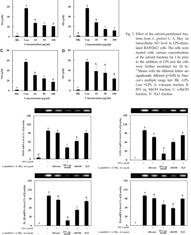
가는갯능쟁이 조추출물을 사용하여 극성순서에 따라 순차
적으로 분획하여 얻은 n-hexane, 85% aq. MeOH, n-BuOH, H2O 분획물의 항염증활성을 관찰하였다(Fig. 3). 세포내 독성 을 나타내지 않는 농도에서 LPS 자극에 의해 활성화된 RAW 264.7 세포를 이용하여 이들 분획물이 NO 생성에 미치는 효과 를 검토한 결과, 시료를 처리하지 않고 LPS를 처리한 대조군 에 비해 모든 분획층에서 유의적으로 높은 NO 생성 억제 효과 를 나타내었다. 특히 85% aq. MeOH 분획에서 가장 높은 활성 을 나타내었으며, 차순으로는 n-BuOH, n-hexane, H2O분획의 순서로 NO 생성 억제효과를 나타내었다. 처리농도 100 μg/
ml에서 NO 생성 억제 정도를 비교한 결과 n-hexane, 85% aq.
MeOH, n-BuOH, H2O 분획은 각각 63.2%, 79.1%, 69.6%, 58.5%
의 저해율을 나타내었다.
최근 건강기능성 소재에 대한 관심의 증가로 기능성 식품 및 신약에 대한 연구가 활발히 이루어지고 있으나, 합성 화합 물의 독성과 부작용의 문제 등으로 천연기능성소재의 발굴에 대한 관심이 증가함에 따라 천연자원으로부터 체내 면역체계 조절 효과를 가지는 생체활성물질(biological response modi- fier, BRM)로서의 효과를 가지는 소재 개발에 대한 연구가 활 발히 이루어지고 있다[20, 22]. iNOS와 COX-2는 pro-in- flammatory agents 의해 발현되어 면역세포의 활성화를 통해 염증반응을 유발하는 물질로 염증상태에서는 NO와 같은 염 증매개물질의 생성을 유도한다[12]. 또한 대표적인 전염증성 사이토카인인 IL-1β 및 IL-6는 활성화된 대식세포에서 과도하 게 생성되어 발열, 조직손상 및 염증반응을 촉진하는 것으로 알려져 있다[2]. 따라서 가는갯능쟁이 조추출물로부터 얻어진 이들 분획물이 염증관련 유전자의 발현에 미치는 영향을 검토 하기 위해 RT-PCR를 수행하였다(Fig. 4). NO 생성 억제능의 측정 결과에서와 같이 대조군에 비해 용매 분획물의 처리에
A B
C D
Fig. 3. Effect of the solvent-partitioned frac- tions from A. gmelinii C. A. Mey. on intracellular NO level in LPS-stimu- lated RAW264.7 cells. The cells were treated with various concentrations of the solvent fractions for 1 hr prior to the addition of LPS and the cells were further incubated for 24 hr.
a-eMeans with the different letters are significantly different (p<0.05) by Dun- can's multiple range test. BK: -LPS, Con: +LPS, A: n-hexane fraction, B:
85% aq. MeOH fraction, C: n-BuOH fraction, D: H2O fraction.
Fig. 4. Effect of solvent-partitioned fractions from A. gmelinii C. A. Mey. on mRNA expression of key inflammation markers in LPS-stimulated RAW264.7 cells. a-eMeans with the different letters are significantly different (p<0.05) by Duncan's multiple range test.
의해 iNOS와 COX-2 및 IL-6와 IL-1β의 발현이 현저히 억제되 었다. 세포내 전사수준에서 분획층 간의 발현 정도를 비교한 결과, H2O 분획을 처리한 군에 비해 85% aq. MeOH, n-BuOH 및 n-hexane 분획을 처리한 군에서 발현 억제율이 높게 나타 났다. 이들 결과로부터 가는갯능쟁이 용매 분획물의 처리는 세포내 전사수준에서의 염증유발 유전자를 조절하여 염증성 물질인 NO의 생성을 효과적으로 저해하는 것으로 사료된다.
특히 항염증 활성이 높은 85% aq. MeOH 분획층에서 염증억 제 효과가 있는 유효 성분의 분리가 이루어질 경우 염증예방 및 면역기능증진에 도움이 되는 기능성 천연물 소재로서의 응용가능성이 있을 것으로 사료된다.
감사의 글
본 연구는 2012년 교육과학기술부의 재원으로 한국연구재 단의 지원을 받아 수행된 기초연구사업(No. 2012R1A1A200 2851)의 연구결과입니다.
References
1. Hamed, K. B., Castagna, A., Salem, E., Ranieri, A. and Abdelly, C. 2007. Sea fennel (Crithmum maritimum L.) un- der salinity conditions: a comparison of leaf and root anti- oxidant responses. Plant Growth Regul. 53, 185-194.
2. Han, M. H., Lee, M. H., Hong, S. H., Choi, Y. H., Moon, J. S., Song, M. K., Kim, M. J., Shin, S. J. and Hwang, H.
J. 2014. Comparison of anti-inflammatory activities among ethanol extracts of Sophora flavescens, Glycyrrhiza uralensis and Dictamnus dasycarpus, and their mixtures in RAW 264.7 murine macrophages. J. Life Sci. 24, 329-335.
3. Hyun, J. O. and Na, H. R. Species Korea, 2011.12.16.
4. Jeong, D. H., Kim, K. B. W. R., Kang, B. K., Jung, S. A., Kim, H. Y., Jeong, H. Y., Bark, S. W. and Ahn, D. H. 2012.
Anti-inflammatory activity of the water extract of Sargassum fulvellum. Biotechnol. Bioprocess. Eng. 27, 325-329.
5. Jin, K. S., Lee, J. Y., Kwon, H. J. and Kim, B. W. 2014.
Antioxidative and anti-inflammatory activities of Ardisia ar- borescens ethanol extract. J. Life Sci. 24, 713-720.
6. Jithesh, M. N., Prashanth, S. R., Sivaprakash, K. P. and Parida, A. K. 2006. Antioxidative response mechanisms in halophytes: their role in stress defence. J. Genet. 85, 237-254.
7. Kang, B. H. 2014. Superintendence of Korean Resource Plant. Richvanilla Seoul 12-16.
8. Kim, M. H., Han, M. S., Kang, K. K., Na, Y. E. and Bang, H. S. 2011. Effects of climate change on C4 plant list and distribution in South Korea: A Review. Kor. J. Agric. For.
Meteorol. 13, 123-139.
9. Kim, Y. J. and Son, D. Y. 2014. Inflammatory mediator regu- lation of the Zizyphus jujube leaf fractions in the LPS-stimu- lated Raw264.7 mouse macrophage. Kor. J. Food Preserv. 21, 114-120.
10. Kim, Y. S., Lee, S. J., Hwang, J. W. Kim, E. H., Park, P.
J. and Jeong, J. H. 2012. Anti-inflammatory effects of extracts
from Ligustrum ovalifolium H. leaves on RAW 264.7 macrophages. J. Kor. Soc. Food Sci. Nutr. 41, 1205-1210.
11. Kong, C. S, Kim, Y. A., Kim, M. M., Park, J. S., Kim, J. A., Kim, S. K., Lee, B. J., Nam, T. J. and Seo, Y. 2008. Flavonoid glycosides isolated from Salicornia herbacea inhibit matrix metalloproteinase in HT1080 cells. Toxicol. In Vitro 22, 1742- 1748.
12. Kong, C. S. 2014. Anti-inflammatory activity of the sol- vent-partitioned fractions from Spergularia marina in LPS- stimulated RAW 264.7 cells. Prev. Nutr. Food Sci. 19, 261-267.
13. Kong, C. S., Lee, J. I., Kwon, M. S. and Seo, Y. 2015.
Inhibitory effect of crude extracts from brown alga Sargas- sum siliquanstrum on 3T3-L1 adipocyte differentiation. Ocean Polar Res. 37, 279-285.
14. Ksouri, R., Falleh, H., Megdiche, W., Trabelsi, N., Mhamdi, B., Chaieb, K., Bakrouf, A., Magne, C. and Abdelly, C. 2009.
Antioxidant and antimicrobial activities of the edible medic- inal halophyte Tamarix gallica L. and related polyphenolic constituents. Food Chem. Toxicol. 47, 2083-2209.
15. Ksouri, R., Megdiche, W., Falleh, H., Trabelsi, N., Boulaaba, M., Smaoui, A. and Abdelly, C. 2008. Influence of biological, environmental and technical factorson phenolic content and antioxidant activities of Tunisian halophytes. Comp. Ren.
Biol. 331, 865-873.
16. Kwon, M. S., Mun, O. J., Bae, M. J., Lee, S. G., Kim, M., Lee, S. H., Yu, K. H., Kim, Y. Y. and Kong, C. S. 2015.
Anti-inflammatory activity of ethanol extracts from Hizikia fusiformis fermented with Lactic acid bacteria in LPS-stimu- lated RAW264.7 Macrophages. J. Kor. Soc. Food Sci. Nutr.
44, 1450-1457.
17. Mun, O. J., Kwon, M. S., Bae, M. J., Ahn, B. N., Fatih, K., Kim, M. H., Lee, S. H., Yu, K. H., Kim, Y. Y., Seo, Y. W.
and Kong, C. S. 2015. Anti-inflammatory activity of Hizikia fusiformis extracts fermented with Lactobacillus casei in LPS- stimulated RAW 264.7 macrophages. Kor. Soc. Biotechnol.
Bioeng. J. 30, 38-43.
18. No, H., Kim, H., Kim, J. A., Karadeniz, F., Ahn, B. N., Seo, Y., Nam, K. H. and Kong, C. S. 2015. Anti-inflammatory effect of byproducts from Haliotis discus hannai in Raw 264.7 cells. J. Chemistry 2015, Article ID 526439, 7 pages.
19. Noh, K. H., Jang, J. H., Min, K. H., Chinzorig, R., Lee, M.
O. and Song, Y. S. 2011. Suppressive effect of green tea seed coat ethyl acetate faction on inflammation and its mecha- nism in RAW264.7 macrophage cell. J. Kor. Soc. Food Sci.
Nutr. 40, 625-634.
20. Son, H. J., Kim, H. J., Chae, J. H., Kwon, H. T., Yeo, H.
S., Eo, S. J., Leem, Y. H., Kim, H. J. and Kim, C. K. 2012.
Effects of arabinoxylan rice bran and exercise training on immune function and inflammation response in lipopoly- saccharide-stimulated Rats. J. Appl. Biol. Chem. 55, 41-46.
21. Woo, K. J. and Kwon, T. K. 2007. Sulforaphane suppresses lipopolysaccharide-induced cyclooxygenase-2 (COX-2) ex- pression through the modulation of multiple targets in COX-2 gene promoter. Int. Immunopharmacol. 7, 1776-1783.
22. Yoon, H. S., Choi, Y. S. and Lee, E. H. 2013. Biologic re- sponse modifiers. J. Kor. Med. Assoc. 56, 135-141.
초록:염생식물 가는갯는쟁이 용매 추출물의 항염증활성 정희정1․김호준2․주은신2․이슬기3․공창숙3․서영완1,2*
(1한국해양대학교 해양과학기술전문대학원 해양과학기술융합학과, 2한국해양대학교 해양과학기술대학 해양환경·
생명과학부, 3신라대학교 의생명과학대학 식품영양학과)
본 연구에서는 염생식물을 이용한 생리활성 소재 개발 연구의 일환으로 가는갯능쟁이의 용매 추출물 및 분획 물을 제조하여 LPS로 염증을 유도한 마우스 유래 macrophages에서의 NO 생성 및 염증관련인자의 발현에 미치 는 영향을 검토하여 가는갯능쟁이의 항염증 소재로서의 이용 가능성을 검토하였다. 가는갯능쟁이는 methylene chloride와 methanol을 이용하여 제조한 조추출물(crude extract, 48.3 mg)을 용매극성에 따라 단계적으로 분획하 여 n-hexane 층(11.5 mg), 85% aqueous methanol 수용액층(85% aq. MeOH, 11.3 mg), n-butanol 층(n-BuOH, 11.2 mg)과 water 층(12.3 mg)을 얻었다. 가는갯능쟁이의 조추출물 및 분획물의 RAW264.7 대식세포에 대한 독성 여부 를 측정하여 세포에 대한 독성이 나타나지 않는 처리 농도에서 추출물 및 분획물의 항염증 활성을 확인하였다.
조추출물은 LPS로 유도된 NO 생성을 유의적으로 억제하는 효과를 나타내었으며, 주요 염증 유발인자인 iNOS와 COX-2의 발현 또한 효과적으로 억제하였다. 분획물의 NO 생성 억제효과는 85% aq. MeOH> n-BuOH >n-hexane
> H2O의 순으로 나타났으며, 세포내 전사수준에서 염증관련 유전자의 발현 억제율은 H2O 분획 처리군에 비해 85% aq. MeOH, n-BuOH 및 n-hexane 분획 처리군에서 높게 나타났다. 이상의 결과로부터 항염증 활성이 가장 높게 나타난 85% aq. MeOH 분획층으로 부터 염증억제 효과가 있는 유효 성분의 분리가 기대된다.