231 책임저자:유호진, 501-759, 광주시 서석동 375
조선대학교 의과대학 약리학교실, 생물신소재학교실 Tel: 062-230-6337, Fax: 062-230-6586
E-mail: [email protected]
접수일:2011년 9월 1일, 1차 수정일:2011년 9월 6일, 2차 수정일:2011년 9월 8일, 게재승인일:2011년 9월 15일
Correspondence to:Ho Jin You
Departments of Pharmacology, Bio-meterials, Chosun University School of Medicine, 375, Seosuk-dong, Gwangju 501-759, Korea
Tel: +82-62-230-6337, Fax: +82-62-230-6586 E-mail: [email protected]
Jeju Ground Water Attenuates Adipogenesis in 3T3-L1 Preadipocytes
Seon-Joo Park1,2, Jin-Won Hyun3 and Ho Jin You1,2
Departments of 1Pharmacology and 2Bio-materials, Chosun University School of Medicine, Gwangju 501-759,
3Department of Biochemistry, College of Medicine, Cheju National University, Jeju 690-756, Korea
To investigated the inhibitory effect of Jeju ground water on adipogenesis, 3T3-L1 preadipocyte cells were cultured and differentiated in media made by Jeju ground water and deionized water (DW) containing a mixture of dexamethasone, isobutylmethylxanthine (IBMX) and insulin. Oil Red O staining showed that lipid accumulation attenuated in adipocyte cells treated with Jeju ground water. The mRNA expression levels of peroxisome-activated receptorγ (PPARγ) and CCAAT-enhancer binding proteinα (C/EBPα), which are a key components of the transcriptional control of adipogenesis, were decreased on adipocyte expose to Jeju ground water. Furthermore, the mRNA expression levels of target genes of these factors, such as adipocyte fatty acid (aP2), lipoprotein lipase (LPL) and Leptin, were also decreased by Jeju ground water treatment compared with control water. These results suggest that Jeju ground water containing vanadium components suppressed 3T3-L1 adipocyte differentiation (Cancer Prev Res 16, 231-237, 2011) Key Words: Jeju ground water, Adipogenesis, Adipocyte differentiation, Lipid
INTRODUCTION
Obesity is a prevalent nutrition-related health risk in world- wide and is mostly associated with a lot of pathological dis- orders, type 2 diabetes, hypertension, cancer, and heart disease.1,2) Obesity results from an excessive accumulation of adipose tissue composed of differentiated adipocyte cells leading to accumulation a larger amount of fat and enlarge adipocyte size.3,4) Adipogenesis has been extensively studied using in vitro model systems in several adipogenic cell lines. The remarkable characteristic of adipogenesis, is the morphological modification in cells as become rounded in shape. Indeed, adipogenesis is accompanied by increased expression of various transcriptional factors and adipocyte-specific genes such as C/EBPα and PPARγ5,6) Cross-regulation between both genes is a key com- ponent of the transcriptional control of adipogenesis. These fac-
tors direct final phase of adipogenesis by activating expression of adipocyte-specific genes, such as regulate adipocyte differ- entiation by modulating the expression of their target genes in a coordinated fashion.7∼10)
Inhibition of adipocyte differentiation is a one of therapeutic trials for prevention and treatment of obesity. Recently, many studies investigates the inhibitory effects of natural product on adipocyte differentiation. To understand the cellular and molec- ular mechanism of function of Jeju ground water on adipocyte differentiation, mouse embryo fibroblast 3T3-L1 cells were dif- ferentiated to adipocytes with adipogenic medium made by Jeju ground water containing insulin, dexamethasone, IBMX. The quantitative real-time PCR and western blot demonstrated that Jeju ground water significantly decreased the expression levels of PPARγ and C/EBPα and other transcriptional factor tar- get genes.
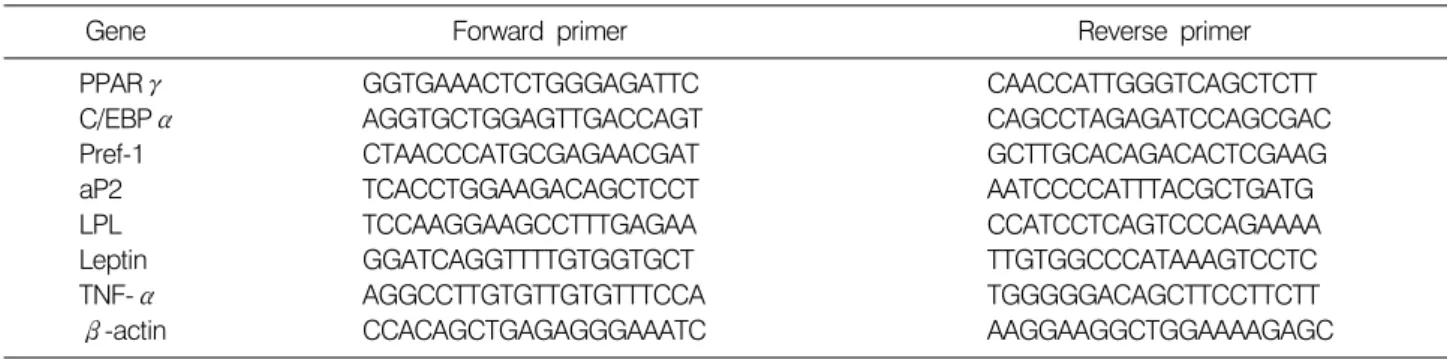
Table 1. Gene-specific primers used for real-time PCR
Gene Forward primer Reverse primer
PPARγ C/EBPα Pref-1 aP2 LPL Leptin TNF-α β-actin
GGTGAAACTCTGGGAGATTC AGGTGCTGGAGTTGACCAGT CTAACCCATGCGAGAACGAT TCACCTGGAAGACAGCTCCT TCCAAGGAAGCCTTTGAGAA GGATCAGGTTTTGTGGTGCT AGGCCTTGTGTTGTGTTTCCA CCACAGCTGAGAGGGAAATC
CAACCATTGGGTCAGCTCTT CAGCCTAGAGATCCAGCGAC GCTTGCACAGACACTCGAAG AATCCCCATTTACGCTGATG CCATCCTCAGTCCCAGAAAA TTGTGGCCCATAAAGTCCTC TGGGGGACAGCTTCCTTCTT AAGGAAGGCTGGAAAAGAGC MATERIALS AND METHODS
1. Reagents
Jeju ground water was provided by the Jeju special self-gov- erning province development corporation (Jeju, Korea). Dexa- methasone, isobutylmethylxanthine, insulin and Oil Red O were purchased from Sigma-Aldrich (St. Louis, MO). TRIzol reagent and SuperScript First-Strand Synthesis System for RT-PCR kit were purchased from Invitrogen (Carlsbad, CA).
Brilliant II SYBR Green QPCR Master Mix was obtained from Stratagene (La Jolla, CA). Antibodies were purchased from Cell Signaling (Beverly, MA) and horseradish peroxidase-conjugated goat anti-rabbit IgG was purchased from Sigma-Aldrich.
2. Cell culture and differentiation
Mouse 3T3-L1 preadipocyte cells were purchased from American Type Culture Collection (Rockville, MD, USA) and cultured in DMEM (Gibco-BRL, Grand Island, NY, USA) con- taining 1% penicillin-streptomycin and 10% bovine calf serum (BCS, Gibco-BRL) at 37oC in a humidified atmosphere of 5%
CO2. For cell viability assay, 3T3-L1 preadipocyte cells were cultured with DW containing 10% BCS. Jeju ground water for 10 passage and then seeded in a 96 well plate at a concen- tration of 1×10⁵ cells/ml, and incubated for an additional 24, 48 h at 37oC. 10μl of MTT solution (5 mg/ml) were added to each well of a total reaction volume of 100μl followed by 3 h. Formazan crystals were dissolved in isopropanol containing hydrochloric acid and measured the absorbance at 570 nm. To induce differentiation, 3T3-L1 cells were grown until reaching confluency, and incubated with a mixture of 0.25 M dex- amethasone, 0.5 mM IBMX and 1 g/ml insulin in DMEM made by DW or Jeju ground water containing 10% FBS. After
2∼3 days, the induction media were replaced by 10%
FBS/DMEM supplemented with 1μg/ml insulin alone for 2 additional days and then the adipocytes were kept in media containing 10% FBS until experimentation.
3. Oil Red O staining
After the induction of differentiation, the cells were washed twice with PBS, fixed with 10% formalin at room temperature for 1 h, washed with 60% isopropanol, completely dried and then stained with Oil Red O solution (six parts 0.5% Oil Red O in isopropanol and four parts water) for 1 h. The images were photographed using microscope (Nikon). The stained lipid droplets were dissolved in isopropanol for 10 min and quanti- fied by measuring the absorbance at 500 nm.
4. Real-time quantitative RT-PCR
Total RNA was extracted from 3T3-L1 adipocytes using Trizol reagent according to the manufacturer’s instruction. The cDNA was synthesized from 1μg total RNA with SuperScript First-Strand Synthesis System for RT-PCR kit. The quantitative real-time PCR was performed with Brilliant II SYBR Green QPCR Master Mix using the MX3000P real-time PCR system (Stratagene). The specific primer sets for adipogenic marker are listed in Table 1. and relative amounts of mRNA levels were calculated using the comparative cycle threshold (CT) method.11)
5. Western blot analysis
3T3-L1 cells were washed twice with PBS and lysed in lysis buffer containing 25 mM Tris-HCl (pH 7.2), 0.1% sodium do- decyl sulfate (SDS), 0.1% Triton X-100, 1% sodium deoxy- cholate, 150 mM NaCl, 1 mM ethylenediaminetetraacetic acid (EDTA), 1 mM sodium orthovanadate, 1 mM phenylmethyl-
Fig. 1. Cytotoxicity of Jeju ground water on 3T3-L1 preadi- pocytes. Cells (1×10⁴ cell/well) were incubated with DW or Jeju ground water for 24 h, 48 h. Cell viability was determined by MTT assay. The result represent the means±SD of data from three experiments.
sulfonylfluoride (PMSF), 10μg/ml aprotinin, and 5μg/ml leu- peptin for 20 min on ice. Equal protein amounts of cell lysate were separated on a 12% SDS-polyacrylamide gel, transferred to a polyvinylidene fluoride (Pall corporation, East Hills, NY).
Antibodies against PPARγ, C/EBPα and adiponectin were used as the primary antibodies, and horseradish peroxidase-con- jugated goat anti-rabbit IgG was used as the secondary antibody. The interesting proteins were visualized using an en- hanced chemiluminescence-based detection system (Amersham- Pharmacia Biotech, Piscataway, NJ, USA). For detect the se- creted adiponectin in the culture media, differentiated 3T3-L1 adipocytes after 10 days of induction were changed with serum free media. Collected culture media was determined the secre- tion of adiponectin using western blot analysis.
6. Statistical analysis
Student’s t test and one-way analysis of variance were used to analyze the differences between values obtained in the vari- ous experimental and control conditions. p<0.05 was consid- ered significant.
RESULTS
1. Cytotoxicity of Jeju ground water
The effect of Jeju ground water on 3T3-L1 preadipocytes cell viability and cytotoxicity were investigated by MTT assay.
The cells were cultured with DW or Jeju ground water for
24, 48 h. As shown in Fig. 1. Jeju waters had no significant cytotoxicity against preadipocytes.
2. Inhibitory effects of Jeju ground water on 3T3-L1 adipocyte differentiation
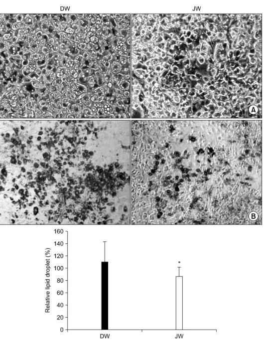
To investigate the effect of Jeju ground water on adipocyte differentiation, 3T3-L1 preadipocyte cells were incubated with culture media made by DW or Jeju ground water each contain- ing dexamethasone, IBMX and insulin for 10 days. The cells were induced differentiation to adipocyte, therefore the mor- phological changes were observed a rounded phenotype due to the accumulation of lipid droplets in adipocytes. As shown in Fig. 2A, Jeju ground water was shown the reduction of lipid droplets accumulation compared with DW. To confirm these observation, differentiated cells were stained with Oil Red O solution, and then quantified by measuring the absorbance. The staining results revealed that Jeju ground water significantly decreased the cell differentiation (Fig. 2B). Theses results showed the inhibitory effects of Jeju waters on 3T3-L1 adipo- cyte differentiation.
3. Effects of Jeju ground water on expression of adipogenic transcription factors
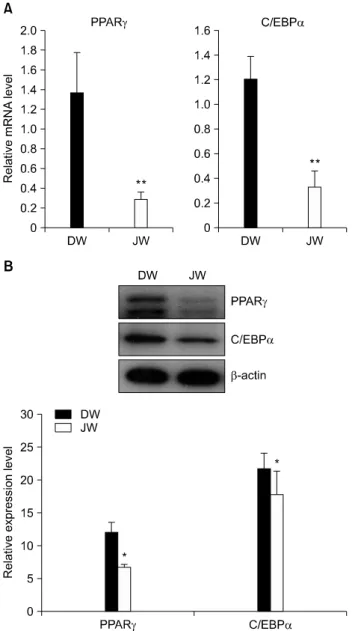
PPARγ and C/EBPα is well known as a adipogenic tran- scriptional factors and show that cross-regulation between both genes is a key component of the transcriptional control of adipogenesis.12,13) The effect of Jeju ground water on the regu- lation of PPARγ and C/EBPα expression was analyzed by quantitative real-time PCR. As shown in Fig. 3A, Expression levels of PPARγ and C/EBPα were reduced in Jeju ground water incubated 3T3-L1 adipocytes. We also examined PPARγ and C/EBPα protein expression level by western blotting. In consist of above results, the expression level of PPARγ and C/EBPα protein were also diminished by Jeju ground water (Fig. 3B). These results suggested that Jeju ground water on differentiation of 3T3-L1 preadipocyte to adipocytes through the down-regulation of PPARγ and C/EBPα.
4. Effect of Jeju ground water on expression of adipogenic transcription factor target genes
We further studied whether Jeju ground water regulates the expression of the adipogenic transcription factor target genes adipocyte fatty acid binding protein (aP2), lipoprotein lipase (LPL) and leptin, as well as the preadipocyte factor-1 (Pref-1)
Fig. 2. Effect of Jeju waters on differentiation of 3T3-L1 adipo- cytes. (A) Morphological changes were photographed the cell at 10 days after induction of differen- tiation with DW or Jeju ground waters, respectively. (B) Cells were stained with Oil Red O stai- ning for intracellular lipid accumu- lation. The results represent the means±SD of data from ten se- parate experiments. *p<0.05 com- pared with cells treated with DW.
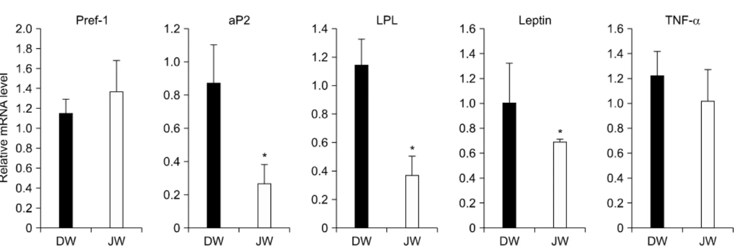
gene. Pref-1 predominantly expressed in undifferentiated 3T3-L1 cells. Whereas the mRNA level of Pref-1 which pre- dominantly expressed in undifferentiated state was up-regu- lated, the mRNA levels of aP2, LPL, Leptin were significantly down-regulated by Jeju ground water during adipocyte differ- entiation (Fig. 4). In addition, adipocytes produce and secrete signaling molecules such as tumor necrosis factor-α (TNFα).
TNFα were also decreased in adipocyte treated with Jeju ground water compared with DW.
5. Effect of Jeju ground water on expression and secretion of adiponectin
Adiponectin is one of multiple adipocytokines secreted by adipose tissue and has been shown to promoters adipocyte dif- ferentiation, insulin sensitivity, and lipid accumulation.14) To determine the expression of adiponectin in adipocyte differ- entiation induce by Jeju ground water, we assessed protein ex- pression level of adiponectin in cell and culture media by west- ern blot analysis. As shown in Fig. 5, the adiponectin concen- tration of the media decreased, consistent with the result of the reduction of cellular adiponectin expression in Jeju ground
Fig. 3. Effect of Jeju ground water on adipogenic transcription factors during differentiation of adipocytes. (A) Expression of PPARγ and C/EBPα mRNA from 3T3-L1 adipocyte cells were measured by quantitative real-time PCR using specific primers. The analysis were performed duplicate experiments (n=9). *p<0.05, **p<0.01 compared with cells treated with DW. (B) Western blot analysis were performed with antibodies against PPARγ and C/EBPα. β-actin was used as loading control for same amounts of protein.
water compared with DW.
DISCUSSION
Obesity is most commonly caused by a combination of ex- cessive dietary calories, lack of physical activity, and genetic susceptibility. The most effective treatment for obesity of diet-
ing and physical exercise. Recently, several reports show that natural products have potential as anti-obesity agents that may not have some harmful side-effects.15∼18)
Vanadium is a well-known antidiabetic metal agent which mimics most of the actions of insulin on mature adipocyte and can also exert anti-tumor effects against chemical carcinogenesis in animals and in various types of malignant cell lines.19) Several researchers have studied about the effect of vanadium in treatment of adipogenesis as anti-adipogenic agents.20∼22) Vanadium was found in various natural sources such as vegeta- bles and food.23)
Vanadium is relatively exist at high concentration (26.0±2.0 μg/l) in Jeju ground water obtained from Jeju island in South Korea. It was reported that the Jeju ground water possessed antioxidant effect via ROS scavenging.24) However, the an- ti-adipogenic mechanisms of Jeju ground water in adipocytes have not yet been investigated. In this study, we demonstrate the effect of Jeju ground water containing vanadium compo- nents on 3T3-L1 adipocyte differentiation. Adipocyte differ- entiation is a multiple process of programmed changes in spe- cific gene expressions. PPARγ and C/EBPα are transiently expressed on early stage for initiating differentiation of preadipocytes. C/EBPα is induced relatively late during adipo- genesis and cross-regulation between PPARγ and C/EBPα is important in maintaining the differentiated state.9,10) Activation of PPARγ induces the expression of genes controlling adipo- cyte fatty acid metabolism, including aP2, LPL.25,26) Here we show that Jeju ground water treatment significantly decreased the expression levels of PPARγ and C/EBPα and their adipo- genic target genes. Adiponectin is expressed by mature adipo- cytes and is the most abundant circulating adipokine. Indeed, adiponectin can promote adipocyte differentiation through au- tocrine effects results in increasing lipid content and insulin sensitivity. Jeju ground water was also shown the down-regu- lation of the expression and secretion of adiponectin on adipo- cyte differentiation.
CONCLUSION
Taken together, these results indicate that suppression of adipocyte differentiation by Jeju ground water treatment is mediated through down-regulation of the adipogenic tran- scription factors, PPARγ and C/EBPα and these target genes. It suggests that the ability of Jeju ground water to in-
Fig. 5. Effect of Jeju ground water on expression of adipo- nectin. The amounts of adiponectin protein in cell and culture media were assessed by western blot analysis. The graph represents the band intensity of secreted adiponectin.
Fig. 4. Effect of Jeju ground water on Pref-1, aP2, LPL, Leptin and TNFα mRNA expression in 3T3-L1. Cells were cultured in the medium containing DW, Jeju ground water. The data represent the mean±SD of three independent experiments. The analysis were performed duplicate experiments (n=9). *p<0.05, **p<0.01 compared with cells treated with DW.
hibit adipocyte differentiation may be contributed as the an- ti-obesity resource.
ACKNOWLEDGEMENT
This study was supported by the Ministry of Knowledge Economy (MKE), the Korea Institute for Advancement of Technology (KIAT) and Jeju Leading Industry Office through the Leading Industry Development for Economic Region.
REFERENCES
1) Haslam DW, James WP. Obesity. Lancet 36, 1197-1209, 2005.
2) Trayhurn P. Endocrine and siganlling role of adipose tissue:
new perspectives on fat. Acta Physiol Scand 184, 285-293, 2005.
3) Kopelma PG. Obesity as a medical problem. Nature 404, 635-643, 2000.
4) Fernyhough ME, Bucci LR, Hausman GJ, Antonio J, Vierck JL, Dodson MV. Gaining a solid grip on adipogenesis. Tissue Cell 37, 335-338, 2005.
5) Gregoire FM. Adipocyte differentiation: from fibroblast to endocrine cell. Exp Biol Med 226, 997-1002, 2001.
6) Koutnikova H, Auwerx J. Regulation of adipocyte differentiation. Ann Med 33, 556-561, 2001.
7) Wu Z, Rosen ED, Brun R, Hauser S, Adelmant G, Troy AE, McKeon C, Darlington GJ, Spiegelman BM. Cross-regulation of C/EBPα and PPARγ controls the transcriptional pathway of adipogenesis and insulin sensitivity. Mol Cell 3, 151-158, 1999.
8) Fonseca-Alaniz MH, Takada J, Alonso-Vale MI, Lima FB.
Adipose tissue as an endocrine organ: from theory to practice.
J Pediatr (Rio J) 83, S192-203, 2007.
9) MacDougald OA, Lane MD. Transcriptional regulation of gene expression during adipocyte differentiation. Annu Rev
Biochem 64, 345-373, 1995.
10) Rosen ED, Walkey CJ, Puigserver P, Spiegelman BM. Trans- criptional regulation of adipogenesis. Genes Dev 14, 1293- 1307, 2000.
11) Livak KJ, Schmittgen TD. Analysis of relative gene expression data using real-time quantitative PCR and the 2(-Delta Delta C(T)) Method. Methods 25, 402-408, 2001.
12) Wang YX. PPARs: diverse regulators in energy metabolism and metabolic diseases. Cell Res 20, 124-137, 2010.
13) Wu Z, Xie Y, Morrison RF, Bucher NL, Farmer SR. PPARγ induces the insulin-dependent glucose transporter GLUT4 in the absence of C/EBPα during the conversion of 3T3 fibroblasts into adipocytes. J Clin Invest 101, 22-32, 1998.
14) Fu Y, Luo N, Klein RL, Garvey WT. Adiponectin promotes adipocyte differentiation, insulin sensitivity, and lipid accumu- lation. Lipid Res 46, 1369-1379, 2005.
15) Sayama K, Lin S, Zheng G, Oguni I. Effects of green tea on growth, food utilization and lipid metabolism in mice. In Vivo 14, 481-484, 2000.
16) Mochizuki M, Hasegawa N. Effects of green tea catechin- induced lipolysis on cytosol glycerol content in differentiated 3T3-L1 cells. Phytother Res 18, 945-946, 2004.
17) Kim JK, So H, Youn MJ, Kim HJ, Kim Y, Park C, Kim SJ, Ha YA, Chai KY, Kim SM, Kim KY, Park R. Hibiscus sabdariffa L. water extract inhibits the adipocyte differentiation through the PI3-K and MAPK pathway. J Ethnopharmacol 114, 260-267, 2007.
18) Hwang HS, Kim SH, Yoo YG, Chu YS, Shon YH, Nam KS,
Yun JW. Inhibitory effect of deep-sea water on differentiation of 3T3-L1 adipocytes. Mar Biotechnol (NY) 11, 161-168, 2009.
19) Evangelou AM. Vanadium in cancer treatment. Crit Rev Oncol Hematol 42, 249-265, 2002.
20) Seale AP, de Jesus LA, Park MC, Kim YS. Vanadium and insulin increase adiponectin production in 3T3-L1 adipocytes.
Pharmacol Res 54, 30-38, 2006.
21) Shukla R, Bhonde RR. Adipogenic action of vanadium: a new dimension in treating diabetes. Biometals 21, 205-210, 2008.
22) Faneca H, Figueiredo VA, Tomaz I, Gonçalves G, Avecilla F, Pedroso de Lima MC, Geraldes CF, Pessoa JC, Castro MM.
Vanadium compounds as therapeutic agents: some chemical and biochemical studies. J Inorg Biochem 103, 601-608, 2009.
23) Barceloux DG. Vanadium. J Toxicol Clin Toxicol 37, 265-278, 1999.
24) Kim AD, Kang KA, Zhang R, Lim CM, Jee Y, Lee NH, You HJ, Ko KS, Hyun JW. Ractive oxygen species scavenging effects of Jeju waters containing vanadium components. Cancer Prev Res 15, 111-117, 2010.
25) Tontonoz P, Graves RA, Budavari AI, Erdjument-Bromage H, Lui M, Hu E, Tempst P, Spiegelman BM. Adipocyte-specific transcription factor ARF6 is a heterodimeric complex of two nuclear hormone receptors, PPAR gamma and RXR alpha.
Nucleic Acids Res 22, 5628-5634, 1994.
26) Tontonoz P, Hu E, Graves RA, Budavari AI, Spiegelman BM.
mPPAR gamma 2: tissue-specific regulator of an adipocyte enhancer. Genes Dev 8, 1224-1234, 1994.