Anti-invasion Effects of Calystegia soldanella Solvent Extracts and Partitioned Fractions on PMA-stimulated Fibrosarcoma Cells
Jaemin Son
1, Junse Kim
1, Hojun Kim
2and Youngwan Seo
1,2*
1
Ocean Science and Technology School, Korea Maritime and Ocean University, Busan 49112, Korea
2
Division of Marine Bioscience, Korea Maritime and Ocean University, Busan 49112, Korea
Received October 25, 2018 /Revised November 12, 2018 /Accepted November 13, 2018
Calystegia soldanella is distributed in coastal sand dunes and has high environmental adaptability; it is also known to be effective for anti-oxidant, anti-pyretic, anti-septic, and diuretic action. This study investigated the effect of crude extracts and organic solvent fractions of C. soldanella on MMP-2 and MMP-9 expression, MMP activity, and cell mobility in phorbol-12-myristate-13-acetate (PMA)-induced fibrosarcoma HT-1080 cells. C. soldanella was twice extracted, once with methylene chloride (MC) and once with methanol (MeOH). After the MC and MeOH extracts were combined, their suppressive effects on MMP-2 and MMP-9 expression, MMP enzymatic activity, and gene and protein expression were measured by gelatin zymography, enzyme-linked immunosorbent assay, reverse-transcription polymerase chain reaction, and western blot method. Cell mobility for the HT-1080 cells was observed by wound healing assay. The combined crude extracts showed a significant suppressive effects on MMP-2 and MMP-9 expression. To explore active inhibitory elements, the combined extracts were fractionated according to polarity into with n-hexane, 85% aqueous methanol, n-butanol, and water.
Across these four solvent fractions, MMP-2 and MMP-9 activity and cell mobility in the HT-1080 cells were all strongly inhibited by the n-hexane fraction. These results suggest that C. soldanella extract and organic solvent fractions could be used as potent MMP inhibitors for effective anti-cancer treatments to suppress cancer invasion and metastasis.
Key words : Anti-invasive, anti-metastasis, Calystegia soldanella, HT-1080, MMP activity
*Corresponding author
Tel : +82-51-410-4328, Fax : +82-51-404-4750 E-mail : [email protected]
This is an Open-Access article distributed under the terms of the Creative Commons Attribution Non-Commercial License (http://creativecommons.org/licenses/by-nc/3.0) which permits unrestricted non-commercial use, distribution, and reproduction in any medium, provided the original work is properly cited.
Journal of Life Science 2019 Vol. 29. No. 3. 287~294 DOI : https://doi.org/10.5352/JLS.2019.29.3.287
서 론
21세기 다수의 새로운 암이 발생하면서 전 세계적으로 암을 극복하기 위한 다양한 연구의 필요성이 대두되고 있다[14]. 현 재 이용되는 다양한 화학요법 및 항암제는 골수에서 형성된 혈액세포, 구강을 포함한 위장관의 상피세포, 머리카락세포 및 생식세포 등에 영향을 주어 백혈구의 혈소판 수 감소, 오심, 구토, 설사, 탈모, 생식기능 장애를 가져오는 부작용이 있다 [20]. 이러한 이유로 부작용을 최소화 할 수 있는 천연물 유래 의 생리활성 물질 개발이 활발하게 진행되고 있다[12].
암세포의 성장 및 전이는 세포외 기질(extracellular matrix, ECM)의 분해와 신생혈관의 형성(angiogenesis) 후 종양세포 의 순환계를 통한 이동, 타 조직에 부착하여 침윤하는 과정을 통해 발생한다[6, 8, 15]. 세포외 기질은 암의 전이 및, 조직형 성, 생체방어에 주요 역할을 하며, 이러한 세포외 기질을 분해
하는 대표적인 유전자가 matrix metalloproteinase (MMPs)이 다[3-5]. MMP는 다른 matrix 단백질과 콜라겐을 분해하여 암 진행에 주요한 역할을 한다[2, 19]. 인체 내부의 26가지 MMP 중 gelatinase인 MMP-2와 MMP-9의 발현이 세포외기질의 주 요 구조 성분인 type IV collagen을 분해하며[7, 10, 17], 기저막 분해 및 신생혈관의 형성에 관여하여 암세포 침윤 및 전이를 유도한다고 알려져 있다[11, 13, 21]. 따라서 암세포의 침윤 및 전이를 효과적으로 억제하기 위해 MMP-2와 MMP-9의 발현 에 영향을 줄 수 있는 천연물 유래의 생리활성 물질 개발이 필요하다.
갯메꽃의 학명은 Calystegia soldanella (L.) Roem. et Schult 으로 메꽃과에 속하는 여러해살이 덩굴식물이다. 잎의 모양은 콩팥과 유사하며, 잎의 크기는 길이 2~3 cm, 폭은 3~5 cm 정도 로 잎바닥은 깊고 잎자루가 잎보다 길다. 해안 사구에 분포하 는 식물로 높은 환경적응력을 가지고 있어 공터나, 자갈해변, 모래해변 근처 공터에서도 잘 자라며 내염성이 강하여 해안가 에서도 잘 자란다[1, 16].
갯메꽃은 해열, 살균, 이뇨작용 등에 효과가 있으며 항산화,
항염증, 항바이러스, 항진균 및 protein tyrosine phosphate 1B
억제 등 다양한 생리활성 있는 것으로 보고 되어 있다. 갯메꽃
으로부터 분리된 화합물로는 nortropane alkaloids, anthocya-
nin, Caffeic acid, courmaric acid 등이 보고되어 있다[9, 18].
위와 같이 겟매꽃의 다양한 생리활성이 보고되었지만 암전 이와 관련된 연구는 아직 보고되지 않았다. 따라서 본 연구진 은 갯메꽃을 추출 및 분획한 후, HT-1080 (human fibrosarco- ma cell)에서 갯메꽃 추출물 및 유기용매 분획물의 MMP-2, MMP-9 발현 억제 효과 및 세포 이동성 억제 효과를 조사하여, 암 침윤 및 전이 억제 활성을 가진 항암소재로서의 활용 가능 성을 제시하고자 한다.
재료 및 방법
시료추출 및 순차분획
본 실험에 사용한 갯메꽃(C. soldanella)은 전라북도 군산시 옥도면 비안도리에서 직접 채취하여 세척하고 그늘에서 자연 건조 후 분쇄한 것을 시료로 사용하였다. 건조 분쇄한 시료를 methylene chloride (MC, Duksan, Korea) 용매에 24시간 추출 한 후 여과하였으며, 이 과정을 2회 실시하였다. 여과 후 남은 시료에 같은 양의 methanol (MeOH, Duksan, Korea) 용매를 첨가하여 동일한 과정을 반복하였고, 얻어진 각각의 추출액을 40°C 수욕 상에서 rotary vacuum evaporator로 농축하여 MC 추출물(2 g)과 MeOH 추출물(12 g)을 얻었다. 각각의 추출물을 합한 조추출물을 용매 극성에 따라 순차적으로 분획 및 감압 농축하여 n-hexane (0.81 g), 85% aq.MeOH (4.07 g), n-butanol (n-BuOH, 1.68 g), H
2O (8.07 g)의 4가지 분획물을 얻었다.
세포 배양
HT-1080 (human fibrosarcoma cell) 세포는 한국 세포주 은행(KCLB, Korean cell line Bank)에서 분양 받아 사용하였으 며, 100 unit/ml의 penicillin-streptomycin과 10% FBS (Fetal Bovine Serum, GenDepot, USA)이 함유된 RPMI 1640 (Gen DEPOT, USA) 배지를 사용하여 5% CO
2, 37℃ incubator (Formascientific, Japan)에서 배양하였다. 배양된 세포는 일주 일에 2~3회 PBS buffer (Gibco-BRL, USA)로 세척 후, 배지를 교환하였다. 세포 배양 6~7일마다 부착된 세포를 0.05% Tryp- sin-0.02% EDTA (Gibco-BRL, USA)를 이용하여 분리하여 원 심분리를 진행한 후 침전된 세포를 T-75 조직 배양 플라스크 (Nunc, Rosklide, Denmark)에 일정 수의 세포를 주입하여 반 복적으로 계대 배양하면서 실험에 사용하였다.
세포 독성(MTT assay) 측정
세포독성은 MTT (3-(4,5-dimethylthiazol-2yl)-2,5-diphenyl- 2H-tetrazolium bromide) assay를 이용하여 확인하였다. HT- 1080 세포를 배양하여 96 well plate에 well 당 1×10
3cells/ml 로 분주하여 37
oC, 5% CO
2incubator에서 24시간 동안 배양한 후, 각각의 시료를 농도별로 처리하고, 대조군은 시료 대신 PBS를 처리하였다. 시료 처리 후 37℃, 5% CO
2incubator에서 24시간 동안 배양하고 세포를 세척하고 100 μl의 MTT용액(1
mg/ml)을 첨가하여 동일한 배양조건에서 4시간 동안 반응시 킨 후 환원으로 생성된 formazan 결정에 100 μl의 DMSO (Duksan, Korea)를 첨가하여 녹여서 ELISA plate reader (Bio- Tek instruments, Winooski, VT)로 540 nm에서 흡광도를 측 정하여 세포 독성에 미치는 효과를 측정하였다.
ELISA (Enzyme-linked immunosorbent) 법을 이용한 MMP activity 측정
ELISA 법을 이용하여 HT-1080세포에서 갯메꽃 추출물과 유기용매 분획물이 MMP-2와 MMP-9 생성에 미치는 영향을 확인하였다. HT-1080 세포를 24 well plate 에 2×10
5cells/well 이 되도록 seeding한 후, FBS를 투입하지 않은 배지를 사용하 여 24시간 동안 안정화하고 추출물과 분획물 시료를 100 μg/
ml 농도로 처리하여 1시간 동안 배양하였다. 그 후 MMP 발현 을 위해 phorbol 12-myristate 13-acetate (PMA, 10 ng/ml, Sigma aldrich, USA)를 각 well에 동량 주입하여 24시간 후 상등액을 얻었다. DuoSet ELISA kit (R&D Systems Inc., Minneapolis, MN USA)의 96 Well plate에 100 μl의 capture antibody를 처리하고 24시간 후, washing buffer (Tween 20 in PBS)로 세척하고 10X Reagent Diluent (1% BSA in PBS)를 희석한 1X Reagent Diluent를 300 μl로 처리하여 1시간 동안 blocking하였다. Washing buffer로 세척한 후, 얻은 상등액을 100 μl씩 처리하여 2시간 동안 반응시킨 뒤 세척한 well에 100 μl의 detection antibody (MMP-2 : 10 ng/ml, MMP-9 : 150 ng/ml)를 처리하여 상온에서 2시간 반응시킨 뒤 세척한 well 에 100 μl의 Streptavidin-HRP (horseradish-peroxidase)를 처 리하여 암실에서 20분 간 반응시키고 세척하였다. 그 후, 100 μl substrate solution (H
2O
2: Tetramethylbenzidine = 1 : 1)로 처리하여 암실에서 20분간 반응시킨 후, stop solution (2 N H
2SO
4)을 첨가하여 반응을 종결한 후 450 nm에서 ELISA reader (Bio-Tek instrument, Winooski, VT)로 흡광도를 측정 하였다.
Gelatin zymography법을 이용한 MMP 발현 억제 활성 측정
HT-1080 세포를 24 well plate 에 2×10
5cells/well이 되도록
seeding한 후, FBS를 투입하지 않은 배지를 사용하여 24시간
동안 안정화하고 추출물과 분획물 시료를 100 μg/ml 농도로
처리하여 1시간 동안 배양하였다. 그 후 MMP 발현을 위해
PMA (10 ng/ml, Sigma aldrich, USA)를 각 well에 동량 주입
하여 24시간 후 배양액을 얻었다. Bradford 단백질 결정법으
로 각 배양액에 함유된 전체 단백질 함량을 정량하여 동량의
단백질을 포함하는 배양액 양을 결정한 후, 1.5 mg/ml gelatin
(Sigma aldrich, USA)이 포함된 10% sodium dodecyl sul-
fate-polyacrylamide gel (비환원조건, Duksan, Korea)에서 전
기영동을 진행하였다. 전기영동 후에 2.5% Triton X-100
Table 1. RT-PCR primer sequence
Gene primer sequence (5'-3')
MMP-2 Forward
Reverse
ATGGCAAGTACGGCTTCTGT ATACTTCTTGTCGCGGTCGT
MMP-9 Forward
Reverse
CTCGAACTTTGACAGCGACA GCCATTCACGTCGTCCTTAT β-Actin Forward
Reverse
AGCCATGTACGTAGCCATCC TCCCTCTCAGCTGTGGTGGT (JUNSEI, Japan)이 함유된 renaturing buffer로 30분씩 2번 씻 은 뒤, 150 mM Tris–HCl, 10 mM CaCl
2,150 mM NaCl을 포함하는 developing buffer로 37℃에서 48시간 반응시켰다.
반응이 완료된 gel은 0.5% Coomassie brilliant blue 250 (LPS solution, Korea)로 30분간 염색한 후 destaining buffer (me- thanol : acetic acid : water = 50 : 10 : 40)로 탈색하여 gelatin 분해 정도를 관찰하였다. 이 때 gelatin 분해로 나타난 투명 band의 면적을 Quantity One (Bionner, Daejeon, Korea) 프로 그램으로 측정하여 그래프로 나타내었다.
Wound healing assay를 이용한 cell migration 관찰 HT-1080 세포를 12 well plate에 seeding 한 후 24시간 동안 37℃, 5% CO
2incubator에서 안정화시킨 후, 2 mm plastic tip 으로 각 well의 중앙을 일자로 그은 뒤 배지를 교환하였다.
배지 교환 후 각 well에 유기용매 분획물 시료를 100 μg/ml 농도로 주입하고, 24시간 배양 후 세포이동성을 관찰하였다.
RT-PCR (Reverse transcription-polymerase chain reaction)에 의한 mRNA 발현
상기의 방법과 동일한 조건에서 배양된 세포를 PBS로 세척 후 Trizol reagent (ambion, life technologies™, USA)로 용해 시켜 RNA를 추출하였다. 추출한 RNA에 DEPC water (Sigma- aldrich, USA)를 20 μl 넣고 이 혼합액 4 μl를 DEPC water 96 μl에 희석하여 260/280 nm에서 흡광도를 측정하여 RNA를 정량하였다. 정량 후 동량의 RNA (2 μg)로 부터 역전사 시스 템 (Promega, Madison, WI, USA)을 이용하여 cDNA를 합성 하였으며, PCR Premix (BIONEER, Daejeon, Korea)에 합성된 cDNA를 MMP-2, MMP-9, β-actin primer (Table 1)와 반응시 킨 후, T100 Thermal Cycler (Bio-Rad, CA, USA)을 이용하여 증폭시켰다.
Western Blot을 이용한 단백질의 발현
상기의 방법과 동일한 조건에서 배양된 세포를 1X PBS로 세척 후 RIPA lysis buffer (50 mM Tris-HCl (pH 8.0), 150 mM NaCl, 1% NP 40, 0.5% sodium deoxycholate, 1 mM phe- nylmethysulfonyl)를 사용하여 단백질을 추출하였다. 추출한 단백질은 Bradford 단백질 결정법을 이용하여 동량의 단백질
이 함유될 수 있도록 양(20 μg)을 정하고, 12% sodium dode- cylsulfate-polyacrylamide gel (비환원조건, Duksan, Korea) 에서 전기영동하여 분리한 후 polyvinylidene fluoride mem- brane (Amersham Pharmacia Biotech, England, UK)로 trans- fer한 후, 5% skim milk로 1시간 동안 blocking하였다. Block- ing후 β-actin, MMP-2, MMP-9 (Cell Signaling Technology Inc., MA, USA)의 1차 항체를 4℃에서 24시간 반응시키고, TBS-T buffer (Tris buffered saline and tween-20, Biosesang, Gyeonggido, Korea)를 사용하여 3회 세척한 뒤, 2차 항체를 처리하여 1시간 동안 반응시켰다. TBS-T buffer로 세척 후 de- tection solution (chemiluminescent ECL kit, GE healthcare, Little Chalfont, UK)에 반응시킨 후 Davinch-Chemi imager
TM(Davinch-K, Seoul, Korea)를 통해 단백질 발현 band를 관찰 하였으며, band의 intensity를 Quantity One (Bionner, Dae- jeon, Korea)프로그램으로 측정하여 그래프로 나타내었다.
통계처리
본 연구의 모든 실험결과는 평균±표준편차(Mean ± Stan- dard deviation, SD)로 나타내었으며, SPSS
+/WIN12.0 (Statis- tical Package for Social Science, version 1 2.0) 통계프로그램을 이용하여 유의성을 검토하였다. 일원배치 분산분석(Oneway Analysis Of Variance: ANOVA)을 통해 집단간의 유의성을 검정하였으며, 각 실 험에서 평균치간의 유의성은 p<0.05 수준 으로 Duncan's multiple range test를 실 시하여 검증하였다.
결과 및 고찰
추출물에 대한 세포독성과 MMP-2 및 MMP-9 생성 억제 효과
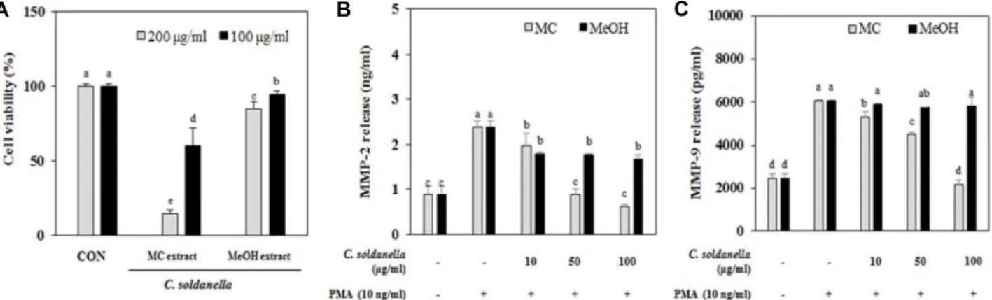
갯메꽃 추출물의 HT-1080 섬유육종세포에 대한 세포독성 에 미치는 효과를 MTT assay를 통해 확인하였다(Fig. 1A). 추 출물을 각각 100, 200 μg/ml 농도로 처리하여 세포 생존율을 확인한 결과, HT-1080 세포에 대하여 MC 추출물은 모든 처리 농도에서 80% 이하의 세포생존율을 보였으나, MeOH 추출물 은 100 μg/ml 농도에서 80% 이상의 생존율을 보였다. MC 추출물도 100 μg/ml 농도에서는 200 μg/ml 농도보다 상대적 으로 세포 생존율이 높았기 때문에 100 μg/ml의 처리농도에 서 MMP 생성 억제 효과를 측정하기 위한 실험을 수행하였다.
선행된 여러 연구를 통해 암의 진행과 MMPs의 발현은 직
접적인 관계가 있다 는 것이 밝혀졌으며 MMP-2와 MMP–9
은 종양세포의 침윤 및 전이에서 주요한 작용을 하는 것으로
알려져 있다[10, 12, 19]. 먼저 갯메꽃 추출물(MC 추출물과
MeOH 추출물)이 HT-1080 세포에서 MMP-2와 MMP-9의 활
성에 미치는 영향을 ELISA kit를 이용한 항원-항체 반응을 통
해 알아보았다. MC 추출물과 MeOH 추출물을 처리한 결과
PMA를 단독 처리한 양성대조군과 비교하였을 때 MC 추출물
A B C
Fig. 1. Effect of methylene chloride (MC) and methanol (MeOH) extracts from C. soldanella on cell cytotoxicity (A) and the production of MMP-2 (B) and MMP-9 (C) in PMA-stimulated HT-1080 human fibrosarcoma cells.
a-eMeans with the different letters are significantly different (p<0.05) by Duncan's multiple range test. Values are means ± SD (n=3).
은 농도 의존적으로 MMP-2와 MMP-9 활성을 억제하는 것을 확인할 수 있었다(Fig. 1B, Fig. 1C). 고농도인 100 μg/ml에서 의 MMP 생성 억제활성은 세포독성에 의한 것으로 사료된다.
MeOH 추출물의 경우 MMP-9보다는 MMP-2의 활성 억제 효 과가 높은 것을 확인할 수 있었다.
조추출물에 대한 HT-1080 세포에서의 MMP-2 및 MMP-9 생성 억제 효과
추출물 단계에서의 MMPs 생성 억제 활성을 검토한 후 MC 추출물과 MeOH 추출물을 합하여 제조한 조추출물을 시료로 하여 HT-1080 세포에서 PMA에 의한 MMP-2와 MMP-9의 분 비에 미치는 효과를 두 효소의 기질 분해능을 이용하여 활성 을 확인하는 방법인 gelatin zymography를 이용하여 측정하 였다. 갯메꽃의 조추출물은 10, 50, 100 μg/ml 농도에서는 약 85% 이상의 생존율을 나타내었으며 이 결과를 바탕으로 실험 을 진행하였다(Fig. 2A). MMP-2와 MMP–9의 gelatin 분해 활성을 측정한 결과, PMA를 단독 처리한 양성대조군과 비교 하였을 때 조추출물 처리에 의해 MMP-2와 MMP-9의 발현이 억제되는 것을 확인할 수 있었다(Fig. 2B).
조추출물의 MMP 생성억제효과가 암세포 전이 관련 인자 에 미치는 영향을 mRNA 발현 및 단백질 발현의 분석을 통해 알아보았다(Fig. 2C, Fig. 2D). 배양된 HT-1080 세포에서 RT- PCR을 이용하여 MMP-2와 MMP-9 mRNA 발현을 분석한 결 과 PMA를 단독으로 처리한 세포의 경우 아무 처리를 하지 않은 세포에 비해 MMP-2와 MMP-9 mRNA 모두 발현이 뚜렷 이 증가하는 경향을 보였지만 PMA와 100 μg/ml 농도의 조추 출물을 동시 처리한 세포에서는 MMP-2와 MMP-9의 발현이 효과적으로 억제하였다(Fig. 2C, Fig. 2D). 암세포 전이와 관련 된 인자의 발현을 단백질 수준에서 측정한 실험에서도 mRNA 의 발현과 유사하게 조추출물의 처리 에 의해 MMP-2와 MMP-9의 발현이 감소하는 것을 확인할 수 있었다(Fig. 2D).
앞의 실험을 종합한 결과 갯메꽃 추출물이 MMP 효소 억제
활성이 있음을 확인 하였고, 활성 성분을 탐색하기 위해 조추 출물을 용매의 극성에 따라 분획 후, 추가 실험을 진행하였다.
유기용매 분획물에 대한 세포독성 및 MMP-2 및 MMP-9 억제활성
갯메꽃의 MC 추출물과 MeOH 추출물을 혼합한 조추출물 을 용매 극성에 따라 n-hexane, 85% aq.MeOH, n-BuOH, H
2O 로 분획하였다. 각각 4가지 유기용매 분획물의 세포독성을 측 정하기 위해 10, 50, 100, 200 μg/ml 농도로 처리하여 세포생존 율을 조사한 결과, n-hexane, n-BuOH, H
2O 분획물은 100 μg/
ml 농도까지 80% 이상의 세포생존율을 나타내었으며, 85%
aq.MeOH 분획물의 경우 67.2%의 세포생존율을 보여 약간의 독성이 있는 것을 확인할 수 있었다(Fig. 3A). 이 결과를 바탕 으로 분획물 단계에서의 MMP 생성 억제 활성을 탐색하기 위해 n-hexane, 85% aq.MeOH, n-BuOH, H
2O의 4가지 분획물 을 100 μg/ml 농도로 PMA와 동시에 처리하여, HT-1080 세포 에서 MMP-2와 MMP-9의 활성에 미치는 영향을 ELISA kit를 이용한 항원-항체반응 분석을 통해 검토하였다. 그 결과 MMP- 2 생성에 대해 n-hexane, 85% aq.MeOH, n-BuOH, H
2O 분획 물이 각각 62.2%, 76%, 42.5%, 57.5%의 억제율을 나타내어 control에 비해 모두 유의적인 억제활성이 있음을 확인하였다 (Fig. 3B). MMP-9 생성에 대하여 n-hexane, 85% aq.MeOH, n-BuOH, H
2O 분획물은 각각 35.8%, 96.9%, 15.4%, 16.4%의 억제율을 나타내었으며, 특히 n-hexane과 85% aq.MeOH에서 높은 억제 활성이 있음을 확인할 수 있었다(Fig. 3C).
유기용매 분획물이 HT-1080 세포의 이동성에 미치는 영향 Wound healing assay를 이용하여 갯메꽃의 n-hexane, 85%
aq.MeOH, n-BuOH, H
2O 유기용매 분획물이 HT-1080 세포의 이동성에 미치는 영향을 알아보았다. 모든 분획물을 100 μg/
ml 농도로 처리하여 24시간 세포 이동성을 측정한 결과 아무
것도 처리하지 않은 대조군에 비해 모든 분획물에서 유의적으
A B
C D
Fig. 2. Effect of crude extracts from C. soldanella on cell cytotoxicity (A), MMPs expression tested by gelatin zymography (B) and mRNA (C) and protein (D) expression in PMA- stimulated HT-1080 human fibrosarcoma cells.
a-dMeans with the different letters are significantly different (p<0.05) by Duncan's multiple range test. Values are means ± SD (n=3).
A
B C
Fig. 3. Effect of solvent-partitioned fractions from C. soldanella on cell cytotoxicity (A) and the production of MMP-2 (B) and MMP-9
(C) in PMA-stimulated HT-1080 human fibrosarcoma cells.
a-eMeans with the different letters are significantly different (p<0.05)
by Duncan's multiple range test. Values are means ± SD (n=3).
A B
Fig. 4. Effect of solvent-partitioned fractions from C. soldanella on cell motility and invasion by wound migration assay (A) and gelatin zymography (B) in HT-1080 human fibrosarcoma cells.
a-dMeans with the different letters at the same concentration are significantly different (p<0.05) by Duncan's multiple range test. Values are means ± SD (n=3).
A B
Fig. 5. Effect of solvent-partitioned fractions from C. soldanella on MMPs expression in mRNA (A) and protein (B) levels in HT-1080 human fibrosarcoma cells.
a-fMeans with the different letters at the same concentration are significantly different (p<0.05) by Duncan's multiple range test. Values are means ± SD (n=3).
로 HT-1080 세포의 이동성이 저해되었음을 확인할 수 있었다 (Fig. 4A). 특히 85% aq.MeOH 과 n-BuOH 분획물에서 세포이 동성이 효과적으로 저해되었는데 이 중 85% aq.MeOH 분획물 은 세포독성에 영향을 받은 것으로 판단되어, n-BuOH 분획물 이 암 세포 전이에 대한 저해 효과가 있음을 확인하였다. 이러 한 결과는 gelatin zymography 법을 이용한 MMP 발현 억제 활성 측정에서도 유사한 결과를 나타내었다(Fig. 4B). MMP-2 발현에 대해 n-hexane, 85% aq.MeOH, n-BuOH, H
2O 분획물 은 48.1%, 63%, 33.1%, 30.5%의 억제율을 나타내었고 MMP-9 발현에 대해 50.7%, 73.4%, 45.7% 26.2%의 억제율을 나타내었다.
MMP-2와 MMP-9의 mRNA 및 protein 발현에 미치는 효과
유기용매 분획물이 암세포 전이와 관련된 인자인 MMP-2
와 MMP-9에 미치는 영향을 mRNA 발현 및 단백질 발현의
분석을 통해 알아보았다(Fig. 5). 배양된 HT-1080 세포에서
MMP-2 및 MMP-9의 mRNA 발현을 측정한 결과 MMP-2에
대해 n-hexane, 85% aq.MeOH, n-BuOH, H
2O 분획물이 각각
15%, 59.6%, 14.1%, 24.5% 의 억제율을 나타내었으며, MMP-9
에 대해 각각 16.2%, 58.5% 37.7%, 36.4%의 억제율을 나타내었
다(Fig. 5A). Western blot법을 이용한 MMP-2, MMP-9의 단백
질 발현에 미치는 효과를 측정한 실험에서는 MMP-2에 대해 n-hexane, 85% aq.MeOH, n-BuOH, H
2O 분획물이 각각 35%, 63%, 39.7%, 10.9%의 억제율을 나타내었고 MMP-9에 대해 각 각 58.7%, 79.4%, 54.4%, 54.3%의 억제율을 나타내었다(Fig.
5B).
본 연구에서는 암의 전이 및 침윤에 가장 밀접하게 관여하 는 MMP-2와 MMP-9의 활성과 발현에 대한 겟매꽃의 효능을 HT-1080 (human fibrosarcoma cell)을 이용하여 조사하였다.
또한 겟메꽃이 세포의 이동성에 미치는 영향을 알아보기 위하 여 HT-1080 세포의 이동을 관찰하였다. 겟메꽃 추출물 수준에 서 MMP-2와 MMP-9에 대한 억제활성을 조사한 결과 두 가지 모두 억제효과를 나타내었고, MMP 억제활성을 보유한 물질 의 특성을 확인하기 위해서 추출물을 용매 극성도에 따라 분 획하여 n-hexane, 85% aq.MeOH, n-BuOH, H
2O 분획물들을 얻었다. 각각의 분획물들에 대한 MMP-2와 MMP-9 억제활성 을 추출물 수준과 동일한 과정들을 통하여 조사하였다. 그 결 과 n-hexane과 85% aq.MeOH 분획물에서 MMP-2 및 MMP-9 생성에 대해 높은 억제효과를 나타내는 것을 확인할 수 있었 다. 추가적으로 Wound healing assay를 이용하여 HT-1080 세포의 이동을 관찰한 결과 85% aq.MeOH 과 n-BuOH 분획물 에서 세포이동성이 효과적으로 저해되었다. 이 중에서 85%
aq.MeOH 분획물의 경우 약간의 세포독성을 보였기 때문에 독성에 영향을 받은 것으로 판단되어 n-hexane 분획물이 MMP- 2 및 MMP-9 발현에 뚜렷한 억제활성이 있음을 확인하였다.
이상의 결과로 85% aq.MeOH 및 n-hexane 분획물에서 MMP 생성 억제활성이 높은 물질이 존재할 것으로 추측되어 항암소 재로서 활용 가능성이 있음을 제시한다.
감사의 글
본 연구는 2016년 교육부의 재원으로 한국연구재단의 지원 을 받아 수행된 기초연구사업(No. 2016R1D1A1B03932769)의 연구결과입니다.
References