41(8), 1079~1085(2012) http://dx.doi.org/10.3746/jkfn.2012.41.8.1079
녹두의 Vitexin이 비만전구세포에서 세포분화 및 아디포사이토카인 분비능에 미치는 영향
위해리1․최문지2․최세림1․김애정3․이명숙1,2†
1성신여자대학교 식품영양학과
2성신여자대학교 비만과학연구소
3경기대학교 대체의학대학원 식품치료전공
Effects of Vitexin from Mung Bean on 3T3-L1 Adipocyte Differentiation and Regulation According to Adipocytokine Secretion
Haeri Wi1, Munji Choi2, Selim Choi1, Ae-Jung Kim3, and Myoungsook Lee1,2†
1Dept. of Food and Nutrition and 2Research Institute of Obesity Sciences, Sungshin Women’s University, Seoul 142-732, Korea
3Nutritional Therapy, The Graduate School of Alternative Medicine, Kyonggi University, Seoul 120-837, Korea
Abstract
Obesity is an important issue worldwide as it may associated with increased prevalence of metabolic diseases.
Mung bean is known as a functional food for decreasing the glycemic index and lipid profile of plasma. The purpose of this study was to investigate the anti-obesity effects of vitexin from mung bean on the regulation of adipocyte differentiation and adipocytokine secretion. When 3T3-L1 adipocytes were treated with vitexin from days 0 to 14 at various levels of 25, 50, 100, and 200 μM, there was no change in cell viability. Vitexin treatment at 50, 100, and 200 μM decreased triacylglycerol levels in cells, but only 100 μM vitexin induced lipolysis. At 200 μM of vitexin, phosphorylation of p38 and ERK, which causes secretion of inflammatory adipo- cytokines, was depressed, whereas there was an increase in expression of PPARγ, the key regulator of adipocyte differentiation. Phosphorylation of AMPK increased at 100 μM vitexin. TNF-α and aP2 mRNA expression in- creased at 25 μM vitexin, whereas only TNF-α mRNA expression increased at 200 μM vitexin. Further, the mRNA levels of TNF-α and aP2 decreased at other concentrations in a dose-dependent manner. Since we ob- served that mRNA expression of C/EBP, SREBP1, and PPARγ did not change upon vitexin treatment, our future studies will investigate other genes such as mTOR, which is related with apoptosis signaling, or SIRT1, which is associated with inhibition of adipogenesis. Our results indicate that vitexin at concentrations between 100 and 200 μM is suitablein vivo for the development of mung bean as an anti-obesity therapy or functional food.
Key words: mung bean, vitexin, adipocytokines, PPARγ, TNF-α
†Corresponding author. E-mail: [email protected]
†Phone: 82-2-920-7211, Fax: 82-2-920-2078
서 론
2011년 세계보건기구(World Health Organization; WHO) 발표에 따르면 전 세계 15억 이상의 성인이 과체중이며 이 중 적어도 5억 이상이 비만환자라고 보고된 바 있다(1). 우리 나라의 국민건강영양조사에 따르면 한국인 성인의 30% 이 상이 과체중 또는 복부비만환자이고 이로 인해 당뇨병 및 고지혈증, 성기능 장애, 관절염, 심혈관계 질환의 발병위험 이 커지기 때문에 조기예방이 반드시 필요하다. 지방세포는 간엽세포(mesenchymal stem cell) 및 전구지방세포(pread- ipocyte)로부터 분화되며, 지질대사기능, 당대사기능, 나아 가 아디포사이토카인[tumor necrosis factor alpha; TNFα,
interleukin 6; IL-6, adiponectin, leptin, resistin, chemo- kines(monocyte chemoattractant protein-1; MCP-1, inter- leukin-8; IL-8)] 분비에 변화를 가져온다(2,3). 비만 환자에 게서 증가하는 TNFα, IL-6, MCP-1 등은 지방세포의 염증 발현으로 지방분화를 촉진하고 기타 성인병 이환율을 증가 시킨다(2). 또한 지방세포의 lipoprotein lipase(LPL) 활성을 저해하고 hormone-sensitive lipase(HSL) 발현 증가를 통해 지방분해를 촉진하여 혈중 유리지방산을 증가시켜 간접적 으로 인슐린저항성을 유도하기도 한다(4-7).
항비만 천연소재들의 기전적 접근은 항산화작용, 항혈소 판 억제작용, 체지방조직의 분화억제작용 등에 집중되어 왔 다. 예를 들면 대두 등에 들어있는 폴리페놀류 이소플라본인
제니스테인(genistein), 다이제인(daidzein) 등은 생체 내 활 성산소(ROS)를 소거시켜 유리기와 과산화지질의 생성을 억 제하며 제니스테인은 지방세포 분화억제 효과가 있다(8,9).
콩과 등의 식물에 배당체로 존재하는 이소플라보노이드를 함유한 천연소재 물질들이 지방세포의 분화조절 및 아디포 사이토카인 분비능에 영향을 준다는 보고가 있다(10). 또 다 른 콩과작물인 녹두에는 vitexin, triacontanol, β-sitosterol, stigmasterol 등의 물질이 존재한다고 보고하였고, vitexin 및 isovitexin은 소현녹두 및 금성녹두에 풍부하며 특히 녹두 의 종실보다는 종피에 다량 존재한다고 보고된 바 있다 (11-13). Vitexin(432.38 g/mol)은 플라본인 apigenin의 배당 체 형태의 플라보노이드로서 3T3-L1 세포에서 지방세포 분 화 촉진 인자인 C/EBPα(CCAAT/ enhancer-binding pro- tein-α) 및 PPARγ(peroxisome profilator-activated re- ceptors-γ)를 억제하였고 세포 내 지방 축적을 감소시켰다 (14). 그러나 vitexin이 지방세포에서의 염증 반응에 미치는 영향을 연구한 결과는 아직까지 없었으며 따라서 지방세포 의 분화 및 염증 반응에 미치는 총체적인 연구가 필요하다.
본 연구에서는 녹두에 풍부한 플라보노이드인 vitexin이 지방세포의 분화조절과 당대사능, 아디포사이토카인 분비 능에 미치는 영향을 검토하여 1차적으로 비만 및 비만으로 유도된 인슐린저항성과 제2형 당뇨 치료제 연구의 기초자료 로 활용할 수 있을 것으로 사료된다.
재료 및 방법 녹두의 vitexin 검출
본 연구에 앞서 사전 실험으로 녹두에서 HPLC analysis를 실시하였다. 녹두는 국내 품종인 금성녹두의 종피를 사용하 였다. 분석 결과 녹두 종피에서 vitexin이 190.8±10.7 mg/g, isovitexin이 211.1 mg/g이 검출되었으며 미확인 물질이 소 량 검출되었으나 결과는 제시하지 않았다. 이러한 결과에 따라 vitexin을 세포에 처리하기로 결정하였으나 녹두 종피 중의 함량이 20% 내외로 매우 소량으로 나타나 chemical 형태의 vitexin(Sigma Chemical Co., St. Louis, MO, USA) 을 3T3-L1 세포에 처치하여 본 연구를 수행하였다.
시약 및 기기
DMEM(Dulbecco’s modified Eagle’s medium), BCS (Bovine calf serum), FBS(Fetal bovine serum), L-gluta- mine, sodium pyruvate, penicillin/streptomycin, trypsin- EDTA, insulin solution(human), 3-isobutyl-1-methyl- xanthin(IBMX), dexamethasone(Dexa)은 Gibco Life Technologies Inc.(Grand Island, NY, USA)에서 구입하였 고 vitexin(apigenin-8-C-glucoside), PBS(phosphate-buf- fered saline), glycerol standard solution, free glycerol re- agent, Oil Red-O, IGEPAL® CA-630는 Sigma Chemical Co.에서 구입하였다. 3-(4,5-Dimethylthiazol-2-yl)-2,5-di-
phenyltetrazolium bromide(MTT)는 Duchefa Biochemie BV(Haarlem, Netherlands)에서 구입하였으며, ReverTra AceⓇ qPCR RT kit와 THUNDERBIRD™SYBRⓇ qPCR Mix(SYBR green)는 Toyobo(Osaka, Japan)에서 구입하였 고 ELISA MAX™ Deluxe Sets Mouse IL-6, TNF-α, MCP-1 kit는 Biolegend(San Diego, CA, USA)에서 구입하 였다. 실험에 사용한 RIPA buffer(pH 8.0)는 150 mM NaCl, 1.0% IGEPAL®CA-630, 0.5% sodium deoxycholate, 0.1%
SDS, 50 mM Tris 등으로 구성되었다. 흡광도는 Multiskan spectrum(Thermo Fisher Scientific, Waltham, MA, USA) 으로 측정하였으며 RNA 정량과 real-time RT-PCR은 각각 Bio-Rad사(Hercules, CA, USA)의 SmartSpec Plus와 MJ Mini를 이용하여 수행하였다.
세포배양 및 처치
본 연구에서는 한국세포주은행(KCLB; Korean Cell Line Bank, 서울대학교 의과대학 암연구소, Seoul, Korea)에서 분 양받은 3T3-L1 전구지방세포를 6 well plate에 5×10⁴cells/
well로 DMEM(10% BCS, 1% penicillin/streptomycin)에 2 일간 배양한 후 100% confluence 상태에서 2일간 더 배양하 였다. Post-confluence 상태에서 0.5 mM IBMX, 1 μM Dexa, 10 μg/mL insulin이 포함된 DMEM(10% FBS, 1%
penicillin/streptomycin)으로 분화를 유도하였고 2일마다 10 μg/mL insulin이 포함된 DMEM(10% FBS, 1% pen- icillin/streptomycin)으로 교체하였다. 분화만을 유도한 세 포를 대조군으로 설정하고 실험군에는 DMSO의 최종 농도 가 0.05% 이하가 되도록 용해시킨 vitexin을 각각의 농도 (25, 50, 100, 200 μM)로 Day 0부터 Day 14까지 이틀마다 처치하였다. 각 실험은 총 5회의 반복실험을 거쳤으며 각각 분석실험을 통하여 결과로 나타내었다.
Cell viability assay
3T3-L1 지방전구세포를 96 well plate에 1×10⁴/well로 접종하고 24시간 후 vitexin을 0, 25, 50, 100, 200 μM로 처리 하여 48시간 동안 배양 후 각각 인산완충용액(PBS, pH 7.4) 에 5 mg/mL의 농도로 녹인 MTT solution을 20 μL씩 처리 하고 4시간 동안 37oC에서 배양하였다. 배양한 세포는 주사 기로 배지 및 MTT solution을 제거한 후 200 μL의 0.1 N HCl-isopropyl alcohol을 주입하고 5분간 약하게 균질화하 여 20분 동안 37oC에 유지시켜준 뒤 570 nm에서 흡광도를 측정하였다(15).
Oil Red O staining 및 free glycerol 측정
Oil Red O solution은 지방만을 염색하기 때문에 지방세 포에 처리하면 축적된 지방을 비교적 쉽게 측정 가능하여 지방세포 연구에서 많이 이용된다(16). 배양 완료된 세포의 배지를 제거한 후 PBS로 2회 세척하고 3% formaldehyde를 200 μL씩 처리하여 30분간 실온에서 고정한다. 0.3% Oil Red O staining solution을 200 μL씩 주입하고 실온에서 30분간
Table 1. Primer sequences of target genes for real time quantitative RT-PCR analysis
Gene Accession No. sense (5'→3')anti-sense (5'→3') GAPDH NG005470 acc aca gtc cat gcc atc ac
tcc acc acc ctg ttg ctg ta PPARγ NM011146 atc tta act gcc gga tcc ac
tgg tga ttt gtc cgt tgt ct CEBPα NM007678 tgt tgg gga ttt gag tct gtg
gga aac ctg gcc tgt tgt aag aP2 NM024406 aac acc gag att tcc ttc aa
agt cac gcc ttt cat aac aca FAS NM007988 cct gct atc atc tga ctt cct ct
agg gtg gtt gtt aga aag atc aa SREBP1α NM011480 act ttt cct taa cgt ggg
gga gag ttg gca cct g TNFα NM013693 ctg agg tca atc tgc cca agt ac
ctt cac aga gca atg act cca aag 반응시킨 후 세척한다. 염색된 지방에 동량의 100% iso-
propyl alcohol을 가하여 용해시킨 후 500 nm에서 흡광도를 측정하였다. 분화완료 세포(Day 14)에 배지 및 vitexin을 각 각의 농도로 처리하고 24시간 배양 후 배지를 취하여 free glycerol 분석에 이용하였다. FBS-DMEM을 blank로 사용 하였으며 glycerol standard solution을 각각의 농도로 희석 한 것과 군별로 수거한 배지를 96 well plate에 20 μL씩 주입 하고 free glycerol reagent를 각각 100 μL씩 처리하여 540 nm에서 흡광도를 측정하였다(17).
Western blot
Transcription factor(PPARγ, C/EBPα) 및 MAPK sig- naling 관련 인자(p38, ERK), AMPK를 단백질 수준에서 확 인하기 위하여 배양된 세포에 RIPA buffer(pH 8.0)를 가하여 시료를 얻은 후 Bradford법으로 단백질을 정량하였다(18).
10~12% SDS-PAGE gel에 60 V로 20분, 90 V로 3시간 동 안 전기영동한 후 20% methanol, 25 mM Tris(pH 7.4), 192 mM glycine이 포함된 완충액에서 300 mA로 80분 동안 transfer한다. 5% BSA로 30분간 blocking하고 세척 후 1차 항체를 3시간 동안 반응시키고 각각의 단백질에 적합한 2차 항체를 1시간 동안 반응시켰다. ECL solution과 필름에 노출 시켜 현상하고 Image J(NIH, Bethesda, NC, USA)를 이용하 여 정량하였다.
Real time quantitative RT-PCR
배양된 세포에 TRI reagent(Molecular Research Center Inc, Cincinnati, OH, USA)를 500 μL/well을 가하여 총 RNA 를 추출하였다(19). 0.2 mL의 chloroform을 가하고 12,000 rpm에서 3분간 원심분리 하여 얻어진 상층액과 동량의 iso- propanol을 넣은 후 상온에서 10분간 반응시켰다. 15분간 원 심분리하고 상층액을 주사기로 제거하여 얻은 RNA pellet 에 70% 에탄올 0.5 mL을 가한 후 1분간 원심분리하고 건조 한다. DEPC water를 15~20 μL씩 가하고 pellet을 용해시켜 얻어진 RNA 용액은 SmartSpec Plus를 이용하여 정량하였 고 ReverTra AceⓇ qPCR RT kit로 cDNA를 합성하였다.
Detector는 SYBR green을 사용하여 real time PCR(MJ Mini, Bio-Rad)을 수행하였고 Ct 값을 분석하였다. 본 연구 에서는 지방세포 분화 관련 유전자(PPARγ, C/EBPα), 분화 마커 유전자(aP2), 지방 합성 및 축적 관련 유전자(FAS, SREBP1α), 염증성 사이토카인 유전자(TNFα) 등과 관련된 primer를 사용하였다(Table 1).
아디포사이토카인 측정
분화 세포의 상층 배지를 이틀마다 수거하여 아디포사이토 카인인 IL-6, TNF-α, MCP-1의 분비량을 각각의 ELISA kit를 사용하여 측정하였다.
통계분석
통계프로그램(SPSS ver18.0, SPSS Inc., Chicago, IL,
USA)을 이용하여 각 집단 간 one way ANOVA와 각 집단 간 independent t-test를 통하여 유의성을 확인하고 각 집단 간의 상관관계를 확인하였다.
결과 및 고찰 3T3-L1 세포생존율
본 실험에서는 vitexin 처리에 따른 3T3-L1 세포생존율 을 MTT assay를 통해 측정하였다(Fig. 1A). 48시간 배양한 결과 대조군에 비하여 25 μM 처리농도에서 생존율이 유의 적으로 높았고 기타 농도는 대조군과 유사하였다. 다른 연구 결과에 따르면 3T3-L1에서 vitexin 25, 50, 100 μM의 처치는 대조군보다 약간 높거나 유사한 세포생존율을 나타냈으며 본 연구에서도 유사한 결과를 보였다(14). 따라서 vitexin은 3T3-L1 세포 내에서 독성을 나타내지 않았다.
Oil Red O staining 및 free glycerol 측정
지방세포에 염색된 Oil Red O를 용해시켜 상대적인 값을 측정한 결과 대조군에 비하여 vitexin을 50, 100, 200 μM 처 리한 군에서 TG 축적량이 각각 0.9, 0.5, 1.1%(p<0.05)로 감 소하였다(Fig. 1B). 3T3-L1에서 천연물 유래 vitexin 100 μM 이 대조군에 비하여 세포 내 지방축적을 37%까지 감소시켰 다는 보고가 있으나 본 연구 결과에서는 대조군과의 차이가 크지 않았다(14). 이것은 천연물에서 유래한 형태와 합성형 의 차이로 생각해볼 수 있으나 정확한 규명을 위한 추후 연 구가 필요하다.
3T3-L1 세포의 24시간 배지에서 vitexin의 처리 농도별 glycerol의 정량을 확인한 결과, 대조군에 비하여 vitexin을 100 μM을 처리한 군에서 glycerol량이 유의적으로 높았으며 50, 200 μM을 처리한 군은 유의적으로 낮은 값을 나타냈다 (Fig. 1C). 지방세포 내에서는 지방합성 촉진 인자에 의해 지방을 합성하거나 분해 인자에 의해 지방을 분해하는 과정 이 모두 일어난다. 본 연구 결과에 따르면 glycerol은 vitexin 처치에 농도 비의존적인 결과를 나타냈으며 이는 세포 내
Cell viability
0 40 80 120
Con 25 50 100 200
Vitexin (μM)
% control of cell viability . b a
b a
b b
c (A)
■ Con
■ 25
□ 50▨ 100
▒ 200
Oil Red-O staining
90 92 94 96 98 100 102
Con 25 50 100 200
Vitexin (μM)
% of control .
b
a ab ab
b (B)
■ Con
■ 25
□ 50▨ 100
▒ 200
Free glycerol
0 40 80 120
Con 25 50 100 200
Vitexin (μM)
g/mL .
b
a c
a b
(C)
■ Con
■ 25
□ 50▨ 100
▒ 200
Fig. 1. Effects of vitexin on 3T3-L1 cell viability, Oil Red-O staining and free glycerol levels. Cell viabilities with treatment of vitexin was determined by MTT assay. (A) The result of 48 h treatment of vitexin. The intracellular accumulation of trigly- ceride was quantified by Oil Red-O staining. (B) TG accumu- lation with treatment of vitexin (p<0.05). The free glycerol in me- dia was measured by glycerol assay. (C) Free glycerol in media with treatment of vitexin (p<0.05).
지방대사 활성의 차이로 생각할 수 있다. 그러나 단순히 gly- cerol만을 측정한 결과로 합성 또는 분해 과정 중 어느 과정 이 활성화되었는가를 판단하기 어려우므로 추후 연구에서 는 지방분해 효소인 HSL(hormone sensitive lipase) 및 지방 분해산물인 free fatty acid의 정량을 수행하여 지방 합성 또는 분해 과정의 활성 정도를 알아보아야 할 것이다(20).
지방세포 분화 및 지방대사 관련 인자 발현
Vitexin 처리에 따른 3T3-L1 지방전구세포에서의 PPAR γ 단백질 발현을 분석하여 각 실험군을 대조군과 비교한
결과, vitexin 25, 50 μM 농도에서는 유의적인 차이가 없었으 나 고농도인 200 μM 농도에서는 오히려 발현량이 증가하였 다(Fig. 2A). C/EBPα와 adipocyte fatty acid binding pro- tein(A-FABP)의 경우 유의적인 차이가 없었으며 결과는 나타 내지 않았다. 3T3-L1 전구지방세포 분화의 과정에는 PPAR γ, C/EBPα 등의 지방세포 특이 전사인자와 단백질이 관여 하고 이에 따른 표현형으로서 세포의 형태 변화와 함께 지방 축적의 결과를 가져온다(21-23). 특히 PPARγ는 지방세포 의 분화과정에서 가장 왕성하게 발현되고 마지막 분화단계 까지 활성이 유지된다(21,24). 그러나 본 연구 결과에서는 vitexin 처치에 따라 PPARγ의 발현량이 증가하는 경향을 나타내었고 농도 의존적으로 지방축적이 감소하는 것과는 반대의 결과로 나타났다. 이러한 결과는 vitexin이 지방세포 의 분화 속도를 지연시켜 vitexin 처치군에서 PPARγ의 발 현이 뒤늦게 증가하도록 자극하는 기전을 고려해볼 수 있으 며 이를 규명하기 위해 분화 시점에 따른 PPARγ 발현에 대한 연구가 필요하다.
Vitexin 처리에 따른 3T3-L1 지방전구세포에서의 aP2 mRNA 발현을 분석한 결과 대조군과 비교했을 때 vitexin을 25 μM 처리한 농도에서 발현이 유의적으로 63.2% 증가하였 다가(p<0.05) 200 μM 처리 시 대조군에 비하여 36.0% 감소 하였다(Fig. 2B). aP2는 지방세포 및 대식세포에서 분비되는 단백질로 세포 내로 지방산을 운반하는 역할을 수행하며 비 만 및 당뇨 환자에서 증가하는 특성을 가진다(25). Vitexin에 의한 aP2의 감소는 세포 내 지방 합성의 감소를 유도하여 궁 극적으로 지방세포 내 지방 축적을 감소시키는 결과를 가져 올 수 있다. PPARγ, CEBP/α, FAS 및 SREBP1α의 경우 vitexin의 농도별 처리에 따른 mRNA 발현의 유의적인 차이 가 없었으며 결과는 나타내지 않았다.
에너지 대사 관련 인자 발현
AMPK(AMP-activated protein kinase)는 세포 내의 에 너지 항상성 유지 센서 역할을 하는 단백질로서 포도당의 결핍, 혈중 산소 감소, ROS 등의 산화적 스트레스에 의해 AMP:ATP 비율이 증가하게 되면 α-subunit의 threonine 172 잔기가 인산화 되어 활성화된다(26). AMPK의 활성화 는 대사조절과 밀접하게 연관되어 여러 가지 대사성 질환의 치료제를 개발하기 위한 표적 단백질로 인식되고 있으나 본 연구에서 분화지방세포 내 vitexin 처리 농도에 따른 AMPK 인산화의 유의적인 차이는 관찰되지 않았다(Fig. 3A).
염증 반응 유도인자 및 염증성 아디포사이토카인 염증성 사이토카인 생산 및 지방세포 분화에 관여하는 신 호전달 체계의 상위단계인 Mitogen Activated Protein Kinase (MAPK)의 작동 여부를 알아보기 위하여 p38과 Extracellular signal-Regulated Kinases(ERK)의 인산화 단백질의 발현 을 측정하였다(Fig. 3-B, C). Vitexin 처리군을 대조군과 비 교하였을 때 p38의 인산화가 유의적으로 감소하여 p-p38/
(A) p-AMPK (Tgr172)
(B)
p-p38 (C)
p-ERK
AMPK p38 ERK
0 40 80 120 160
Con 25 50 100 200
Vitexin (μM)
ρ-AMPK (Thr172) (% of control) . P=N.S
0 30 60 90 120
Con 25 50 100 200
Vitexin (μM)
phopho-p38 (% of control) . b
a a a a
0 20 40 60 80 100 120 140
Con 25 50 100 200
Vitexin (μM) phospho-ERK (p42/44) . (% of control) .
c
a cd d b
Fig. 3. Protein expression for MAPK signalings and AMPK according to various levels of vitexin treatment. The expression of protein with treatment of vitexin was measured by western blotting. (A) expression of phospho-p38 (p<0.05), (B) expression of phospho-ERK (p<0.05), (C) expression of phospho-AMPK.
aP2 mRNA
0 50 100 150 200 250
Con 25 50 100 200
Vitexin (μM)
% of control .
a
a a
a b
(B)
TNFα mRNA
0 50 100 150 200 250
Con 25 50 100 200
Vitexin (μM)
% of control .
a (C)
ab a a
b
(A)
PPARγ β-actin
0 50 100 150 200 250
Con 25 50 100 200
Vitexin (μM)
PPAR gamma (% of control) .
a
b
a ab a
Fig. 2. Expression of PPARγ protein, aP2 mRNA and TNFα mRNA according to various levels of vitexin treatment. (A) The expression of PPARγ protein with treatment of vitexin was measured by western blotting (p<0.05). The expression of mRNA was measured by real time RT-PCR (p<0.05). (B) aP2 mRNA. (C) TNFα mRNA.
p38 비율이 감소되는 것으로 나타났다(p<0.01). p-ERK/
ERK(42/44)의 경우 대조군에 비하여 25, 200 μM 농도에서 각각 8%, 32% 감소하였지만 50 μM 농도에서는 11% 증가하 여 농도에 비의존적인 경향을 나타냈다. Vitexin 처리에 따 른 3T3-L1 지방전구세포에서의 TNFα mRNA 발현 정도는 25, 200 μM에서 63.1%, 36.8%(p<0.05)로 유의적으로 증가하 였다(Fig. 2C). MAPK 신호체계는 성장인자 등에 의해 활성 화되어 세포막으로부터 핵으로 신호를 전달함으로써 세포 의 성장과 분화를 조절한다(27). 또한 포유동물에서 세포의
종류에 따라서 다양한 MAPK가 존재하며 주로 외부 자극, 염증성 사이토카인에 의해서 활성화된다(28). 본 연구에서 염증성 사이토카인인 TNF-α가 vitexin 처리에 따라 유의하 게 증가하였음에도 불구하고 지방전구세포 자체의 p38 활성 이 감소된 것으로 보아 vitexin이 염증성 스트레스 자극과 관련되어 있을 것으로 생각된다.
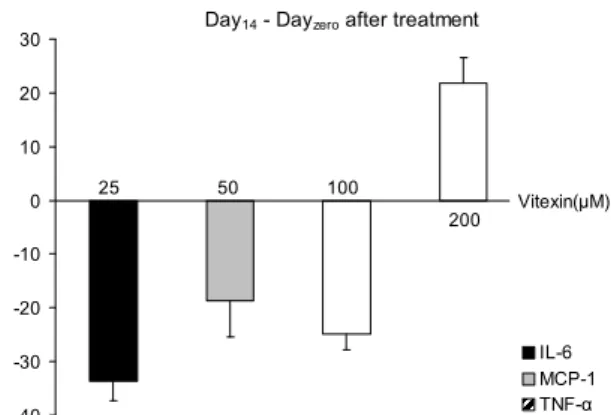
분화 중 이틀마다 배지를 수거하여 지방세포가 분비하는 TNF-α, IL-6, MCP-1의 양을 ELISA법으로 측정하였으며 최종분화일인 Day 14에서 Day 0의 차이를 결과로 나타냈다
Day14 - Dayzero after treatment
-40 -30 -20 -10 0 10 20 30
% of control .
IL-6 MCP-1 TNF-α 25 50 100
200 Vitexin(μM)
Fig. 4. Changes of cytokines (IL-6, MCP-1, TNF-a) com- pared to the control on the differences from Day14to Dayzero
in 3T3L-1 cells were dependent on the levels of vitexin. IL-6 were significantly reduced to almost 30% of control with 25/100 uM vitexin, and MCP-1 was reduced to 20% at 50 μM vitexin compared to control (p=0.01). However, no declined cytokines were found by 200 μM of vitexin, otherwise, TNF-α was in- creased to 123% of control at 200 μM vitexin.
(Fig. 4). IL-6 분비량은 vitexin을 25, 100 μM 처치한 군에서 대조군에 비하여 30%까지 유의적으로 감소하였으며 MCP- 1은 vitexin을 50 μM 처치한 군에서 20%가 유의적으로 감소 하였다(p=0.01). 그러나 200 μM의 vitexin에서는 감소하는 결과가 없었으며 특히 TNF-α는 대조군과 비교하여 123%
까지 증가하였다.
TNF-α는 염증반응에서 중요한 작용을 하는 세포분비물 질이며(29), IL-6는 염증성 및 항염증성 사이토카인 양쪽의 역할을 모두 담당하는 인터루킨으로 외상, 화상 및 조직 손 상에 의해 자극받은 T 세포와 대식세포 및 지방세포에서 분비된다(30,31). MCP-1은 염증성 케모카인으로서, 지방세 포에서 분비되어 비만, 인슐린저항성, 동맥경화증에 영향을 미치는 것으로 알려져 있다(32). 일반적으로 분화된 3T3-L1 세포에서 분비되는 IL-6 수준은 본 연구결과와 유사하거나 높았으며 MCP-1 수준 역시 유사한 것으로 나타났다(33-35).
TNF-α의 경우 본 연구결과의 150배 이상 분비되었다는 결 과가 존재하였으나 1/5배 수준의 분비량을 나타낸 연구결과 도 존재하므로 분화만을 유도한 3T3-L1 세포에서 측정된 값을 절대값으로 비교하였다(36,37).
본 연구 결과에 따르면 Day 14과 Day 0의 차이로 나타낸 결과에서 TNF-α의 경우 오히려 대조군보다 증가하였지만 Day 0부터 Day 14까지의 누적 분비량에서는 vitexin 처치에 따라 농도 의존적으로 감소되는 경향을 보였다. 그러나 대조 군과 유의적인 차이를 보이지 않았으며 결과는 나타내지 않 았다. 이를 확인하기 위해 TNF-α의 mRNA량을 RT-PCR 을 수행하였으며 결과는 위에서 설명한 내용과 같다. 이처럼 대조군보다 감소하는 TNF-α, IL-6 및 MCP-1의 경향은 vitexin 농도에 따라 각기 다른 것으로 나타났으며 각각의 아디포사이토카인 조절을 위한 적절한 vitexin의 농도가 존 재할 수 있으며 그것을 밝히기 위한 연구가 필요할 것으로 사료된다.
요 약
본 연구는 녹두묵을 이용한 항비만 임상선행연구를 바탕 으로 녹두에 풍부한 vitexin의 항비만 효과의 기작을 살펴보 고자 하였다. 녹두 vitexin은 200 μM 농도까지 3T3-L1 지방 전구세포의 독성효과는 없었으며 50, 100, 200 μM 농도에서 지방합성이 억제되었고 100 μM 농도에서 지방분해효과로 추측할 수 있는 결과가 나타났다. 또한 200 μM에서 염증성 아디포사이토카인 유전자발현을 증폭시키는 상위신호체계 (p38, ERK)가 억제된 반면 AMPK의 경우는 100 μM 농도까 지 증가하였고 PPARγ 단백질 발현도 200 μM에서 증폭하 였으며 TNF-α와 aP2 mRNA 발현은 25 μM에 증가하였다 가 감소하였다. 이러한 결과를 바탕으로 결론을 내리면 vi- texin 100~200 μM 사이의 농도가 항비만 기작을 규명하는 in vivo 실험을 위한 적절한 농도로 여겨진다. 그러나 본 연 구에서는 vitexin 처리에 따른 C/EBP, SREBP1, PPARγ mRNA 농도의 변화는 관찰되지 않아 3T3-L1 세포자살 (apoptosis) 신호체계 및 mTOR와의 연계성체계 혹은 기타 adipogenesis 억제 유전자로 SIRT1 등의 연구가 향후에 더 필요할 것으로 사료된다.
감사의 글
이 논문은 2011년도 성신여자대학교 학술연구조성비 지 원에 의하여 연구되었습니다. 녹두의 Vitexin 검출에 도움을 주신 농촌진흥청 국립식량과학원 기능성작물부 두류유지작 물과의 하태정 박사님께 감사드립니다.
문 헌
1. WHO. 2011.Obesity and overweight.World Health Organi- zation, Geneva, Switzerland.
2. Guzik TJ, Mangalat D, Korbut R. 2006. Adipocytokines-novel link between inflammation and vascular function?J Physiol Pharmacol 57: 505-528.
3. Rondinone CM. 2006. Adipocyte-derived hormones, cyto- kines, and mediators. Endocrine 29: 81-90.
4. Kawakami M, Pekala PH, Lane MD, Cerami A. 1982. Lipo- protein lipase suppression in 3T3-L1 cells by an endotox- in-induced mediator from exudate cells. Proc Natl Acad Sci U S A 79: 912-916.
5. Fried SK, Zechner R. 1989. Cachectin/tumor necrosis factor decreases human adipose tissue lipoprotein lipase mRNA levels, synthesis, and activity.J Lipid Res30: 1917-1923.
6. Feingold KR, Grunfeld C. 1992. Role of cytokines in induc- ing hyperlipidemia. Diabetes41: 97-101.
7. Greenberg AS, Nordan RP, Mclntosh J, Calvo JC, Scow RO, Jablons D. 1992. Interleukin 6 reduces lipoprotein lipase ac- tivity in adipose tissue of micein vivoand in 3T3-L1 adipo- cytes: a possible role for interleukin 6 in cancer cachexia.
Cancer Res 52: 4113-4116.
8. Pietta PG. 2000. Flavonoids as antioxidants. J Nat Prod 63: 1035-1042.
9. Park HJ, Della-Fera MA, Hausman DB, Rayalam S, Ambati S, Baile CA. 2009. Genistein inhibits differentiation of pri- mary human adipocytes. J Nutr Biochem 20: 140-148.
10. Pinent M, Espinel AE, Delgado MA, Baiges I, Bladé C, Arola L. 2011. Isoflavones reduce inflammation in 3T3-L1 adipocytes. Food Chem 125: 513-520.
11. Shin SH, Kang SS, Kwon KS. 1990. Studies on the compo- nents of the seeds ofPhaseolus radiatus.Yakhak Hoeji34:
282-286.
12. Kim DK, Son DM, Choi JK, Chon SU, Lee KD, Rim YS.
2010. Growth property and seed quality of mungbean culti- vars appropriate for labor saving cultivation. Korean J Crop Sci 55: 239-244.
13. Kim DK, Chon SU, Lee KD, Kim JB, Rim YS. 2008. Variation of flavonoids contents in plant parts of mungbean.Korean J Crop Sci 53: 279-284.
14. Kim JP, Lee IS, Seo JJ, Jung MY, Kim YH, Yim NH, Bae KH. 2010. Vitexin, orientin and other flavonoids from Spirodela polyrhiza inhibit adipogenesis in 3T3-L1 cells.
Phytother Res 24: 1543-1548.
15. Ait Mbarek L, Ait Mouse H, Elabbadi N, Bensalah M, Gamouh A, Aboufatima R, Benharref A, Chait A, Kamal M, Dalal A, Zyad A. 2007. Anti-tumor properties of black- seed (Nigella sativaL.) extracts. Braz J Med Biol Res 40: 839-847.
16. Ramirez-Zacarias JL, Castro-Munozledo F, Kuri-Harcuch W. 1992. Quantitation of adipose conversion and trigly- cerides by staining intracytoplasmic lipids with Oil red O.
Histochemistry97: 493-497.
17. Clark AM, Sousa KM, Jennings C, MacDougald OA, Kennedy RT. 2009. Continuous-flow enzyme assay on a microfluidic chip for monitoring glycerol secretion from cultured adipocytes. Anal Chem 81: 2350-2356.
18. Bradford MM. 1976. A rapid and sensitive method for the quantitation of microgram quantities of protein utilizing the principle of protein-dye binding.Anal Biochem72: 248-254.
19. Boney CM, Gruppuso PA, Faris RA, Frackelton AR Jr. 2000.
The critical role of Shc in insulin-like growth factor-I- mediated mitogenesis and differentiation in 3T3-L1 pre- adipocytes. Mol Endocrinol 14: 805-813.
20. Ahmadian M, Duncan RE, Sul HS. 2009. The skinny on fat: lipolysis and fatty acid utilization in adipocytes.Trends Endocrinol Metab 20: 424-428.
21. Gregoire FM, Smas CM, Sul HS. 1998. Understanding adi- pocyte differentiation. Physiol Rev 78: 783-809.
22. Smas CM, Sul HS. 1995. Control of adipocyte differentiation.
Biochem J 309: 697-710.
23. Wu Z, Rosen ED, Brun R, Hauser S, Adelmant G, Troy AE, McKeon C, Darlington GJ, Spiegelman BM. 1999. Cross- regulation of C/EBP alpha and PPAR gamma controls the transcriptional pathway of adipogenesis and insulin sensi-
tivity. Mol Cell 3: 151-158.
24. Tontonoz P, Hu E, Spiegelman BM. 1994. Stimulation of adipogenesis in fibroblasts by PPAR gamma 2, a lipid-ac- tivated transcription factor. Cell 79: 1147-1156.
25. Xu A, Wang Y, Xu JY, Stejskal D, Tam S, Zhang J, Wat NM, Wong WK, Lam KS. 2006. Adipocyte fatty acid-bind- ing protein is a plasma biomarker closely associated with obesity and metabolic syndrome. Clin Chem 52: 405-413.
26. Hurley RL, Anderson KA, Franzone JM, Kemp BE, Means AR, Witters LA. 2005. The Ca2+/calmodulin-dependent protein kinase kinases are AMP-activated protein kinase kinases. J Biol Chem 280: 29060-29066.
27. Herlaar E, Brown Z. 1999. p38 MAPK signalling cascades in inflammatory disease. Mol Med Today5: 439-447.
28. Seger R, Krebs EG. 1995. The MAPK signaling cascade.
FASEB J 9: 726-735.
29. Dandona P, Aljada A, Bandyopadhyay A. 2004. Inflamma- tion: the link between insulin resistance, obesity and dia- betes. Trends Immunol 25: 4-7.
30. Gabay C. 2006. Interleukin-6 and chronic inflammation.
Arthritis Res Ther 8: S3.
31. Xing Z, Gauldie J, Cox G, Baumann H, Jordana M, Lei XF, Achong MK. 1998. IL-6 is an antiinflammatory cytokine re- quired for controlling local or systemic acute inflammatory responses. J Clin Invest 101: 311-320.
32. Sartipy P, Loskutoff DJ. 2003. Monocyte chemoattractant protein 1 in obesity and insulin resistance.Proc Natl Acad Sci U S A 100: 7265-7270.
33. Candida Deoliveira C, Coghetto Acedo S, Martins Ferreira Gotardo E, de Oliveira Carvalho P, Rocha T, Pedrazzoli J Jr, Gambero A. 2012. Effects of methotrexate on in- flammatory alterations induced by obesity: anin vivoand in vitrostudy.Mol Cell Endocrinol[Epub ahead of print].
34. Yin X, Tu L, Yang H. 2007. Effect of simvastatin on IL-6 and adiponectin secretion and mRNA expression in 3T3-L1 adipocytes.J Huazhong Univ Sci Technolog Med Sci27:
248-251.
35. Zoico E, Garbin U, Olioso D, Mazzali G, Fratta Pasini AM, Di Francesco V, Sepe A, Cominacini L, Zamboni M. 2009.
The effects of adiponectin on interleukin-6 and MCP-1 se- cretion in lipopolysaccharide-treated 3T3-L1 adipocytes:
role of the NF-kappaB pathway.Int J Mol Med 24: 847- 36. Bradley RL, Fisher FF, Maratos-Flier E. 2008. Dietary fatty851.
acids differentially regulate production of TNF-alpha and IL-10 by murine 3T3-L1 adipocytes.Obesity16: 938-944.
37. Berg AH, Lin Y, Lisanti MP, Scherer PE. 2004. Adipocyte differentiation induces dynamic changes in NF-kappaB ex- pression and activity.Am J Physiol Endocrinol Metab287:
E1178-E1188.
(2012년 4월 16일 접수; 2012년 7월 18일 채택)