396
Copyright © 2017 The Korean Society of Fisheries and Aquatic Science pISSN:0374-8111, eISSN:2287-8815
서 론
어류는개체발생동안자어기에서치어기로변태하기직전에 형태적
,
생리적,
행동적으로극심한변화를겪으므로발생학적 지식은이러한측면뿐만아니라어업생물학적및양식업에도 매우중요하다(Koumoundouros et al., 1999).
또한해부학의 발달은유어기의형태발달에서기능적발달양상과환경에대한 선호도를이해하는데도움을준다(Fukuhara, 1992).
어류의자 치어기골격발달과정에대한관심과지식은사육초기에골격 이상의탐지와제거를위해필수적이며(Koumoundouros et al.,
1997a,b),
유어기의계통분류학적기초형질을제공하고있어이분야를연구하는열기가높다
(Sfakianakis et al., 2004).
얼록 동사리(Odontobutis interrupta)
는 농어목(Perciformes),
동사 리과(Odontobutidae),
동사리속(Odontobutis)
에 속하는 어류 로물의흐름이비교적느린하천의중류에서식하며,
바위틈에 은신하여소형어류를섭식하는육식성어류이다.
주로영산강 북쪽서해로흐르는하천에분포하는우리나라고유어종으로얼록동사리를비롯한동사리
(O. platycephala),
남방동사리(O.
obscura)
및좀구굴치(Micropercops swinhonis)
등2
속4
종이 분포하는것으로알려져있다(Kim et al., 2005).
얼록동사리에관한연구로는산란행동및초기생활사
(Choi and Na, 2000; Park et al., 2014)
등주로초기생활사에대한 연구가이루어졌으며,
같은동사리과어류에대한연구로는산 란행동(Mashiko, 1976),
분포(Iwata et al., 1985; Sakai et al., 1999),
자어발달(Iwata et al., 1988; Voskoboinikova and Pav- lov, 2006), O. hikimius
의배체발달(Doi and Aoyama, 2006),
유전적비교(Sakai et al., 1993)
및생식주기(Lee, 1998; Lee
and Yang, 1998)
등이수행되었으나골격발달에관한연구는잘알려져있지않다
.
또한,
동사리과어류는주변환경에따라 보호색을띠며,
다양한체색과몸의형태가유사하여외부형태 적분류에다소어려움이있다.
따라서본연구에서는얼록동사 리자치어의발육단계중에나타나는골격발달특징을관찰하 여유사종과의차이점을밝히기위한기초자료를마련하고,
내 부형태적특성을파악하여같은동사리과어류종동정을위한한국산 얼록동사리(Odontobutis interrupta) 자치어의 골격발달
박재민·한지형
1·윤성민
2·한경호
1*
경상북도 토속어류산업화센터, 1전남대학교 해양기술학부, 2경상북도 민물고기연구센터
Early Osteological Development of Larvae and Juveniles in the Korean Spotted Sleeper Odontobutis interrupta from Korea
Jae Min Park, Ji Hyeong Han
1
, Seong Min Yun2
and Kyeong Ho Han1
*Gyeongsangbuk-Do Native Fish Business Center, Uiseong 37366, Korea
1Marine Technology Undergraduate, Chonnam National University, Yeosu 59626, Korea
2Gyeongsangbuk-Do Research Center for Freshwater Fishes, Uljin 36332, Korea
We observed the osteological development in larvae and juveniles of Korean spotted sleeper Odontobutis interrupta bred in the laboratory in April 2014. Immediately after hatching, the prelarvae, which were about 4.27 mm long, showed ossification of the premaxillary bones in the upper jaw and the dentary and articular bones in the lower jaw.
At 7.11 mm, the larvae showed complete fusion of the post-cleithra and ossification of the scapulae with the appear- ance of one hole. At 8.65 mm, the larvae showed ossification of seven ribs from the third abdominal vertebra and an increase in the length of the neural spine and hemal spine. The number of caudal fin rays increased to 19. At 11.9 mm, the juveniles showed ossification of three procurrent rays on the side of the parhypural bone as well as ossification of two procurrent rays on the side of the epural bone, indicating the complete ossification of all spicules.
Key words: Juvenile, Larvae, Osteological, Odontobutis interrupta
This is an Open Access article distributed under the terms of the Creative Commons Attribution Non-Commercial Licens (http://creativecommons.org/licenses/by-nc/3.0/) which permits unrestricted non-commercial use, distribution, and reproduction in any medium, provided the original work is properly cited.
https://doi.org/10.5657/KFAS.2017.0396 Korean J Fish Aquat Sci 50(4) 396-405, August 2017 Received 29 May 2017; Revised 16 June 2017; Accepted 6 July 2017
*Corresponding author: Tel: +82. 61.659.7163 Fax: +82. 61.659.7169
E-mail address: [email protected]
분류학적연구자료로제공하고자한다
.
재료 및 방법
채집 및 동정
2014
년4
월전남여수시소라면에위치한소라천(
지방2
급하 천)
에서바위틈에산란된수정란과주변을보호하는행동을보 이는개체를채집하여연구실로운반하였다.
종동정은Kim et al. (2005)
를이용하였다.
수정란 관리 및 자치어 사육
수정란은 사각수조
(50×30×20 cm)
에수용하였고,
수온은18.5-19.5℃(
평균19.0±1.0℃)
를 유지하였으며,
용존산소는5.94-7.10 mg/L(
평균6.52 mg/L)
였다.
자치어의사육환경은수 정란과동일한조건이었고,
수조내스펀지여과기를설치하여 순환여과식으로사육하였다.
먹이는난황흡수후부터30
일까 지Artemia sp. nauplius
유생을공급하였고,
이후에는실지렁 이및냉동장구벌레(Blood warm Hikari, Japan)
를공급하였다. 골격염색
발육단계에 따른자치어의골격발달 관찰을위해매일
5
마 리씩5%
중성포르말린에고정하였고, Walker and Kimmel
(2007)
의이중염색법에따라내부골격을염색후KOH 0.1%
와
Glycerol 50%
에보존하였다.
염색한자치어는실체현미경(Nikon SMZ18, Japan)
을이용하여각부위를관찰하고스케 치하였다.
골격의각부위명칭은Kang et al. (2012)
에따랐다.
결 과
종 동정
바위에산란된수정란의주변에서보호행동을보인개체는몸 이원통형으로꼬리쪽으로갈수록측편형에가깝고
,
제1
등지 느러미전반부에서후반부까지와,
제2
등지느러미후반부및꼬 리지느러미기부에검은색띠가있으며,
계수형질에서는제1
등지느러미8
개,
제2
등지느러미9
개뒷지느러미8
개,
측선비늘 수40
개를가져얼록동사리로동정되었다(Kim et al., 2005).
자치어 골격발달
얼록동사리 자치어의 골격발달은 두개골
(cranium),
내장골(visceral skeleton)
견대골(pectoral girdle bone),
미골(caudal skeleton),
척추골(vertebral column)
과 지느러미(fin rays)
및 담기골(pterygiophore)
로구분하여관찰하였다.
두부골격
두개골과내장골로이루어진얼록동사리의두부골격발달은
Table 1,2
와같았다.
부화직후자어의전장은3.87-4.53 mm(
평균
±SD, 4.27±0.35 mm, n=5)
로위턱을구성하는전상악골
(premaxillary)
과 아래턱을지지하는 치골(dentary)
및관절 골(articular)
이골화하였다.
두개골의기저를형성하는부설골(parasphenoid)
과 앞쪽에는 안전골(preorbital=lucrymal)
이 골 화하였고,
눈을둘러싸고있는안하골(suborbital)
이골화하였으 며,
상후두골(supraoccipital),
익이골(pterotic),
설이골(sphen- otic),
전이골(prootic)
및액골(frontal)
등이골화하였다.
구개 부에는설악골(hyomandibular),
접속골(sympletic)
및내익상 골(endopterygoid)
등이골화하였고,
설궁부에는하설골(hypo- hyal),
각설골(ceratohyal),
상설골(epihyal),
간설골(interhyal)
및5
개의새조골(branchiostegal rays)
이골화하였으며,
새개부 에는 전새개골(preopercle),
주새개골(opercle)
이 골화하였다(Fig. 1A).
부화후
13
일째자어의전장은6.84-7.38 mm(
평균7.11±0.26 mm, n=5)
로 두개골에노정골(parietal),
익설골(alisphenoid),
상이골(epiotic),
중사골(mesethmoid)
및 외후두골(exoccipi- tal)
등이골화하였고,
위턱을구성하는주상악골(maxillary)
이 골화하였으며,
새개부에는 하새개골(subopercle)
과 간새개골(interopercle)
이골화하였다(Fig. 1B).
부화후
18
일째자어의전장은8.49-8.93 mm(
평균8.65±0.20 mm, n=5)
로 두개골에 기저설골(basisphenoid),
기저후두골(basioccipital),
후이골(opisthotic),
서골(vomer)
및측사골(lat- eral ethmoid),
구개부에구개골(palatine),
방골(quadrate),
외익 상골(ectopterygoid)
및후익상골(metapterygoid)
이 골화하였 고,
아래턱관절골에는각골(angular)
이골화하였다(Fig. 1C).
부화후
30
일째치어의전장은11.7-12.2 mm(
평균11.9±0.23 mm, n=5)
로두개골에비골(nasal)
이골화하였고,
설궁부의새 조골은6
개로골화하면서두부골격의모든골화가완료되었다(Fig. 1D).
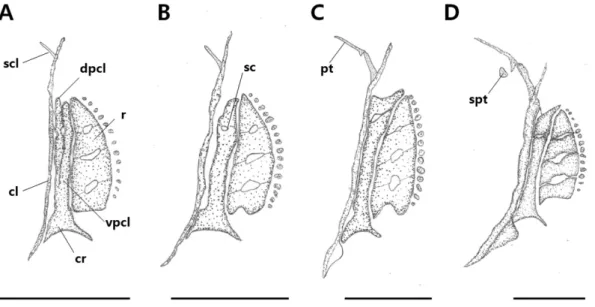
견대골
견대골은가슴지느러미를지지하는골격으로후측두골
(post- temporal)
에 의해두개골과관절되었다(Fig. 2, Table 3).
부 화 직후자어의전장은3.87-4.53 mm(
평균4.27±0.35 mm, n=5)
로 쇄골(cleithrum)
을 중심으로 위쪽에는 상쇄골(supra- cleithrum),
옆에는2
개의후쇄골(postcleithrum)
이골화하였는 데후쇄골은양옆으로접합되었고,
아래쪽의오훼골(coracoid)
과접합되어있었다.
후쇄골옆쪽에는사출골(actinost=radial)
이골화하였고,
사출골은3
개로나누어졌으며, 3
개의구멍을형 성하였다(Fig. 1A).
부화후13
일째자어의전장은6.84-7.38 mm(
평균7.11±0.26 mm, n=5)
로2
개의후쇄골은접합이완 료되었고, 1
개의구멍이생기면서견갑골(scapula)
이골화하였 다(Fig. 2B).
부화후18
일째자어의전장은8.49-8.93 mm(
평 균8.65±0.20 mm, n=5)
로상쇄골위쪽에후측두골(posttem-
poral)
이 골화하였고,
오훼골은 쇄골과 접합하기 시작하였다(Fig. 2C).
부화후30
일째치어의전장은11.7-12.2 mm(
평균11.9±0.23 mm, n=5)
로후측두골아래에상측두골(suprapost-
temporal)
이골화하기시작하였고,
오훼골과쇄골은접합이완료되면서견대부골격의골화가완료되었다
(Fig. 2D).
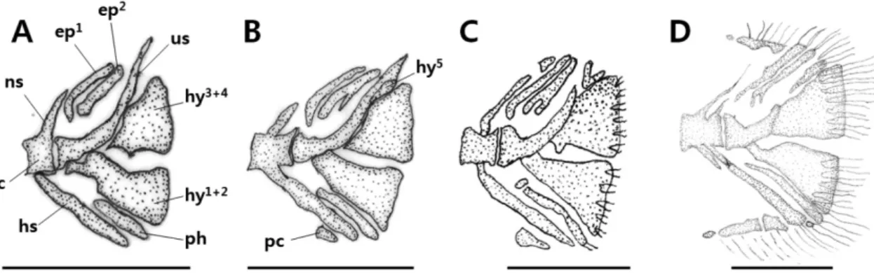
미골
꼬리지느러미를지지하는미골은몇개의추골과골편으로된 미골복합체
(caudal complex)
를이룬다(Fig. 3, Table 3).
부화 직후자어의전장은3.87-4.53 mm (
평균4.27±0.35 mm, n=5)
로추체(centrum)
를중심으로위쪽에는신경극(neural spine),
아래쪽에는 혈관극(hemal spine)
이 골화되었고,
미부봉상골(urostyle)
은45°
로휘어져있었으며,
신경극과미부봉상골사 이에는2
개의상미축골(epural bone)
이골화하였다.
하미축골(hypural bone)
은미부봉상골에융합되어있었고,
하단부에하 미축골1+2
가융합되었으며,
상단부에는하미축골3+4
가융 합되었다.
그아래에는1
개의준하미축골(parhypural bone)
이 골화하기시작하였다(Fig. 3A).
부화후13
일째자어의전장은6.84-7.38 mm (
평균7.11±0.26 mm, n=5)
로5
번째하미축골 이골화하였고,
준하미축골옆에는1
개의부속줄기(procurrent)
가골화하였다(Fig. 3B).
부화후18
일째자어의전장은8.49- 8.93 mm (
평균8.65±0.20 mm, n=5)
로상미축골옆에1
개의 부속줄기가골화하였고(Fig. 3C),
부화후30
일째치어의전장 은11.7-12.2 mm (
평균11.9±0.23 mm, n=5)
로준하미축골옆 에3
개의부속줄기가골화하였으며,
상미축골옆에는2
개의부 속줄기가골화하였다(Fig. 3D).
척추골
몸의중축을이루는척추골은척수와혈관을보호하며
,
발달 과정은Table 3
과같았다.
부화직후자어의전장은3.87-4.53 mm (
평균4.27±0.35 mm, n=5)
로척추골이완전히골화되지 않았고,
척색(notochord)
으로이루어져있었으며,
아래쪽에는 Table 1. The development process of cranium and orbital region of Korean spotted sleeper Odontobutis interruptaDays after hatching 0 3 6 10 13 15 18 21 30
Total length (mm) 4.27 6.20 6.43 6.69 7.11 8.60 8.65 9.71 11.9
Cranium
Parasphenoid Basioccipital Nasal Frontal Sphenotic Pterotic Lateral ethmoid Alisphenoid Parietal Prootic Epiotic Supraoccipital Exoccipital Opisthotic Mesethmoid Basisphenoid Vomer
Orbital region Preorbital Suborbital
Fig. 1. Development of the cranium and visceral skeleton in Korean spotted sleeper Odontobutis interrupta. A: 4.27 mm, TL; B: 7.11 mm, TL; C: 8.65 mm, TL; D: 11.9 mm, TL. ar, articular; al, lisphenoid; an, angular; bas, basisphenoid; bao, basioccipital; brs, branchiostegal rays; ch, ceratohyal; d, dentary; ecp, ectopterygoid; eh, epihyal; ep, epiotic; enp, endopterygoid; exo, exoccipital; fr, frontal; h, hypohyal; hy, hyomandibular; in, interhyal; iop, interopecle; le, lateral ethmoid; lu, lucrimal; me, mesethmoid; mpt, metapterygoid; mx, maxillary; na, na- sal; op, opercle; opi, opisthotic; par, parietal; pal, palatine; pop, preopercle; pmx, premaxillary; ps, parasphenoid; pro, prootic; pto, pterotic;
q, quadrate; soc, supraoccipital; so, suborbital; sop, subopercle; sph, sphenotic; sy, symplectic; v, vomer. Scale bars=1.0 mm.
Fig. 2. Development of the pectoral girdle in Korean spotted sleeper Odontobutis interrupta. A: 4.27 mm, TL; B: 7.11 mm, TL; C: 8.65 mm, TL; D: 11.9 mm, TL. cl, cleithrum; cr, coracoid; dpcl, dorsal post cleithrum; pt, posttemporal; r, radial; sc, scapula; spt, supraposttemporal;
scl, supracleithrum; vpcl, ventral post cleithrum. Scale bars=1.0 mm.
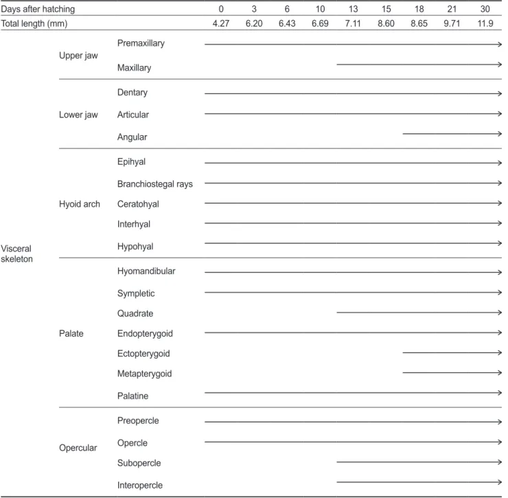
Table 2. The development process of visceral skeleton of Korean spotted sleeper Odontobutis interrupta
Days after hatching 0 3 6 10 13 15 18 21 30
Total length (mm) 4.27 6.20 6.43 6.69 7.11 8.60 8.65 9.71 11.9
Visceral skeleton
Upper jaw Premaxillary Maxillary
Lower jaw
Dentary Articular Angular
Hyoid arch
Epihyal
Branchiostegal rays Ceratohyal Interhyal Hypohyal
Palate
Hyomandibular Sympletic Quadrate Endopterygoid Ectopterygoid Metapterygoid Palatine
Opercular
Preopercle Opercle Subopercle Interopercle
4
개의측돌기(parapophysis)
와15
개의혈관극이 골화하였다.
추체의위쪽에는 미골부앞쪽에2
개의신경극이골화하였다.
제1
등지느러미는막으로되어있었고,
아래에는5-7
개의신경 간극(interneural spine)
이골화하였다.
제2
등지느러미는9-10
개의줄기가발달하였고,
아래에9-10
개의신경간극이골화하 였다.
아래쪽에는뒷지느러미줄기가6
개발달하였고, 6
개의혈 관간극(interhemal spine)
이골화하여신경간극과혈관간극의 개수에따라지느러미줄기가발달하였으며,
꼬리지느러미줄 기수는15
개였다(Fig. 4A).
부화후6
일째자어의전장은6.25-
6.56 mm (
평균6.43±0.16 mm, n=5)
로척색으로이루어져있 던척추골이추체(centrum)
로골화하였고,
복추골에서부터미 추골방향으로7
개의추체가골화하였으며,
추체가골화함에따 라신경극이골화하였다.
제1
등지느러미에는7
개의줄기가발 달하였고,
미부봉상골이골화하기시작하였다(Fig. 4B).
부화후
13
일째자어의전장은6.84-7.38 mm (
평균7.11±0.26
mm, n=5)
로제1
등지느러미의신경간극이7-8
개로증가하면서줄기수도같이
7-8
개로발달하였다.
뒷지느러미의혈관간 극은7-8
개로증가하였고,
줄기수도같이7-8
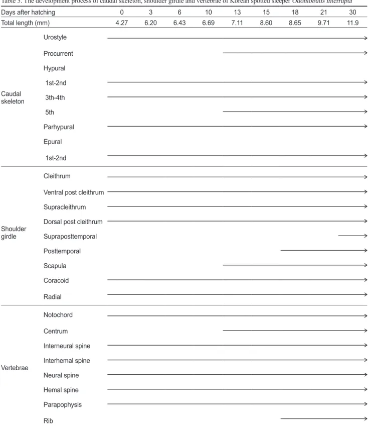
개로발달하였다Table 3. The development process of caudal skeleton, shoulder girdle and vertebrae of Korean spotted sleeper Odontobutis interrupta
Days after hatching 0 3 6 10 13 15 18 21 30
Total length (mm) 4.27 6.20 6.43 6.69 7.11 8.60 8.65 9.71 11.9
Caudal skeleton
Urostyle Procurrent Hypural 1st-2nd 3th-4th 5th Parhypural Epural 1st-2nd
Shoulder girdle
Cleithrum
Ventral post cleithrum Supracleithrum Dorsal post cleithrum Supraposttemporal Posttemporal Scapula Coracoid Radial
Vertebrae
Notochord Centrum Interneural spine Interhemal spine Neural spine Hemal spine Parapophysis Rib
(Fig. 4C).
부화후18
일째자어의전장은8.49-8.93 mm (
평균8.65±0.20 mm, n=5)
로3
번째복추골부터7
개의늑골(rib)
이골화하기 시작하였고
,
신경극과혈관극의 길이가신장하였으 며,
꼬리지느러미줄기수는19
개로증가하였다(Fig. 4D).
고 찰
얼록동사리의부화직후자어는평균전장
4.27 mm
일때두 개골에기저를형성하는부설골을비롯하여안전골,
안하골,
상 후두골및익이골등이골화되었고,
구개부에설악골,
접속골,
내익상골,
설궁부에하설골,
각설골,
상설골및간설골이골화 하였으며,
새개부에는전새개골및주새개골이골화하였다.
일 반적으로농어목어류는부화이후에두개골과지느러미의발 달이관찰된다고알려져있으나얼록동사리의경우골격발달 이진행된상태로부화되어난내에서골격이발달하는 것으 로알려진연어과어류와유사하다고판단되어 난발생과정 중골격발달정도를면밀히관찰할필요가있을것으로생각된 다(Kendall et al., 1984; Matsuoka, 1985; Koumoundouros et al., 1997b; Faustion and Power, 1999; Koumoundouros et al., 2001a, 2001b; Sfakianakis et al., 2004, 2005).
다른어류의최초골격발달을살펴보면구굴무치과에속하는 발기
, Perccottus glenii (Voskoboinikova and Pavlov, 2006)
는 부설골,
기저후두골,
외후두골 및상후두골이골화하였고,
농 어목에속하는꺽지, Coreoperca herzi (Han et al., 2017)
는부 설골,
전상악골,
치골및견대부쇄골이골화하였으며,
점농어, Lateolabrax maculatus (Kang et al., 2012)
는부설골,
전상악골 및치골,
잿방어, Seriola dumerili (Liu, 2001)
는전상악골,
주상 악골,
치골및전새개골등이골화하였다.
능성어, Epinephelus septemfasciatus (Park et al., 2015)
와저울베도라치, Entomac- rodus stellifer lighti (Kim et al., 1992)
는부설골및쇄골,
복어 목인졸복, Takifugu pardalis (Han et al., 2005)
은악골이동시 에골화하였다.
농어목어류의골격발달은두부골격의기저를 형성하는부설골과호흡및섭이에관련된기능을하는악골,
가 슴지느러미를지지하고유영능력에영향을미치는쇄골이각 부위별골격가운데우선적으로발달하는공통적인경향을나 타냈으며,
이러한현상은생존을위한이들의진화적특성과연관되는것으로생각된다
.
얼록동사리의견대부골격중견갑골에는
1
개의체공(fora-
Fig. 3. Development of the caudal skeleton in Korean spotted sleeper Odontobutis interrupta. A: 4.27 mm, TL; B: 7.11 mm, TL; C: 8.65 mm, TL; D: 11.9 mm, TL. c, centrum; ep, epural bone; hy, hypural bone; hs, hemal spine; pc, procurrent; ph, parhypural bone; ns, neural spine;us, urostyle. Scale bars=1.0 mm.
Fig. 4. Development of the vertebrae column in Korean spotted sleeper Odontobutis interrupta. A: 4.27 mm, TL; B: 6.43 mm, TL;
C: 7.11 mm, TL; D: 8.65 mm, TL. c: centrum; hs, hemal spine; ins, interneural spine; his, interhemal spine; n, notochord; ns, neural spine; pp, parapophysis; r, rib. Scale bars=1.0 mm.
men)
이형성되는것이관찰되는데꺽지(Han et al., 2017),
점농 어(Kang et al., 2012),
참돔, Pagrus major (Matsuoka, 1987),
황돔, Dentex tumifrons (Koumoundouros et al., 2001b),
자리 돔류(Emery, 1973),
쏨뱅이목어류(Kim and Han, 1991; Kim et al., 1997; Han et al., 2001)
등다른분류군에서도형성되 는것이관찰되어대부분의어류에서형성되는전형적인형질 로보인다.
척추골은몸의중축을이루는골격으로얼록동사리는부화직후평균전장
4.27 mm
일때척추의골화는이루어지지않았고
,
척색으로이루어져있었으며,
부화후6
일평균전장
6.43 mm
일때골화가진행되었고,
추체가발달하면서부화후
13
일평균전장7.11 mm
일때척추를이루었다.
골격의골 화방향은복추골에서미추골,
꼬리방향으로진행되었고,
추체 의골화가완전히이루어지기전에측돌기,
혈관극,
혈관간극 및신경간극이우선적으로골화되기시작하였으며,
또한추체 가골화하면서동시에미추골과미부봉상골이골화하기시작 하였다.
다른분류군과비교하였을때이러한현상은쏨뱅이목 어류(Kim and Han, 1991; Kim et al., 1993; Kim et al., 1997;
Han et al., 2001; Byun et al., 2012),
농어목의능성어(Park et al., 2015),
점농어(Kang et al., 2012),
꺽지(Han et al., 2017),
동갈치목인날치, Prognichthys agoo (Park and Kim, 1987)
등 에서도나타나어종별로발달양상이유사한것으로판단된다.
구굴무치과에속하는발기(Voskoboinikova and Pavlov, 2006)
는부화직후복추골,
미추골및신경극,
혈관극이골화한상태 였고, 5
개의늑골이3
번째추체부터발달하였으며,
척추골의골 화방향은얼록동사리와유사하였으나골격의발달이보다많이 이루어진상태에서부화하여차이를보였다.
농어목어류의지느러미발달양상은후방의등지느러미
,
뒷지 느러미가먼저발달하고전방의등지느러미가발달한다고알려 져있다(Johnson, 1984; Faustino and Power, 1999).
얼록동사 리는후방의등지느러미와뒷지느러미가발달한뒤전방의등 지느러미가발달하여일반적인농어목어류의발달양상과일치 하였다.
각부위별기조수는부화후13
일평균전장7.11 mm
일때제1
등지느러미8
개,
제2
등지느러미9
개,
뒷지느러미8
개 로종동정한개체와기조수가일치하였다.
지느러미를지지하 는담기골의골화는먼저담기골이골화한뒤지느러미가형성 되었고,
골화방향은전방에서후방쪽으로이루어졌다.
담기골 은추체가골화되기전에발달하기시작하였고,
추체가완전히 골화되었을때담기골의형태가완전한모습을갖추었다.
척추 골의골화와지느러미의줄기발달이완성된후에담기골의골 화가완료되는것은어류의유영능력에있어추진력을증가시 키는것과관련이있는것(Lee et al., 2001)
으로보여지고,
얼 록동사리의경우지속적인유영보다는대부분을바닥에서생 활하기때문에지느러미줄기보다담기골이먼저골화하는것 으로추정되며,
유영하는어류는담기골보다지느러미줄기의 발달이우선적으로이루어져유영형태에따라발달정도에차 이를나타냈다.
꼬리지느러미를지지하는 미골부는 상미축골
,
하미축골 및 미부봉상골 등으로이루어져 있으며,
얼록동사리는 부화 직후평균전장
4.27 mm
일때미부봉상골이45°
로휘어져있었다
.
미끈날망둑, Chaenogobius laevis (Kim and Han, 1989)
미 끈망둑, Luciogobius guttatus (Kim et al., 1992),
큰미끈망둑, Luciogobius grandis (Yoon, 2004)
의부화자어는성장함에따 라미부봉상골이휘어지면서골화하였고,
쏨뱅이목어류(Kim and Han, 1991; Kim et al., 1993; Kim et al., 1997; Han et al., 2001; Byun et al., 2012),
농어목의꺽지(Han et al., 2017),
능 성어(Park et al., 2015),
점농어(Kang et al., 2012)
또한성장하 면서발달하여부화직후부터미부가휘어진얼록동사리와차 이를나타냈다.
미골부의하미축골은얼록동사리가부화후
13
일평균전장7.11 mm
일때하미축골(1+2, 3+4, 5)
이 융합되었고,
외관상3
개의 하미축골을 형성하였으며,
쏨뱅이목의 황점볼락, Se- bastes oblongus (Byun et al., 2012),
동갈치목의학공치, Hy- porhamphus sajori (Lee at al., 2001)
가이와같은발달양상을 보였다.
망둑어과어류인미끈날망둑(Kim and Han, 1989),
미 끈망둑(Kim et al., 1992),
큰미끈망둑(Yoon, 2004)
은외관상2
개의하미축골이골화하였고,
다른농어목어류인꺽지(Han et al., 2017),
쏘가리와꺽저기(Park, 2001)
및방어류(Kohno, 1997; Liu, 2001)
는외관상2
개인하미축골(1+2, 3+4+5),
능성 어(Park et al., 2015)
는외관상3
개의하미축골(1+2, 3, 4+5)
을 나타내하미축골의외관상개수와융합형태가종에따라다르 게나타나차이를보였다.
하미축골의융합현상은계통학적연 구의기초자료로서중요하다고생각되며,
자치어시기의골격 발달양상은종묘생산과정에서발생되는기형발생원인파악,
유사종과의식별이나생태학적으로중요한자료로활용될수 있어향후이분야에대한지속적인연구가필요할것으로생 각된다.
References
Byun SG, Kang CB, Myoung JG, Cha BS, Han KH and Jung CG. 2012. Early osteological development of the larvae and juveniles in Sebastes oblongus (Pisces: Scorpaenidae). Ko- rean J Ichthyol 24, 67-76.
Choi SS and Na YU. 2000. The spawning behavior and egg de- velopment of Odontobutis interrupta Iwata and Jeon, 1985.
Korean J Envion Biol 18, 323-330.
Doi T and Aoyama S. 2006. Embryonic larval and juvenile morphologies of the freshwater goby Odontobutis hikimius reared in an aquarium were observed and described. Japan J Ichthyol 53, 63-70.
Emery AR. 1973. Ecology and functional osteology damselfish (Pisces; Pomacentridae) at Alligator reef, Florida Keys. Bull Mar Sci 23, 649-770.
Faustino M and Power DM. 1999. Development of the pectoral,
pelvic, dorsal and anal fins in cultured sea bream. J Fish Biol 54, 1094-1110.
Fukuhara O. 1992. Study on the development of functional mor- phology and behavior of the larvae of eight commercially valuable teleost fishes. Contr Fish Res Jpn Sea Block 25, 1-122.
Han KH, Cho JK, Lee SH, Hwang SY, Yoon SM, Seo WI and Kim CC. 2005. Osteological development of the larvae and juveniles of Takifugu pardalis (Teleostei: Tetraodontodae).
Korean J Ichthyol 17, 29-35.
Han KH, Lim SK, Kim KS, Kim CW and Yoo DJ. 2001. Osteo- logical development of the larvae and juveniles of Sebastis-
cus tertius (Barsukov et Chen) in Korea. Korean J Ichthyol
13, 63-68.Han KH, Park JT, Jin DS, Yoo DJ and Park JM. 2017. Osteolog- ical development of the larvae and juvenile in Coreoperca
herzi (Perciformes: Centropomidae). Korean J Ichthyol 29,
32-40.Iwata A, Jeon SR, Mizuno N and Choi KC. 1985. Arevision of the eleotrid goby genus Odontobutis in Japan, Korea and China. Japan J Ichthyol 31, 373-388.
Iwata A, Jeon SR, Mizuno N and Choi KC. 1988. Larval de- velopment of a gobiid fish, Odontobutis obscura obscura in comparison with that of O. interrupta and of O. platy-
cephala. Japan J Ichthyol 35, 371-381.
Johnson GD. 1984. Percoidei: development and relationship. In:
Moser HG, Richards WJ, Cohen DM, Fahay MP, Kendall AW and Richardson SL (eds.), Ontogeny and systematics of fishes. American Society of Ichthyologists and Herpetolo- gists, Lawrence KS, U.S.A., 464-498.
Kang CB, Myoung JG, Kim YU and Kim HC. 2012. Early Os- teological development and squamation in the spotted sea bass Lateolabrax maculatus (Pisces: Lateolabracidae). Ko- rean J Fish Aquat Sci 45, 271-282.
Kendall AW and Vinter B. 1984. Development of Hexagram- mids (Pisces: Scorpaeniformes) in the northeastern Pacific Ocean. US Dep Commer NOAA Tech Rep NMFS 2, 44.
Kim IS, Choi Y, Lee CL, Lee YJ, Kim BJ and Kim JH. 2005. Il- lustrated book of Korean fishes. Kyo-Hak Publishing Seoul, Korea, 417-418.
Kim YU and Han KH. 1989. Early life history of the marine animals 1. Egg development larvae and juveniles of Chae-
nogobius laevis (Steindachner). Bull Korean Fish Soc 22,
317-331.Kim YU, Han KH, Kang CB and Ryu JW. 1992. Early life his- tory and spawning behavior of the gobiid fish, Luciogobius
guttatus Gill. Korean J Ichthyol 4, 1-13.
Kim YU, Han KH and Kang CB. 1992. Morphology and skel- etal development of larvae and juveniles of Entomacrodus
stellifer lighti (Herre). Korean J Ichthyol 4, 31-43.
Kim YU and Han KH. 1991. The early life history of rockfish
Sebastes schlegeli. Korean J Ichthyol 3, 67-83.
Kim YU, Han KH and Byun SK. 1993. The early life history of the rockfish, Sebastes inermis 2. Morphological and skeletal development of larvae and juveniles. Bull Korean Fish Soc 26, 465-476.
Kim YU, Han KH, Kang CB, Kim JK and Byun SK. 1997. The early life history of the rockfish, Sebastiscus marmoratus 2.
Morphology and skeletal development of larvae and juve- nile. Korean J Ichthyol 9, 186-194.
Kohno H. 1997. Osteological development of the caudal skel- eton in the carangid, Seriola lalandi. Ichthyol Res 44, 219- Koumoundouros G, Divanach P and Kentouri M. 1999. Osteo-221.
logical development of the vertebral column and of the cau- dal complex in Dentex dentex. J Fish Bio 54, 424-436.
Koumoundouros G, Gagliardi F, Divanach P, Boglione C, Cataudella S and Kentouri M. 1997a. Normal and abnormal osteological development of caudal fin in Sparus aurata L.
fry. Aquaculture 149, 215-226.
Koumoundouros G, Oran G, Divanach P, Stefanakis S and Kentouri M. 1997b. The opercular complex deformity in in- tensive gilthead sea bream (Spartus aurata L.) larviculture.
Moment of apparition and description. Aquaculture 156, 165-177.
Koumoundouros G, Sfakianakis DG, Maingot E, Divanach P and Kentouri M. 2001a. Osteological development of the vertebral column and of the fins in Diplodus sargus (Tele- ostei: Perciformes: Sparidae). Mar Biol 139, 853-862.
Koumoundouros G, Divanach P and Kentouri M. 2001b. Osteo- logical development of Dentex dentex (Osteichthyes: Spari- dae): dorsal, anal, paired fins and squamation. Mar Bio1 38, 399-406.
Lee WK. 1998. Annual reproductive cycle and changes in plas- ma levels of sex steroid hormones of the female Korean dark sleeper, Odontobutis platycephala (Iwata et Jeon). J Korean Fish Soc 31, 599-607.
Lee WK and Yang SW. 1998. Testicular development and serum levels of gonadal steroids hormone during the annual repro- ductive cycle of the male Korean dark sleeper, Odontobutis
platycephala (Iwata et Jeon). J Aquacultule 11, 475-485.
Lee SJ, Kim YU and Han KH. 2001. Osteological development of larvae and juveniles of Hyporhampus sajori (Teleostei:
Hemiramphidae). Korean J Ichthyol 13, 173-180.
Liu CH. 2001. Early osteological development of the yellow tail
Seriola dumerili (Pisces: Carangidae). Zool Stud 40, 289-
298.Matsuoka M. 1987. Development of the skeletal tissues and skeletal muscles in the red sea bream. Bull Seikai Red Fish Res Lab 65, 1-14.
Matsuoka M. 1985. Osteological development in the red sea bream, Pagrus major. Japan J Ichthyol 32, 35-51.
Mashiko K. 1976. Reproductive behavior of an eleotrid goby
Odontobutis obscurus in aquaria. Japan J Ichthyol 23, 69-78.
Park JM, Han KH, Kim NR, Yoo DJ, Yun SM and Han JH.
2014. Egg development and early life history of Korean endemic species Korean spotted sleeper, Odontobutis inter-
rupta (Pisces: Odontobutidae). Dev Reprod 18, 259-266.
http://dx.doi.org/10.12717/DR.2014.18.4.259.
Park JT. 2001. Phylogenetic study of the centropomidae (Pisces, Perciformes) in Korea. Ph. D. Dissertation, University of Yosu National, Yosu, Korea, 29-56.
Park JY, Hong CG, Cho JK, Son MH, Han KH and Park JM.
2015. Early osteological development of the larvae and ju- veniles in sevenband grouper, Epinephelus septemfasciatus (Pisces: Serranidae). Korean J Ichthyol 27, 189-198.
Park YS and Kim YU. 1987. Studies on the larvae and juveniles of flying fish, Prognichthys agoo (Temminck et Schlegel) (Pisces, Exocoetidae) II. Osteological development of lar- vae and juveniles. Bull Korean Fish Soc 20, 447-456.
Sakai H, Iwata A and Jeon SR. 1993. Genetic evidence support- ing the existence of three distinct species in the genus Odon-
tobutis (Gobiidae) from Japan and Korea. Japan J Ichthyol
40, 61-64.Sakai H, Tanaka Y, Tsujii H, Iwata A and Ikeda I. 1999. Distribu- tion pattern of two genetically different groups of Odonto-
butis obscura in Takatsu river and its vicinity. Japan J Ich-
thyol 46, 109-114.Sfakianakis DG, Doxa CK, Kouttouki S, Koumoundouros G, Maingot E, Divanach P and Kentouri M. 2005. Osteologi- cal development of the vertebral column and of the fins in
Diplodus puntazzo (Cetti, 1777). Auqaculture 250, 36-46.
Sfakianakis DG, Koumoundouros G, Divanach P and Kentouri M. 2004. Osteological development of the vertebral column and of the fins in Pagellus erythrinus (L. 1758). Tempera- ture effect on the developmental plasticity and morpho-ana- tomical abnormalities. Auqaculture 232, 407-424.
Voskoboinikova OS and Pavlov DA. 2006. Larval development of the Amur sleeper Perccottus glenii (Perciforms, Gobioi- dei, Odontobutidae) and the origin of fish of the suborder Gobioidei. J Ichthyol 46, 826-841.
Walker MB and Kimmel CB. 2007. A two-color acid-free carti- lage and bone stain for zebrafish larvae. Biotech Histochem 82, 23-28.
Yoon SM. 2004. Early life history of the nake-head goby, Lu-