port
2
Department of Oral and Maxillofacial Radiology and Research Institute of Oral Science, College of Dentistry, Gangneung-Wonju National University, Gangneung, Korea
Jin-Woo Han
Glandular odontogenic cyst in the posterior mandible: A case report
Glandular odontogenic cyst in the posterior mandible: A case report Department of Oral and Maxillofacial Radiology and Research Institute of Oral Science, College of Dentistry,
Gangneung-Wonju National University, Gangneung, Korea Jin-Woo Han
The glandular odontogenic cyst (GOC) is a rare cyst derived from odontogenic epithelium with a spectrum of characteristics including salivary gland features. It occurs more commonly in the mandible and most often in the anterior mandible.
Radiographically, most cases present a well-defined unilocular or multilocular radiolucency with a cortical boundary. Despite no unique or pathognomonic clinical or radiographic features, the lesion shows potentially aggressive behavior. A 76-year-old male was referred to Gangneung-Wonju National University Dental Hospital with a chief complaint of slight swelling of the right mandible. Cone-beam computed tomography examination revealed a unilocular radiolucent lesion involving impacted third molar at the right posterior mandible. Slight lingual cortical thinning with suspected perforation was also shown. Histopathologically, multiple areas of cyst epithelium showed a glandular differentiation, resulting in mucoid-filled secretory cells and microcyst.
Based on these findings, the final diagnosis was determined to be GOC.
Key words : Cone-beam computed tomography, Mandible, Odontogenic cysts ABSTRACT
Ⅰ. Introduction
The glandular odontogenic cyst(GOC) is a
relatively rare-developmental cyst of the jaws presenting potential aggressiveness on histopathology. In 1987, Padayachee and Van Corresponding Author
Jin-Woo Han
Department of Oral and Maxillofacial Radiology, College of Dentistry, Gangneung-Wonju National University, 123 Jibyun-Dong, Gangneung, Gangwon-do, 210-702, Korea
Tel : +82-33-640-3135, Fax: +82-33-640-3113, E-mail : [email protected]
Wyk first reported as a case of mandibular cystic lesion with a glandular element and designated it as sialodontogenic cyst1). In 1992, the World Health Organization accepted GOC as a distinct pathological entity and classified it as a developmental odontogenic cyst lacking evidence of salivary gland origin2). GOC is now the preferred term because of lack of evidence for salivary gland origin. However, the etiology and pathogenesis of GOC still remains uncertain.
The mandible is the most common site of this lesion(85%), especially in the anterior region, followed by the anterior region of the maxilla. It has a slight male predilection and occurs primarily among middle-aged patients. The most predominant clinical finding is the presence of a painless swelling3). Radiographically, GOC does not display specific or pathognomonic features.
It may present as a multilocular or unilocular radiolucency with well-defined borders4). Cortical integrity was compromised in half of the reported cases with cortical perforation, thinning, or erosion of the cortical plate5). However, these features are difficult to find in radiographs3, 4, 6). The recognition of this cyst is practically impossible on clinical and radiographic examination. Histopathological examination alone, allows for confirmed diagnosis of the cyst5).
Histopathologically, GOC shows a cyst wall lining of cuboidal to thin squamous through pseudostratified focally ciliated columnar epithelium with duct or gland-like spaces5~7). Fine-needle aspiration, electrophoresis, and exfoliative cytological examination of the cyst contents might help to differentiate glandular
odontogenic cysts from other odontogenic cysts, as previously reported7). In this article, we report that a case of GOC occurring at the posterior mandible which is not a usual occurring site.
Such study will add some knowledge about this rare entity.
Ⅱ. Case Report
A 76-year-old male was referred to the Gangneung-Wonju National University Dental Hospital from a local-dental clinic under a tentative diagnosis of dentigerous cyst on the right posterior mandible. The clinical examination showed a mild swelling on the retromolar area of the right mandible.
The patient underwent a panoramic radiograph and cone-beam computed tomography(CBCT).
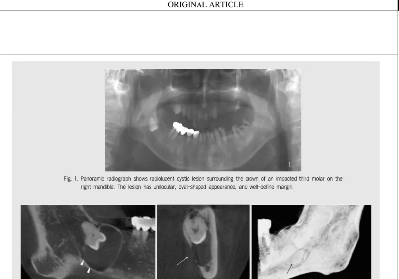
Panoramic radiograph showed a radiolucent shadow surrounding the crown of an impacted third molar on the right posterior mandible(Fig. 1).
CBCT images showed a well-defined margin of lesion with thinning of the lingual cortex adjacent to impacted third molar crown and partial discontinuity suspected as perforation on the lingual cortex(Fig. 2). Dentigerous cyst was the presumptive diagnosis based on the clinical and radiographic examinations.
Enucleation of the lesion was performed with extraction of the impacted tooth and bone graft under general anesthesia. Excisional biopsy was conducted intra-operatively and submitted for histopathologic analysis. Histopathologic findings showed that the lesion had a variable
port
thickness of the cyst lining with glandular differentiation, resulting to mucoid-filled secretory cells and microcysts(Fig. 3). The final diagnosis was confirmed as GOC.
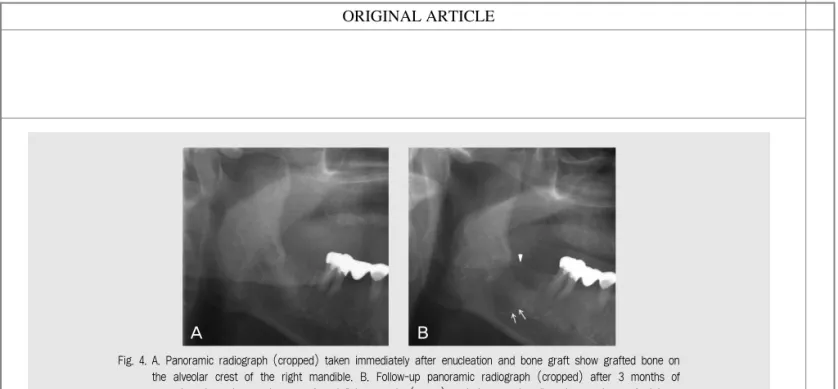
At the 3 month follow-up after surgery, decreased radiopacity of grafted bone and persistence of definite margin were found on panoramic radiography(Fig. 4). The patient was
Fig. 1. Panoramic radiograph shows radiolucent cystic lesion surrounding the crown of an impacted third molar on the right mandible. The lesion has unilocular, oval-shaped appearance, and well-define margin.
Fig. 2. A, B, C Parapanoramic, cross-sectional, and 3D cone-beam computed tomographic images show thinning and partial discontinuity of lingual cortex suspected as perforation(arrow). Mandibular canal shows inferior deviation (arrow head) due to lesion.
A B C
Fig. 3. A, B Photomicrographs of biopsy specimen show multiple areas of cyst epithelium of glandular differentiation, resulting in mucoid-filled secretory cells (arrow) and microcysts (arrow head). (H&E stain, A. ×100, B. ×400)
A B
diagnosed with recurring GOC. Meticulous curettage was performed on the inferior area of the lesion with nerve retraction. The patient has been on periodic check-ups; and a 1 year follow- up radiograph after operation showed no evidence of recurrence.
Ⅲ. Discussion
Although the pathogenesis of GOC remains uncertain, several case reports and short series have been reported in the past 2 decades4~8). There is some agreement that GOC has 2 clinically important attributes i.e., display of aggressive growth potential and a high recurrence rate.
Correct diagnosis is of major clinical importance because of aggressive potential, a high incidence of cortical perforation, and a relatively high rate of recurrence, especially in cases treated with a conservative approach4~6, 8).
GOC does not display pathognomonic radiological features; moreover, recognition of
this cyst is practically impossible on physical and radiographic examination. Histopathological examination alone, allows for a confirmed diagnosis of GOC5). Radiographically, the lesion is round to oval shaped with a smooth or scalloped margin. It shows unilocular or multilocular radiolucent lesion, usually with well-defined borders4, 6, 8, 9). Expansion is observed in majority of cases, with thinning, erosion, or perforation of the cortical plates in 67% of cases5,
10). Furthermore, it may be associated with impacted teeth and resorption, and tooth displacement is common11). Sizes vary from less than 1 cm in diameter to larger dimensions4).
Treatment is controversial, varying from conservative methods to block excision. Initial biopsy, enucleation with peripheral ostectomy for unilocular cases, and marginal resection for multilocular lesion are recommended as a treatment of choice according to several studies.
However, some suggest conservative surgery followed by long-term follow-up3, 11, 12).
Kaplan et al classified the histological
Fig. 4. A. Panoramic radiograph (cropped) taken immediately after enucleation and bone graft show grafted bone on the alveolar crest of the right mandible. B. Follow-up panoramic radiograph (cropped) after 3 months of operation show the persistence of a definite margin (arrow) and decreased radiopacity on the grafted bone (arrow head).
A B
port characteristics of GOC into major and minor
categories13). Major criteria include squamous epithelial lining, variations in thickness of the lining with or without epithelial whorl, cuboidal eosinophilic cells, mucous cells with interepithelial mucous pools, and interepithelial glandular microcystic or duct-like structures12, 13). The specific microscopic features more helpful in differentiating GOC from dentigerous cyst were demonstrated by Fowler et al. They suggested that the presence of microcysts, clear cells, epithelial spheres, and variations in thickness of the lining are of much value in problematic cases in diagnosis14).
In this case, we reported a rare lesion of GOC occurring in an unusual site with similar radiological features of dentigerous cyst. The lesion was incorrectly diagnosed through CBCT examination, despite the distinct thinning, suspected perforation on adjacent cortical bone, and relatively small size. Because of its
proximity to the mandibular canal, the patient was treated conservatively with enucleation.
Histopathological findings included multiple areas of cyst epithelium with glandular differentiation, resulting in mucoid-filled secretory cells and microcyst. Based on these findings, the lesion was confirmed as GOC. After 3 months, the lesion recurred, and hence, we performed meticulous curettage with nerve retraction. Since then, the lesion has been checked periodically with no sign of recurrence for 12 months.
In conclusion, GOC is a rare and aggressive lesion with a high recurrence rate that requires careful clinical and radiological evaluation. It is crucial that radiograph modality provides cross- sectional information concerned with fine differences in extent of expansion, perforation, and thinning. Thorough histopathological examination coupled with radiographs is the effective diagnostic method for GOC.
1. Padayachee A, Van Wyk CW. Two cystic lesions with features of both the botryoid odontogenic cyst and the central mucoepidermoid tumour: sialo- odontogenic cyst J Oral Pathol 1987; 16 : 499?504.
2. Kramer IR, Pindborg JJ, Shear M. The WHO hisological typing of odontogenic tumours. A commentary on the second edition. Cancer 1992; 70 : 2988-94.
3. Lyrio MC, de Assis AF, Germano AR, de Moraes M.
Treatment of mandibular glandular odontogenic cyst with immediate reconstruction: case report and 5- year follow-up. Br J Oral Maxillofac Surg 2010; 48 : 651-3.
4. Chung GC, Han WJ, Kim EK. A huge glandular odontogenic cyst occurring at posterior mandible.
Korean J Oral Maxillofac Radiol 2004; 34 : 209-13.
5. Kaplan I, Anavi Y, Hirshberg A. Glandular odontogenic cyst: a challenge in diagnosis and treatment. Oral Dis 2008; 14 : 575-81.
6. Oliveira JX, Santos KC, Nunes FD, Hiraki KR, Sales MA, Cavalcanti MG, et al. Odontogenic glandular cyst: a case report. J Oral Sci 2009; 51 : 467-70.
7. Zhang L, Sun ZJ, Chen XM, Chen Z.
Immunohistochemical expression of SHH, PTC, SMO and GLI1 in glandular odontogenic cysts and dentigerous cysts. Oral Dis 2010; 16 : 818-22 8. Krishnamurthy A, Sherlin HJ, Ramalingam K,
Natesan A, Premkumar P, Ramani P, et al.
Glandular odontogenic cyst: report of two cases and review of literature. Head Neck Pathol 2009; 3 : 153-8.
9. Manor R, Anavi Y, Kaplan I, Calderon S.
Radiological features of glandular odontogenic cyst.
Dentomaxillofac Radiol 2003; 32 : 73-9.
10. Araújo de Morais HH, José de Holanda Vasconcellos R, de Santana Santos T, Guedes Queiroz LM, Dantas da Silveira ?J. Glandular odontogenic cyst: case report and review of diagnostic criteria. J Craniomaxillofac Surg 2012; 40 : 46-50.
11. Boffano P, Cassarino E, Zavattero E, Campisi P, Garzino-Demo P. Surgical treatment of glandular odontogenic cysts. J Craniofac Surg 2010; 21 : 776-80.
12. Kaplan I, Gal G, Anavi Y, Manor R, Calderon S.
Glandular odontogenic cyst: treatment and recurrence. J Oral Maxillofac Surg 2005; 63 : 435-41.
13. Kaplan I, Anavi Y, Manor R, Sulkes J, Calderon S.
The use of molecular markers as an aid in the diagnosis of glandular odontogenic cyst. Oral Oncol 2005; 41 : 895-902.
14. Fowler CB, Brannon RB, Kessler HP, Castle JT, Kahn MA. Glandular odontogenic cyst: analysis of 46 cases with special emphasis on microscopic criteria for diagnosis. Head Neck Pathol. 2011; 5 :364-75.
참 고 문 헌