ISSN 0378-6471 (Print)⋅ISSN 2092-9374 (Online)
https://doi.org/10.3341/jkos.2018.59.1.87
Case Report
안와에 발생한 연골종 1예
A Case of Orbital Chondroma
최재환1⋅이동철1⋅김정희1⋅배현식2⋅서연림2⋅우경인1⋅김윤덕1
Jae Hwan Choi, MD1, Dong-Cheol Lee, MD1, Jeong Hee Kim, MD1, Hyunsik Bae, MD2, Yeon Lim Suh, MD, PhD2, Kyung In Woo, MD, PhD1, Yoon-Duck Kim, MD, PhD1
성균관대학교 의과대학 삼성서울병원 안과학교실1, 성균관대학교 의과대학 삼성서울병원 병리학교실2 Department of Ophthalmology, Samsung Medical Center, Sungkyunkwan University School of Medicine1, Seoul, Korea
Department of Pathology, Samsung Medical Center, Sungkyunkwan University School of Medicine2, Seoul, Korea
Purpose: To report a case of orbital chondroma.
Case summary: A 15-year-old male presented with an 8-month history of left hypertropia. The best-corrected visual acuity was 20/20 in both eyes. The exophthalmometry showed no exophthalmos, with 13 mm in both eyes. There was a hard palpable mass at the superonasal orbit of the left eye. Orbital computed tomography showed a heterogenous soft tissue shadow at the super- onasal orbit of the left eye, and orbital magnetic resonance imaging revealed a 25 × 16 × 20 mm well-defined mass with low sig- nal intensity in the T1-weighted image, high signal intensity in the T2-weighted image, and heterogenous enhancement in the contrast enhanced T1-weighted image. The mass was surgically removed with anterior orbitotomy. A 27 × 17 mm well-capsu- lated lobular mass was found, and histopathological examination revealed hyaline cartilage and chondrocyte. The mass was di- agnosed as a chondroma.
Conclusions: A chondroma is a benign tumor, which usually occurs in long bones and the small bones of the hands and feet. It is very rare in the facial and pelvic bones. The sites of chondroma occurring in the head and neck include the ethmoid sinus and maxilla, but it is extremely rare in the orbit.
J Korean Ophthalmol Soc 2018;59(1):87-92 Keywords: Chondroma, Orbital tumor
■Received: 2017. 10. 19. ■ Revised: 2017. 10. 29.
■Accepted: 2017. 12. 20.
■Address reprint requests to Yoon-Duck Kim, MD, PhD Department of Ophthalmology, Samsung Medical Center, #81 lrwon-ro, Gangnam-gu, Seoul 06351, Korea
Tel: 82-2-3410-3548, Fax: 82-2-3410-0074 E-mail: [email protected]
*Conflicts of Interest: The authors have no conflicts to disclose.
ⓒ2018 The Korean Ophthalmological Society
This is an Open Access article distributed under the terms of the Creative Commons Attribution Non-Commercial License (http://creativecommons.org/licenses/by-nc/3.0/) which permits unrestricted non-commercial use, distribution, and reproduction in any medium, provided the original work is properly cited.
연골종은 손가락, 발가락 소골 및 장관골에서 주로 발 생하며, 드물게 두개골 및 골반골에 발생하는 양성종양으 로, 종괴의 성장속도가 빠르거나, 통증, 관절의 기능적 장 애, 그리고 주변 신경과 혈관을 압박하는 경우 수술적인 제거를 고려하는 질환이다.
두개골 내에서는 주로 사골동, 상악골 및 비중격에서 발생하나, 안와 내에서 발생한 경우는 매우 드물게 보고되 어 있는 질환으로, 저자들은 좌안의 안구운동장애 및 복시 환자에서 좌안 상비측에 주변조직과 경계가 명확한 조직 을 발견하여 절제생검술을 시행하였고, 조직검사상 안와 연골종으로 진단하였다. 해외에서는 안와의 활차오목(tro- chlear fossa)에서 발생한 몇 증례만이 보고되어 있고, 국 내에서는 보고된 적이 없는 드문 질환으로 이를 경험하였 기에 보고하고자 한다.
증례보고
15세 남자 환자가 8개월 전부터 시작된 좌상사시를 주
A B
C
Figure 1. Preoperative photographies and the Hess screen test of the patient. (A) Clinical photograph demonstrates inferior dystopia
of the left eye. (B) In primary position, the patient has left hypertropia of 25 prism diopters, and exotropia of 4 prism diopters. Note secondary overaction of the left inferior oblique muscle and mild limitation of depression when looking down. (C) The Hess screen test also shows secondary overaction of the left inferior oblique muscle with a limitation of inferior gaze of the left eye.소로 내원하였다. 안과적 과거력은 없으며, 본원 초진 시 최대교정시력 양안 모두 1.0이었으며, 골드만 압평안압계 로 측정한 안압은 우안 13 mmHg, 좌안 14 mmHg였다.
안구돌출계 검사에서 우안 13 mm, 좌안 13 mm로 안구돌 출은 관찰되지 않았으며, 촉진 시 좌안 상비측에 딱딱하게 만져지는 종괴가 촉진되었다. 좌안은 하방편위(inferior dystopia) 되어 있었으며(Fig. 1A), 안구운동검사 및 헤스 스크린 검사(Hess screen test)에서 좌안의 하전 장애, 상사 근 기능저하 및 상대적 하사근 기능항진 소견이 관찰되었 다(Fig. 1B, C). 프리즘 교대 가림 검사에서 25프리즘디옵 터의 좌상사시 및 4프리즘디옵터의 외사시가 관찰되었으 며, 골드만 시야검사에서 중심에서 40° 이상 주시시 복시 가 관찰되었다. 동공반사 및 상대구심성동공운동은 양안 모두 정상이었다.
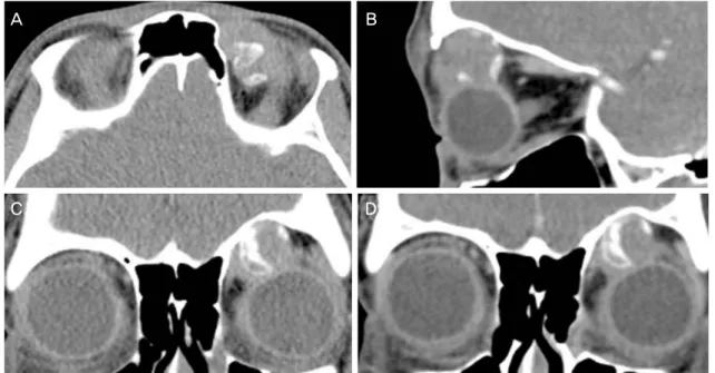
안와 전산화단층촬영을 시행하였으며, 좌안 상비측에 비균질한 연부조직 및 골음영의 종괴가 관찰되었다(Fig.
2). 종괴에 의한 안와 상벽의 변형이 관찰되었으며, 조영
증강은 관찰되지 않았다. 안와 자기공명영상에서 좌안 상 비측으로 25 × 16 × 20 mm 크기의 주변조직과 경계가 뚜 렷한 종괴가 관찰되었다. 종괴는 T1 강조영상에서 전체적 으로 저신호강도 음영을 보였으며, 중심 일부에서 고신호 강도를 보였다(Fig. 3A, B). T2 강조영상에서는 종괴는 전 반적으로 고신호강도를 보였으며 T1 강조영상에서 고신 호강도를 보이던 부분은 저신호강도를 보였다(Fig. 3C, D). 조영증강 T1 강조영상에서 종괴 내부의 비균질한 조 영증강이 나타났다(Fig. 3E, F).
좌안 안와종괴에 대해 절제생검술을 시행하였다(Fig.
4A). 종괴는 상사근을 둘러싸는 형태로 존재하였으며, 종 괴를 주변조직과 박리하여 완전히 제거하였다. 종괴는 27
× 17 mm 크기였으며, 표면은 회백색 내지 엷은 분홍색을 띠었고, 매끄럽고 광택이 있었다(Fig. 4B).
종괴의 병리 조직검사를 시행하였으며, 저배율에서 종 괴는 섬유피막에 둘러싸인 분엽성 조직이었고 골조직 및 골수 조직을 일부 포함하고 있었으나, 대부분에서 풍부
A B
C D
Figure 2. The patient's computed tomography images. Orbital computed tomography images taken preoperatively demonstrate an
orbital mass at the superonasal orbit of the left eye which has a bony component (A, B). Superior orbital wall remodeling is found (B, C), and the eyeball is placed inferolaterally (C). The mass shows no enhancement in the contrast-enhanced image (D).A B
C D
E F
Figure 3. The patient's orbit magnetic resonance images. They show 25 × 16 × 20 mm-sized mass at the superomedial portion of
the left orbit. Mass showing low signal intensity with small high signal intensity portion in the T1-weighted images (A, B), and high signal intensity with small low signal intensity portion in the T2-weighted images (C, D). Gadolinium-enhanced T1-weighted axial (E) and coronal (F) images demonstrate heterogeneous enhancement.A B
C D
Figure 4. Intraoperative findings and Histopathological images. (A) Through a superomedial sub-brow incision, orbital mass exci-
sional biopsy was done. The mass is located at the superomedial orbit, and surrounds superior oblique muscle. (B) Gross image of mass shows 27 × 17 mm-sized mass with pearly white and pinkish surface. (C) Histopathological image shows well-differentiated chondrocytes which is clustered as lobulated manner. The mass is hypocellular with cartilaginous matrix and shows no atypia. The mass contains small portion of bone marrow tissue and calcification (arrow) is seen at between the chondroid tissue and the bone marrow tissue (hematoxylin-eosin, ×40). (D) The chondrocytes are placed in the lacunar spaces with ovoid nuclei without nuclear pleomorphism or mitosis (hematoxylin-eosin, ×200)Table 1. Clinical features, treatment, and outcomes of orbital chondromas
First author (year) Age(year) Sex (M/F)
Symptom onset (months)
Clinical features Location Therapy Recurrence
Jepson CN (1966)4 50 M 30 Palpable mass Superomedial Orbit Local excision No
Albert DM (1982)3 14 M 12 Proptosis, Inferior displacement of eyeball
Superomedial Orbit Local excision No
Faber W (1992)9 19 M 12 Proptosis, Inferotemporal
displacement of eyeball
Superomedial Orbit Local excision No
Harrison A (2006)10 9 M 6 Ptosis Superomedial Orbit Local excision No
Kabra RS (2015)5 27 M 3 Palpable mass, Ptosis Superomedial Orbit Local excision No
M = male; F = female.
한 기질(matrix) 내에 고르게 분포한 소강(lacuna)이 관찰 되며, 소강 내에 타원형의 핵(nuclei)을 가진 연골세포 (chondrocyte)가 관찰되었다(Fig. 4C). 고배율에서 연골세 포의 핵은 다형성(plemorphism)을 보이지 않고 일정한 타 원형으로 존재하였고, 세포분열(mitosis)은 일어나지 않는
양성 연골성 종괴 소견으로(Fig. 4D), 연골종(chondroma) 으로 진단되었다.
수술 후 외래 추적관찰을 시행하였으며, 수술 후 7일째 시행한 사시각 검사에서 정면 주시 시 15프리즘디옵터의 좌상사시가 관찰되었으나, 수술 4주 후부터는 정면 및 상
하좌우 주시 시 정위였으며, 안구운동검사 및 복시검사 모 두 정상이었다. 수술 6개월 뒤 안와 전산화단층촬영을 시 행하였으며, 종양의 재발은 관찰되지 않았다.
고 찰
연골종은 손과 발의 소골 및 장관골에서 주로 발생하는 양성종양으로, 특히 손의 소골에서 발생하는 양성종양 중 가장 많은 비율을 차지하며, 전 연령대에 걸쳐서 나타나나 주로 20대에서 40대 사이에 호발하는 것으로 알려져 있 다.1 안와에 발생한 양성 연골성 종양(benign cartilaginous tumor)은 매우 드문 질환으로, 연골종(chondroma), 골연골 종(osteochondroma), 섬유연골종(fibrchondroma) 및 연골 성 과오종(cartilaginous hamartoma)이 보고되어 있다.2-7 Selva et al6 및 Shields et al7은 각 3,340명 및 645명의 안와 종양 환자에서 연골종 한 증례를 보고하였고, Bonavolontà et al2은 2,480명의 안와종양 환자에서 연골육종(chon- drosarcoma) 2증례를 보고하였으나, 연골종은 발견되지 않았다고 보고하였으며, 국내에서는 안와에 발생한 골연 골종(osteochondroma) 1예가 보고되어 있으나8, 연골종은 아직까지 보고되지 않았다.
본 증례에서 연골종은 안와 상내측에 존재하였다. 종괴 로 인한 안와 상벽의 변형이 관찰되었으나, 연골종은 섬유 피막에 둘러싸여 있었으며, 주변 안와골 및 상사근을 침범 하지 않고 경계가 명확하게 구분되어 있어, 안와의 활차오 목의 연골조직이나 주변 연부조직에서 기원한 것으로 보인 다. 조직학적으로 안와의 활차오목(trochlear fossa)에 연골 조직이 존재하기 때문에, 안와의 연골종은 이곳에서 기원 하는 것으로 보이며, 안와 연골종을 보고한 다른 논문에서 도 활차오목이 존재하는 안와 상내측에서 연골종이 발생하 였다(Table 1).3-5,9,10 안와의 연골종은 연골 조직 외에도 골 조직, 점액종성 조직(myxomatous tissue) 등 다른 조직과 동반되어 나타나는 경우가 흔한 것으로 보고되어 있으며10, 본 증례에서도 골조직 및 골수 조직이 함께 존재하였다.
연골종은 골피질(cortex)을 침범하지 않기 때문에, 장관 골에 단독으로 발생한 경우에는 병적골절(pathologic frac- ture)을 거의 일으키지 않으므로 외과적 제거가 필요하지 는 않으나, 악성변화가 의심되는 경우에는 종양의 완전절 제가 필요하다. 소골에 발생한 경우에는 병적골절을 일으 킬 가능성이 있기 때문에 외과적인 제거가 필요하다. 안와 에 발생한 경우에는 보고된 증례가 적어 표준화된 치료가 확립되어 있지는 않으나, 보고된 증례에서 환자들은 종양 에 의한 안구돌출 및 위치 이상, 안구운동장애가 동반되어 있었으며,4,5,11 본 증례 또한 좌안의 안구운동제한 및 하방
편위를 동반하고 있어, 종양에 의한 안과적 증상을 동반할 경우 수술적 제거가 필요할 것으로 보인다.
연골종은 악성변화가 매우 드문 질환이나,12 종괴 일부 에 저등급의 연골육종(low grade chondrosarcoma)이 동반 된 경우 정상 연골세포와 영상학적 및 조직학적으로 구분 하기 힘든 경우가 많아 양성 연골종으로 오진되는 경우가 많고,13 두경부에 발생한 연골육종의 경우 공격적인 성향을 보이나, 종양을 완전히 절제한 경우 상대적으로 좋은 장기 예후를 보이기 때문에,14,15 안와 내에서 발생한 연골종 또 한 종양 전체를 완전하게 제거해야 할 것으로 생각된다.
연골종은 재발률이 매우 낮은 양성종양으로,16 안와에 발생한 연골종의 경우 수술적 제거 후 종양이 재발한 보 고는 현재까지 없으며, 본 증례 또한 수술적 제거 6개월 뒤 시행한 안과 검사 및 안와 전산화단층촬영에서 종양의 재발 소견은 관찰되지 않은 상태로 정기적으로 추적관찰 중으로, 국내에서는 보고된 바가 없어 본 증례를 보고하는 바이다.
REFERENCES
1) Unni KK, Inwards CY. Dahlin’s Bone Tumors: General Aspects and Data on 10,165 Cases, 6th ed. Philadelphia: Lippincott Williams & Wilkins, 2009; 22-39.
2) Bonavolontà G, Strianese D, Grassi P, et al. An analysis of 2,480 space-occupying lesions of the orbit from 1976 to 2011. Ophthal Plast Reconstr Surg 2013;29:79-86.
3) Albert DM, Ni C, Sebag J, Renna T. Rare orbital tumors. Int Ophthalmol Clin 1982;22:183-205.
4) Jepson CN, Wetzig PC. Pure chondroma of the trochlea. A case report. Surv Ophthalmol 1966;11:656-9.
5) Kabra RS, Patel SB, Shanbhag SS. Orbital Chondroma: A rare mesenchymal tumor of orbit. Indian J Ophthalmol 2015;63:551-4.
6) Selva D, White VA, O'Connell JX, Rootman J. Primary bone tu- mors of the orbit. Surv Ophthalmol 2004;49:328-42.
7) Shields JA, Bakewell B, Augsburger JJ, Flanagan JC.
Classification and incidence of space-occupying lesions of the orbit. A survey of 645 biopsies. Arch Ophthalmol 1984;102:
1606-11.
8) Han SW, Lee JK, Kim KS. A case of orbital osteochondroma. J Korean Ophthalmol Soc 1985;26:551-6.
9) Faber W, Kock E, Landau I, Tengroth B. Para-trochlear chondroma of the orbit. Klin Monbl Augenheilkd 1992;200:138-9.
10) Harrison A, Loftus S, Pambuccian S. Orbital chondroma. Ophthal Plast Reconstr Surg 2006;22:484-5.
11) Pasternak S, O'Connell JX, Verchere C, Rootman J. Enchondroma of the Orbit. Am J Ophthalmol 1996;122:444-5.
12) Sridhar H, Vijaya M, Clement W, Srinivas C. Chondrosarcoma arising in an enchondroma of the metacarpal bone - a case report. J Clin Diagn Res 2014;8:142-3.
13) Randall RL, Gowski W. Grade 1 chondrosarcoma of bone: a diag- nostic and treatment dilemma. J Natl Compr Canc Netw 2005;3:
= 국문초록 =
안와에 발생한 연골종 1예
목적: 안와에 발생한 연골종(chondroma) 1예를 경험하였기에 이를 보고하고자 한다.
증례요약: 15세 남자 환자가 8개월 전부터 발생한 좌상사시를 주소로 내원하였다. 교정시력은 양안 모두 1.0이었으며, 안구돌출계 검 사에서 우안 13 mm, 좌안 13 mm로 좌안의 돌출은 관찰되지 않았으나, 촉진 시 좌안 상비측으로 딱딱한 종괴가 촉지되었다. 안와 전산화단층촬영에서 안와 상비측에 비균질한 연부조직음영이 관찰되었으며, 안와 자기공명영상에서 경계가 명확하면서 T1 강조영상 에서 저신호강도, T2 강조영상에서 고신호강도를 보이며, 조영증강 T1 강조영상에서 불균질한 조영증강을 보이는 25 × 16 × 20 mm 크기의 종괴가 관찰되었다. 안와종괴 절제생검술을 시행하였으며 조직검사에서 27 × 17 mm 크기의 피막화가 잘된 분엽성 종괴 내에 유리 연골 및 연골세포가 관찰되었으며 연골종으로 진단되었다.
결론: 연골종은 주로 손가락, 발가락 소골 및 장관골에서 주로 발생하며, 드물게 두개골 및 골반골에 발생하는 양성종양이다. 두개골 내에서는 사골동 및 상악골에서 주로 발생하며, 안와에 발생하는 경우는 매우 드물며, 해외에서 몇 증례가 보고되어 있으나 국내에서 는 보고된 사례가 없었다.
<대한안과학회지 2018;59(1):87-92>
149-56.
14) Hong P, Taylor SM, Trites JR, et al. Chondrosarcoma of the head and neck: report of 11 cases and literature review. J Otolaryngol Head Neck Surg 2009;38:279-85.
15) Pellitteri PK, Ferlito A, Fagan JJ, et al. Mesenchymal chon-
drosarcoma of the head and neck. Oral Oncology 2007;43:970-5.
16) Bauer HC, Brosjö O, Kreicbergs A, Lindholm J. Low risk of re- currence of enchondroma and low-grade chondrosarcoma in extremities. 80 patients followed for 2-25 years. Acta Orthop Scand 1995;66:283-8.