ISSN 0378-6471 (Print)⋅ISSN 2092-9374 (Online)
https://doi.org/10.3341/jkos.2018.59.9.819
Original Article
유리체절제술과 백내장 병합수술에서 공기충전술을 시행한 경우 후낭절제술이 굴절력에 미치는 영향
Postoperative Refractive Errors after Air Tamponade with Posterior Capsulectomy during Combined Vitrectomy and Cataract Surgery
채수혜1⋅김상원2⋅윤희성1
Sue Hey Chae, MD1, Sang Won Kim, MD2, Hee Seong Yoon, MD1
성모안과병원1, 부평성모안과2 Sungmo Eye Hospital1, Busan, Korea Bupyung St. Mary’s Eye Clinic2, Incheon, Korea
Purpose: To evaluate the postoperative refractive errors after air tamponade with posterior capsulectomy during combined vi- trectomy and cataract surgery.
Methods: Patients who underwent combined vitrectomy, cataract surgery, and air tamponade with or without posterior capsu- lectomy were reviewed. All patients were followed for 4 months after surgery. Preoperative characteristics such as anterior chamber depth, axial length, and refractive error were analyzed and refractive errors after the surgery were evaluated. The dif- ference between the target refractions and final refractive errors after the surgery according to the biometry method, and intra- ocular lens power calculations, were observed.
Results: Fourteen eyes of 14 patients who had combined vitrectomy and cataract surgery with posterior capsulectomy and air tamponade were classified as group A, and 10 eyes of 10 patients who had combined vitrectomy and cataract surgery with only air tamponade were classified as group B. The target refraction of group A measured with A-scan biometry using the Sanders-Retzlaff-Kraff/Theoretical (SRK/T) calculation was -0.21 ± 0.22 diopters (D), and the final refractive error at 9.5 (± 2.20) months after the surgery was -0.52 ± 0.54 D. The mean difference between the two was -0.32 ± 0.44 D. The target refraction of group B measured with A-scan biometry using the SRK/T calculation was -0.33 ± 0.29 D, and the final refractive error at 9.5 (±
2.20) months after the surgery was -0.27± 0.39 D. The mean difference between the two was 0.06 ± 0.53 D.
Conclusions: Posterior capsulectomy during combined vitrectomy and cataract surgery with air tamponade led to myopic shifts compared with no posterior capsulectomy with air tamponade during combined vitrectomy and cataract surgery. Performing poste- rior capsulectomy with air tamponade during combined vitrectomy and cataract surgery should, therefore, be carefully considered.
J Korean Ophthalmol Soc 2018;59(9):819-826
Keywords: Air tamponade, Cataract surgery, OA-2000 optical biometry, Posterior capsulectomy, Vitrectomy
■Received: 2018. 4. 12. ■ Revised: 2018. 6. 5.
■Accepted: 2018. 8. 28.
■Address reprint requests to Hee Seong Yoon, MD
Sungmo Eye Hospital, #409 Haeun-daero, Haeundae-gu, Busan 48064, Korea
Tel: 82-51-743-0775, Fax: 82-51-743-0776 E-mail: [email protected]
*Conflicts of Interest: The authors have no conflicts to disclose.
ⓒ2018 The Korean Ophthalmological Society
This is an Open Access article distributed under the terms of the Creative Commons Attribution Non-Commercial License (http://creativecommons.org/licenses/by-nc/3.0/) which permits unrestricted non-commercial use, distribution, and reproduction in any medium, provided the original work is properly cited.
유리체절제술은 망막질환을 치료하는 중요한 술기인데 유리체절제술 그 자체만으로도 백내장을 발생시키거나 그 진행을 빠르게 만들 수 있다.1-3 유리체절제술 후 백내장수 술 시 수정체 소대 손상이나 후낭 파열 등 합병증 또한 증 가하여 백내장수술 자체의 난이도가 높아지는 것으로 알려 져 있다.4 이러한 이유들로 유리체망막질환에서 유리체절 제술과 백내장을 동시에 수술하는 경우가 많이 있다.
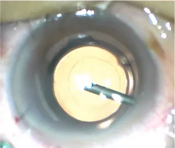
Figure 1. Clinical intraoperative photograph of the anterior
segment showing the center of the posterior capsule removed using vitreous cutter. The posterior capsule was removed cur- vilinearly and completely.후낭혼탁은 백내장수술 후 발생하는 가장 대표적인 합병 증으로 백내장 단독수술과 백내장수술과 유리체절제술을 병 행하여 수술한 경우를 비교하여 살펴보면 유리체절제술을 함께 시행한 경우 후낭혼탁의 발생률이 더 높은 것으로 보고 되었다.5 Neodymium:yttrium-aluminum-garne (Nd:YAG) 레 이저후낭절개술(posterior capsulotomy)은 가장 대표적인 치료법으로 많이 시행되고 있는데, 이러한 레이저후낭절개 술은 안압 상승, 인공수정체 위치이탈, 황반부종, 망막박리 등의 부작용을 발생시킬 수 있다.6 이러한 합병증을 예방하 고 술 후 발생하는 후낭혼탁에 의한 시력 저하를 막기 위하 여 유리체절제술과 백내장 병합수술 중에 유리체절제기를 이용하여 후낭절제술(posterior capsulectomy)을 시행하기 도 한다. 이러한 유리체절제술과 백내장병합수술 중에 시 행하는 예방적 후낭절제술이, 수술 후 시행하는 Nd:YAG 레이저후낭절개술과 비교하여도 굴절력 변화 등의 합병증 없이 좋은 치료 효과를 나타냄이 보고된 바 있다.7
눈 속 충전물 주입술은 황반원공, 망막박리 등 유리체망 막수술 후 망막 유착이 필요하거나 다량의 망막하액 제거 후 발생할 수 있는 저안압 상태를 해결하는 목적으로도 사 용되는데, 유리체절제술과 백내장 병합수술과 후낭절제술 을 함께 시행한 후 눈 속 충전물 주입술을 시행하는 경우, 눈 속 충전물을 사용하지 않은 군에 비해 근시의 이행을 보 인다는 보고가 있었다.8 하지만 충전물로 인하여 생성되는 후방으로부터의 압력이 후방절제술을 시행한 경우와 시행 하지 않은 경우 인공수정체의 위치이동에 의한 전방 깊이 변화로 실제 술 후 굴절력에 얼마나 영향을 미치는지에 대 하여 비교 연구한 보고는 없었다. 본 연구에서는 유리체절 제기를 이용한 후낭절제술 후 시행한 공기주입술이 술 전 의 안축장을 이용한 인공수정체 도수계산에 따른 예측 굴 절력과 술 후 실제 굴절력 간의 차이에 미치는 영향에 대하 여 알아보았다.
대상과 방법
2015년 12월부터 2017년 4월까지 망막질환과 백내장을 진단받고 유리체절제술과 백내장수술을 동시에 시행받은 환자들을 대상으로 하였으며 술 중 후낭절제술과 공기충전 술을 모두 시행한 14안을 A군, 후낭절제술 없이 공기충전 술만을 시행한 10안을 B군으로 분류하고 후향적으로 의무 기록을 분석하였다. 모든 진단과 수술은 동일한 의사에 의 해서 시행되었다. 술 후 최소 6개월 이상 경과관찰이 되었 던 환자들을 포함시켰으며 이전 각막질환 또는 각막 이식 등의 과거력으로 정확한 인공수정체 도수계산이 어려웠던 환자들, 술 중 필요에 따라 공기충전술 외에 SF6, C3F8 등
의 가스 충전술이나 오일 주입술을 시행한 경우, 섬모체 소 대 약화 등의 이유로 섬모체고랑에 인공수정체를 위치시킨 경우, 술 후 최종 경과관찰 기간 이전에 인공수정체의 기계 적인 합병증으로 재수술이 필요하였던 경우는 모두 제외하 였다. 후낭절제술은 모두 동일하게 4 mm 크기로 시행하였 다(Fig. 1). A군은 남자 4명 4안, 여자 10명 10안, B군은 남 자 3명 3안, 여자 7명 7안이 포함되었다. A군은 평균 연령 이 68세, B군은 65세였다. A군에는 황반원공 5안, 표층황 반원공 4안, 망막전막 환자에서 막 제거 중 심한 견인이 발 생한 경우 4안, 상부의 망막열공으로 인한 열공망막박리 1 안이 포함되었다. B군은 황반원공 3안, 표층황반원공 4안, 망막전막 환자에서 막 제거 중 심한 견인이 발생한 경우 3 안이 포함되었다. 수술에는 동일한 인공수정체 enVista™
(Bausch & Lomb, Inc., Rochester, NY, USA)를 사용한 경우 만을 포함시켰고, 눈생체계측은 초음파 장비인 Axis Nano™
(Quantel Medical™, Clermont-Ferrand, France) (A-scan)과 OA-2000® (Tomey, Nagoya, Japan)을 이용하여 측정하였 고, 기대 술 후 굴절도는 A-scan에 내장되어 있는 Sanders -Retzlaff-Kraff/Theoretical (SRK/T) 공식과 OA-2000®에 내 장되어 있는 SRK/T 공식과 Haigis 공식을 이용하여 계산하 고 비교해 보았다. 굴절검사는 Autorefractor (KR-8900®, Topcon Corp, Tokyo, Japan)를 사용하였고, 수술 후 1, 2, 4, 6개월째, 그리고 마지막 경과관찰 시점인 평균 9.5 (±2.20)개월째 실제 굴절력과 예측 굴절력의 차이를 계산하 였다. 굴절력은 구면렌즈대응치(spherical equivalent, SE)로 계산하였다.
Characteristics Group A (n = 14) Group B (n = 10) p-value*
Number of eyes 14 eyes 10 eyes -
Gender (male:female) 4:10 3:7 -
Age (years) 67.6 ± 5.8 64.9 ± 5.8 0.378
Duration of follow-up (months) 6.37 ± 1.98 8.27 ± 4.26 0.349
Preoperative UCVA (logMAR) 0.56 ± 0.19 0.54 ± 0.26 0.662
Preoperative BCVA (logMAR) 0.47 ± 0.21 0,43 ± 0.26 0.389
Central macular thickness (μm) 379.7 ± 79.8 414.5 ± 65.1 0.235
Preoperative sphere (diopter) 0.35 ± 1.02 1.34 ± 1.54 0.051
Preoperative cylinder (diopter) -1.07 ± 0.71 -1.43 ± 0.47 0.112
Preoperative spherical equivalent (diopter) -0.17 ± 1.13 0.62 ± 1.68 0.161
K1 (keratometry, diopter) 44.3 ± 1.4 44.2 ± 1.8 0.953
K2 (keratometry, diopter) 44.4 ± 1.5 43.9 ± 1.9 0.428
Corneal astigmatism (diopter) -0.86 ± 0.5 -0.81 ± 0.41 0.929
Anterior chamber depth (mm) 3.054 ± 0.28 3.18 ± 0.42 0.364
Axial length (mm) 23.53 ± 0.65 23.08 ± 0.91 0.169
Values are presented as mean ± SD unless otherwise indicated.
UCVA = uncorrected visual acuity; BCVA = best corrected visual acuity; K = keratometry.
*Statistical analysis performed using Mann-Whitney test for categorical variables.
Table 1. Baseline characteristics of posterior capsulectomy with air tamponade (group A) and only air tamponade (group B)
통계학적 분석은 SPSS ver. 20.0 (IBM Corp., Armonk,NY, USA)을 사용하였고 시력의 경우 logarithm of minimal angle of resolution (logMAR) 값으로 변환하여 분석하였으 며, 두 군 간의 나이, 수술 전 굴절력, 수술 전 망막두께 등 은 Mann-Whitney test를 이용하여 분석하였다. 각각의 생 체계측법과 계산 공식에 따른 측정값의 두 그룹 간의 비교 에서는 Mann-Whitney test를 이용하고 동일 그룹에서의 생 체계측법에 따른 굴절 오차값의 비교, 계산 공식에 따른 굴 절 오차값의 비교에서는 Wilcoxon signed ranks test를 이용 하였다. p-value가 0.05 미만인 경우를 통계적으로 유의한 것으로 정의하였다. 본 연구는 헬싱키선언에 입각한 성모안 과병원의 연구 윤리 심의위원회(institutional review board, IRB)의 승인하에 진행되었다(승인번호: 2018-BM-01).
결 과
유리체절제술과 백내장 병합수술을 시행하고 후낭절제 술과 공기충전술을 함께 시행받은 A군의 평균 연령은 67.6 세, 평균 경과관찰 기간은 6.37개월이었으며 술 전 최대교 정시력은 0.47 ± 0.21 logMAR, 중심망막두께는 379.7 ± 79.8 μm였다. 유리체절제술과 백내장 병합수술을 시행하고 후낭절제술 없이 공기충전술만을 시행받은 B군의 평균 연 령은 64.9세, 평균 경과관찰 기간은 8.27개월이었으며 술 전 최대교정시력은 0.43 ± 0.26 logMAR, 중심망막두께 (central macular thickness, CMT)는 414.5 ± 65.1 μm였다.
모든 값에서 두 군 간의 유의한 차이는 없었다(p=0.378, p=0.349, p=0.389, p=0.235). 술 전 구면렌즈대응치는 A군
-0.17 ± 1.13디옵터(D), B군 0.62 ± 1.68 D로 측정되었으며 두 군 간의 유의한 차이는 없었다(p=0.161). 술 전 평균 앞 방깊이(anterior chamber depth)는 A군 3.05 ± 0.28 mm, B 군 3.18 ± 0.42 mm, 안축장은 A군 23.53 ± 0.65 mm, B군 23.08 ± 0.91 mm로 모두 두 군 간의 유의한 차이는 없었다 (p=0.364, p=0.169) (Table 1). A-scan에 내장된 SRK/T 공 식을 이용한 경우, 술 전 예측 굴절력은 A군 -0.21 ± 0.22 D, B군 -0.33 ±0.29 D, OA-2000® (Tomey)에 내장된 SRK/T를 이용한 경우, 술 전 예측 굴절력은 A군 -0.12 ± 0.28 D, B군 -0.23 ± 0.51 D, OA-2000® (Tomey)에 내장된 Haigis를 이용한 경우, 술 전 예측 굴절력은 A군 -0.49 ± 0.32 D, B군 -0.35 ± 0.29 D로 각각의 경우에 두 군 간에 유의한 차이는 없었다(p=0.151, p=0.992, p=0.357). 이렇게 예측된 굴절값에 의하여 결정된 인공수정체는 A군에서 22.2 ± 1.91 D, B군에서 22.5 ± 1.79 D였으며 두 군 간에 유의한 차이는 없었다(p=0.563) (Table 2, Fig. 2). 술 후 1, 2, 4, 6개월째 그리고 마지막 경과관찰 시점인 평균 9.5 (±2.20)개월째 확인된 최종 굴절력은 A군에서 1개월째 -0.59 ± 0.54 D, 2개월째 -0.41 ± 0.41 D, 4개월째 -0.44 ± 0.51 D, 6개월째 -0.51 ± 0.67 D, 최종 -0.52 ± 0.54 D로 나 타났으며, B군에서는 1개월째 -0.28 ± 0.67 D, 2개월째 -0.37 ± 0.52 D, 4개월째 -0.41 ± 0.39 D, 6개월째 -0.31 ± 0.43 D, 최종 -0.27 ± 0.39 D로 측정되었다(Table 3). 두 군 간의 술 전, 술 후 CMT를 비교해 본 결과 술 전, 술 후 모 두 A군과 B군 간의 CMT는 통계학적으로 유의한 차이를 보이지 않았으며(p=0.235, p=0.084), 각각의 군에서 수술 전과 수술 후의 CMT를 비교해 본 결과 두 군 모두에서 수
A B
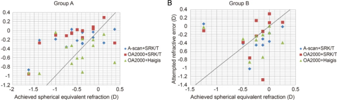
Figure 2. Comparison of spherical equivalent correction of two different groups. The results of the group A (capsulectomy + air
tamponade) (A) and group B (only air tamponade) (B) at last visit (mean postoperative 9.5 ± 2.20) months compared with predictive values from three different biometry devices and formulas combinations. SRK/T = Sanders-Retzlaff-Kraff/Theoretical.Group A (n = 14) Group B (n = 10) p-value*
Preoperative target diopter using A-scan (IOL calculation formula: SRK/T)
-0.21 ± 0.22 -0.33 ± 0.29 0.151
Preoperative target diopter using OA-2000 (IOL calculation formula: SRK/T)
-0.12 ± 0.28 -0.23 ± 0.51 0.992
Preoperative target diopter using OA-2000 (IOL calculation formula: Haigis)
-0.49 ± 0.32 -0.35 ± 0.29 0.357
IOL power (diopter) 22.2 ± 1.91 22.5 ± 1.79 0.563
Values are presented as mean ± SD unless otherwise indicated.
SRK/T = Sanders-Retzlaff-Kraff/Theoretical.
*Statistical analysis performed using Mann-Whitney test for categorical variables.
Table 2. Predicted postoperative refraction values measuring with different biometry machines and formula, and actual inserted intra-
ocular lens (IOL) power in posterior capsulectomy with air tamponade (group A) and only air tamponade (group B)Group A (n = 14) Group B (n = 10) p-value*
Postoperative SE (diopter)
1 month -0.59 ± 0.54 -0.28 ± 0.67 0.086
2 months -0.41 ± 0.41 -0.37 ± 0.52 0.837
4 months -0.44 ± 0.51 -0.41 ± 0.39 0.993
6 months -0.51 ± 0.67 -0.31 ± 0.43 0.506
Final -0.52 ± 0.54 -0.27 ± 0.39 0.141
Values are presented as mean ± SD unless otherwise indicated.
SE = spherical equivalent.
*Statistical analysis performed using Mann-Whitney test for categorical variables.
Table 3. Actual postoperative refraction values measuring at 1,2,4,6 months in phacovitrectomy and posterior capsulectomy with air
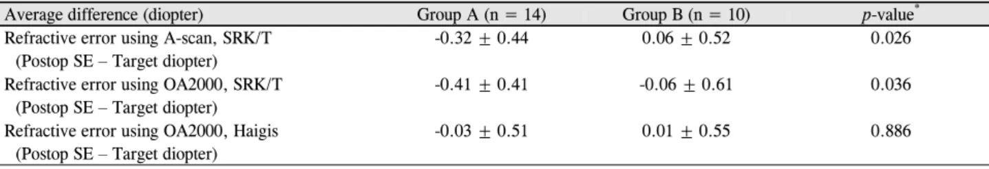
tamponade (group A) and phacovitrectomy with only air tamponade (group B)술 전과 수술 후 유의한 차이를 보였다(p<0.05, in both) (Table 4). 최종 관찰시점인 평균 9.5 (± 2.20)개월째 측정된 실제 구면렌즈대응값과 술 전 예측 굴절력과의 차이를 계 산해 보았다. A-scan에 내장된 SRK/T 공식을 이용한 경우, A군은 -0.32 ± 0.44 D로 B군에서 보인 +0.06 ± 0.53 D만큼의 차이에 비해 유의한 근시 이행을 보였다(p=0.026). OA-2000® (Tomey)에 내장된 SRK/T를 이용한 경우, A군은 -0.41 ±
0.42 D로 B군에서 보인 -0.06 ± 0.61 D만큼의 차이에 비하여 통계학적으로 유의한 근시 이행을 보였다(p=0.036). OA-2000® (Tomey)에 내장된 Haigis를 이용한 경우, A군은 -0.03 ± 0.52 D, B군은 +0.01 ± 0.55 D만큼 차이를 보였고 두 군 간의 차이는 통계학적으로 유의한 값은 아니었다(p=0.886) (Table 5). A-scan을 사용한 경우와 OA-2000® (Tomey)을 사용한 경우의 예측 굴절력과 실제 굴절력과의 오차를 구
Group A (n = 14) Group B (n = 10) p-value*
preoperative mean CMT (μm) 379.7 ± 79.8 414.5 ± 65.1 0.235
postoperative mean CMT (μm) at 6 months 320.5 ± 70.1 357.8 ± 57.1 0.084
p-value† 0.04 0.028
Values are presented as mean ± SD unless otherwise indicated. No significant differences related to the mean CMT value was seen between group A and group B, preoperatively or postoperatively (p > 0.05, in both). In both groups, the mean CMT was significantly reduced at 6 months visit compared with preoperative values (p < 0.05, in both).
*Statistical analysis performed using Mann-Whitney test for categorical variables; †Statistical analysis performed using Wilcoxon signed ranks test for continuous variables.
Table 4. Preoperative and postoperative mean central macular thickness (CMT) measurements of the patients in two groups
Average difference (diopter) Group A (n = 14) Group B (n = 10) p-value*
Refractive error using A-scan, SRK/T (Postop SE – Target diopter)
-0.32 ± 0.44 0.06 ± 0.52 0.026
Refractive error using OA2000, SRK/T (Postop SE – Target diopter)
-0.41 ± 0.41 -0.06 ± 0.61 0.036
Refractive error using OA2000, Haigis (Postop SE – Target diopter)
-0.03 ± 0.51 0.01 ± 0.55 0.886
Values are presented as mean ± SD unless otherwise indicated. Average difference of preoperative target diopter and actual postoperative re- fraction values measuring at last visit 9.5 (±2.20) months in phaco-vitrectomy and posterior capsulectomy with air tamponade (group A) and phaco-vitrectomy with only air tamponade (group B).
SRK/T = Sanders-Retzlaff-Kraff/Theoretical; SE = spherical equivalent.
*Statistical analysis performed using Mann-Whitney test for categorical variables.
Table 5. Average difference of preoperative target diopter and actual postoperative refraction values (group A) and (group B)
Comparison in ocular biometry Group A (n = 14) Group B (n = 10)
Refractive error using A-scan, SRK/T (Postop SE – Target diopter)
-0.32 ± 0.44 0.06 ± 0.52
Refractive error using OA2000, SRK/T (Postop SE – Target diopter)
-0.41 ± 0.41 -0.06 ± 0.61
p-value* 0.655 0.653
Values are presented as mean ± SD unless otherwise indicated.
SRK/T = Sanders-Retzlaff-Kraff/Theoretical; SE = spherical equivalent
*Statistical analysis performed using Wilcoxon signed ranks test for continuous variables.
Table 6. Comparison in A-scan ocular biometry and OA2000 of phaco-vitrectomy and posterior capsulectomy with air tamponade
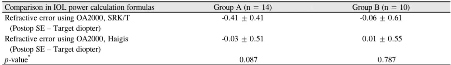
(group A) and phaco-vitrectomy with only air tamponade (group B)해보았으며 A, B 두 군 모두에서 장비에 따른 굴절력의 차이 는 보이지 않았다(p=0.655, p=0.653) (Table 6). 또한 인공수 정체 도수계산법에 따른 차이를 알아보기 위하여 OA-2000® (Tomey)에 내장된 SRK/T를 이용한 경우와 OA-2000® (Tomey)에 내장된 Haigis의 결과를 비교하였을 때 두 군 간 모두에서 두 공식 간에 통계적으로 유의한 차이가 없는 예측 굴절력 값을 얻을 수 있었다(Table 7).
고 찰
유리체절제술과 백내장 동시 수술에서 목표 굴절력을 설 정할 때 고려되어야 할 여러 가지 요소들 중 가스 주입술
시행과 수정체 후낭 제거의 동시 시행은 술 후 최종 굴절력 에 영향을 미치는 매우 중요한 요소라고 할 수 있다.9
수정체의 후낭은 전방을 후방으로부터 구분시켜 주는 중 요한 역할을 한다고 볼 수 있는데 후방에 가스를 넣거나 실 리콘 기름을 넣을 때 전방의 침범 없이 깨끗하고도 효과적 으로 시행할 수 있는 것은 후낭이 물리적인 장벽 역할을 하 기 때문이다.9 또한 후낭절제술을 시행하고 나면 방수와 유 리체 사이의 장벽이 사라져 혈관내피세포성장인자들이 후 방에서 전방으로 이동할 수 있는 경로가 되어 허혈성망막 질환에서는 전방 내 신생혈관형성에 영향을 미칠 가능성도
있다.10,11 따라서 유리체절제술과 백내장 병합수술 중 후낭
절제술의 시행은 신중하게 고려되어야 한다.
Comparison in IOL power calculation formulas Group A (n = 14) Group B (n = 10) Refractive error using OA2000, SRK/T
(Postop SE – Target diopter)
-0.41 ± 0.41 -0.06 ± 0.61
Refractive error using OA2000, Haigis (Postop SE – Target diopter)
-0.03 ± 0.51 0.01 ± 0.55
p-value* 0.087 0.787
Values are presented as mean ± SD unless otherwise indicated.
SRK/T = Sanders-Retzlaff-Kraff/Theoretical; IOL = intraocular lens; SE = spherical equivalent.
*Statistical analysis performed using Wilcoxon signed ranks test for continuous variables.
Table 7. Comparison in SRK/T and Haigis formula of phacovitrectomy and posterior capsulectomy with air tamponade (group A) and
phacovitrectomy with only air tamponade (group B)후낭 혼탁은 백내장수술 후 시력저하를 일으키는 대표적 인 합병증으로 안압 상승, 앞유리체혼탁, 낭포황반부종, 망 막박리, 안내염 등의 합병증을 발생시키기도 하며 유리체 절제술과 백내장 병합수술 시행 후에는 백내장 단독수술에 비하여 술 후 발생 빈도가 높은 것으로 알려져 있다.7 유리 체절제술과 백내장 동시수술 시행 중에 유리체절제기를 이 용하여 시행하는 후낭절제술은 술 후 발생하는 가장 대표 적인 합병증인 후낭혼탁을 사전에 방지할 수 있고 그로 인 한 합병증을 미연에 방지할 수 있으며 비교적 간단한 방법 으로 제거할 수 있다는 장점이 있다.7,12-14 Kim et al7은 유 리체절제술과 백내장 병합수술 중에 후낭절제술을 시행한 결과를 시행하지 않은 군과 비교하여 보았을 때 통계적으 로 유의한 수준은 아니었지만 후낭절제술을 함께 시행한 군에서 약간의 근시성 변화를 보이는 경향이 나타났다고 보고하였다. Jeoung et al15은 유리체절제술과 백내장 병합 수술 후 최종 굴절력에 영향을 미치는 요인들을 알아보기 위한 연구에서 수술 중에 시행한 가스나 오일 유리체주입 술은 술 후 굴절력에 의미있는 변화를 미치지 못하였으며 단지 술 전 안축장의 길이가 긴 경우 유의하게 술 후 근시 경향을 많이 보였다고 보고하였다. Wagenfeld et al16 역시 유리체절제술과 백내장 병합수술과 함께 시행한 가스 충전 술이 술 후 최종 굴절력에 미치는 영향을 평가하기 위한 연 구를 시행하였으며 가스 충전의 여부는 술 후 굴절력에 유의 한 영향을 미치지는 않았다고 보고하였다. Falkner-Radler et al17은 유리체절제술과 백내장 병합수술과 함께 가스 충전 술을 시행한 경우 가스 충전술을 시행하지 않은 환자들에 비하여 예측 굴절값보다 좀 더 근시로 이행하는 경향을 보 였다고 보고하였다. Suzuki et al8은 유리체절제술과 백내장 동시수술 시행 후 굴절력이 백내장 단독수술 후에 비하여 더 근시 변화를 보이는 경향을 나타낸다고 하였고 유리체 절제술과 백내장 동시수술을 진행하고 가스 주입술을 함께 시행한 경우와 시행하지 않은 경우를 비교하였을 때는 가 스 주입술을 시행한 군에서 더 근시값을 보이는 결과를 나 타냈다고 보고하였다. 본 연구에서도 통계학적으로 유의한
차이는 아니었지만 유리체절제술과 백내장 동시수술에서 후낭절제술과 공기주입술을 함께 시행한 경우에서 후낭절 제술을 시행하지 않고 공기주입술을 시행한 군에 비해서 결과적으로 예측 굴절력보다 근시로 이행하는 경향을 보였 다.
예측 굴절력과 술 후 굴절력이 차이를 보이는 데 영향을 주 는 요인 중에 한 가지로 인공수정체 도수 결정을 위한 안구길 이 측정을 위하여 사용된 장비에 따른 부분을 들 수 있는데, A-scan은 접촉식 초음파를 이용하여 각막 전면에서 내경계막 까지를 안구길이로 측정하는 반면, OA-2000® (Tomey)은 적 외선 레이저 광선을 통하여 반사되어 오는 간섭 신호를 이 용하여 각막전면에서부터 망막색소상피층까지를 안구길이 로 측정한다. 이러한 측정 장비간의 기준차이로 인하여 안 구길이 측정의 오차가 0.1-0.5 mm 정도 생길 수 있으며, 안 구길이 100 μm 차이가 있을 경우 술 후 굴절력은 0.28 D 정도의 오차를 만들 수 있다고 한다. 그래서 본 연구에서는 A-scan을 사용한 경우와 OA-2000® (Tomey)을 사용한 경 우의 예측 굴절력과 실제 굴절력과의 오차를 구해보았으며 장비에 따른 굴절력의 유의한 차이는 보이지 않았다(Table 6). Drexler et al18은 A-scan과 IOL master를 이용한 경우의 비교에서 술 후 굴절력의 차이가 유의하게 확인되었다고 보고한 바 있으며 Haigis et al19과 Lim et al20은 앞선 연구 에서 술 후 굴절력은 각각의 장비에서 비교해 본 결과 통계 적으로 유의한 차이는 보이지 않았다고 보고한 바 있다. 검 사 장비에 따른 굴절력의 차이에 대한 뚜렷한 결과는 더 발 전된 연구가 필요할 것으로 생각된다.
4세대 Haigis 공식은 술 전 전방 깊이를 실제로 측정한 값을 사용하여 효과적인 렌즈 위치(effective lens position) 를 결정하였고, 따라서 더 정확한 인공수정체안 전방 깊이 를 얻을 수 있었으며, 더 정확한 인공수정체 도수 결정이 가능하게 되었다. Haigis 공식과 다른 인공수정체 도수계산 공식을 비교한 이전의 연구들을 살펴보면, Haigis21는 990명 을 대상으로 한 연구에서 Haigis 공식이 SRK-II, SRK/T, Holladay 1, Hoffer Q 공식보다 정확하게 수술 후 굴절력을
예측하고 있다고 보고하였지만, Kim et al22은 81명을 대상 으로 한 연구에서 술 후 굴절력 예측에 있어서 공식들 간의 유의한 차이는 없다고 보고하였다. Yi et al23 연구에서도 전체 환자를 대상으로는 평균 실제 오차 및 평균 절대 오차 가 각 공식들 사이에 통계적으로 유의한 차이는 없었다. 본 연구에서는 SRK/T를 이용한 경우와 비교하였을 때 Haigis 공식을 사용한 결과 두 군 간에 통계적으로 유의한 차이가 없는 예측 굴절력 값을 얻을 수 있었다(Table 5). 하지만 같 은 군 내에서 두 공식을 사용한 예측 굴절력과 실제 굴절력 과의 오차를 비교해 본 결과 두 군 모두 통계적으로 유의한 차이를 보이지는 않았다(Table 7). 본 연구에서는 전방 깊 이의 측정이나 인공수정체 위치 변화에 대한 직접적인 분 석이 이루어지지 않았기에 대상 환자에서 보이는 술 후 근 시의 이행이 후낭절개와 공기주입술을 시행하여 발생한 후 방 압력에 의한 것인지 다른 원인에서 발생된 문제인지 정 확하게 파악하는 것이 어려웠다는 한계점이 있다. 또한 대 상 환자들이 동일한 진단명에 대하여 시행받은 수술이 아 니었으므로 예측 굴절력 역시 중심망막두께 등 환자들의 질환별로 나타날 수 있는 특이점들이 모두 배제된 상황이 아니라는 면이 또 다른 한계점이 될 수 있겠다. Lim et al20 이 발표한 논문에서 특발성망막앞막과 황반원공 환자의 유 리체절제술과 백내장 동시수술에서 술 후 굴절값 오차를 비교해 본 결과 유의한 상관관계를 보이지는 않았다는 보 고가 있었다.
결론적으로 유리체절제술과 백내장 동시 제거수술을 한 환자들에서 후낭절제술과 공기주입술을 함께 시행한 경우 공기주입술만 시행한 경우에 비하여 근시 이행을 보이는 경향을 나타내었으나 이는 통계적으로 유의한 차이는 아니 었다. 각각의 서로 다른 장비로 측정한 결과 A-scan과 OA-2000® (Tomey)은 예측 굴절력과 실제 굴절력과의 차 이가 A, B 두 군 모두에서 통계적으로 유의하게 나타나지 않았다. 인공수정체 도수계산 공식 간의 비교에서는 4세대 Haigis 공식을 사용하였을 때 SRK/T 사이 예측 굴절력과 실제 굴절력과의 차이가 A, B 두 군 모두에서 통계적으로 차이가 없는 것으로 나타났고 공기충전술이 필요한 환자에 서 후발백내장 동시 제거를 목표로 하였을 때는 Haigis 공 식을 이용하는 것이 효과적일 수 있겠다.
REFERENCES
1) Hsuan JD, Brown NA, Bron AJ, et al. Posterior subcapsular and nuclear cataract after vitrectomy. J Cataract Refract Surg 2001;27:437-44.
2) Melberg NS, Thomas MA. Nuclear sclerotic cataract after vi- trectomy in patients younger than 50 year of age. Ophthalmology 1995;102:1466-71.
3) Thompson JT. The role of patient age and intraocular gas use in cat- aract progression after vitrectomy for macular holes and epiretinal membranes. Am J Ophthalmol 2004;137:250-7.
4) Grusha YO, Masket S, Miller KM. Phacoemulsification and lens implantation after pars plana vitrectomy. Ophthalmology 1998;105:
287-94.
5) Toda J, Kato S, Oshika T, Sugita G. Posterior capsule opacification after combined cataract surgery and vitrectomy. J Cataract Refract Surg 2007;33:104-7.
6) Bhargava R, Kumar P, Phogat H, Chaudhary KP. Neodymium-yt- trium aluminium garnet laser capsulotomy energy levels for poste- rior capsule opacification. J Ophthalmic Vis Res 2015;10:37-42.
7) Kim JH, Han SB, Lee SJ, Kim MS. Postoperative refractive errors after posterior capsulectomy during combined vitrectomy and cat- aract surgery. J Korean Ophthalmol Soc 2015;56:709-14.
8) Suzuki Y, Sakuraba T, Mizutani H, et al. Postoperative refractive er- ror after simultaneous vitrectomy and cataract surgery. Ophthalmic Surg Lasers 2000;31:271-5.
9) Ohrloff C, Schalnus R, Rothe R, Spitznas M. Role of the posterior capsule in the aqueous-vitreous barrier in aphakic and pseudo- phakic eyes. J Cataract Refract Surg 1990;16:198-201.
10) Weinreb RN, Wasserstrom JP, Parker W. Neovascular glaucoma following neodymium-YAG laser posterior capsulotomy. Arch Ophthalmol 1986;104:730-1.
11) Poliner LS, Christianson DJ, Escoffery RF, et al. Neovascular glau- coma after intracapsular and extracapsular cataract extraction in di- abetic patients. Am J Ophthalmol 1985;100:637-43.
12) Gimbel HV, Neuhann T. Development, advantages, and methods of the continuous circular capsulorhexis technique. J Cataract Refract Surg 1990;16:31-7.
13) Gimbel HV. Posterior continuous curvilinear capsulorhexis and optic capture of the intraocular lens to prevent secondary opacifica- tion in pediatric cataract surgery. J Cataract Refract Surg 1997;23 Suppl 1:652-6.
14) Galand A, van Cauwenberge F, Moosavi J. Posterior capsulorhexis in adult eyes with intact and clear capsules. J Cataract Refract Surg 1996;22:458-61.
15) Jeoung JW, Chung H, Yu HG. Factors influencing refractive out- comes after combined phacoemulsification and pars plana vi- trectomy: Results of a prospective study. J Cataract Refract Surg 2007;33:108-14.
16) Wagenfeld L, Hermsdorf K, Stemplewitz B, et al. Refractive predict- ability in eyes with intraocular gas tamponade - results of a prospective controlled clinical trial. Clinical Ophthalmology 2017:11:993-8.
17) Falkner-Radler CI, Benesch T, Binder S. Accuracy of preoperative biometry in vitrectomy combined with cataract surgery for patients with epiretinal membranes and macular holes: results of a prospective controlled clinical trial. J Cataract Refract Surg 2008;34:1754-60.
18) Drexler W, Findl O, Menapace R, et al. Partial coherence inter- ferometry: a novel approach to biometry in cataract surgery. Am J Ophthalmol 1998;126:524-34.
19) Haigis W, Lege B, Miller N, Schneider B. Comparison of im- mersion ultrasound biometry and partial coherence interferometry for intraocular lens calculation according to Haigis. Graefes Arch Clin Exp Ophthalmol 2000;238:765-73.
20) Lim HC, Kim KH, Shin MK, et al. Accuracy of predictive refraction in combined vitrectomy-cataract surgery for epiretinal membrane and macular hole. J Korean Ophthalmol Soc 2015;56:219-27.
= 국문초록 =
유리체절제술과 백내장 병합수술에서 공기충전술을 시행한 경우 후낭절제술이 굴절력에 미치는 영향
목적: 유리체절제술과 백내장 병합수술에서 공기충전술을 시행한 경우 후낭절제술이 술 후 굴절력에 미치는 영향에 대해 알아보고자 하였다.
대상과 방법: 유리체절제술과 백내장 병합수술에서 공기충전술과 후낭절제술을 받고 6개월 이상 경과관찰이 가능하였던 환자들을 대상으로 하였다. 대조군으로 후낭절제술 없이 공기충전술을 시행받은 환자들이 포함되었다. 술 전 인자들 중 전방깊이, 안축장, 술 전 굴절력, 목표굴절력을 알아보고 술 후 최종 경과관찰 시 굴절력을 알아보았다. 생체계측법에 따른 측정값과 인공수정체 도수 결정 을 위하여 사용한 계산 공식에 따른 측정값을 최종 굴절력과 비교하였다.
결과: 술 중 후낭절제술과 공기충전술을 모두 시행한 14명, 14안 A군을 후낭절제술을 시행하지 않고 공기충전술을 시행한 환자 10명, 10안 B군과 비교해 본 결과, 접촉식 초음파장비 A-scan에서 Sanders-Retzlaff-Kraff/Theoretical (SRK/T) 공식을 이용한 경우, 후낭 절제술을 시행한 A군에서 목표 굴절력은 -0.21 ± 0.22 D, 최종 경과관찰 시점인 9.5 (±2.20)개월째 술 후 굴절력은 -0.52 ± 0.54 D였 고, 평균 오차는 -0.32 ± 0.44 D였다. 후낭절제술을 시행하지 않은 B군에서 목표 굴절력은 -0.33± 0.29 D, 최종 경과관찰 시점인 9.5 (±2.20)개월째 술 후 굴절력은 -0.27± 0.39 D였고, 평균 오차는 0.06 ± 0.53 D였다. 두 군 간의 평균 오차는 통계적으로 유의한 차이를 보였다(p=0.026).
결론: 유리체절제술과 백내장 병합수술에서 공기충전술을 시행한 경우, 후낭절제술을 시행하지 않은 군과 비교하여 후낭절제술을 시 행한 군에서 통계학적으로 유의한 근시이행을 보였고, 공기충전술을 계획한 수술에서 후낭절제술 시에 이에 대한 고려가 필요할 것으 로 사료된다.
<대한안과학회지 2018;59(9):819-826>
채수혜 / Sue Hey Chae
성모안과병원 Sungmo Eye Hospital 21) Haigis W. Intraocular lens power calculations. Thorofare: Slack
Inc., 2003; 41-57.
22) Kim DY, Kim MJ, Kim JY, Tchah H. Comparison of formulas for intraocular lens power calculation installed in a partial coherence
interferometer. J Korean Ophthalmol Soc 2009;50:523-8.
23) Yi CH, Choi SH, Chung ES, Chung TY. Accuracy of the Haigis for- mula based on axial length and anterior chamber depth. J Korean Ophthalmol Soc 2011;52:175-81.