© 2020 The Korean Ophthalmological Society
This is an Open Access article distributed under the terms of the Creative Commons Attribution Non-Commercial License (http://creativecommons.org/licenses /by-nc/3.0/) which permits unrestricted non-commercial use, distribution, and reproduction in any medium, provided the original work is properly cited.
173
Importance of Head Position after Gas Tamponade in Bilateral De- scemet’s Membrane Detachment Following Cataract Surgery
Dear Editor,
Descemet’s membrane detachment (DMD) is a rare complication of cataract surgery. It is associated with inad- equate surgery techniques and pre-existing anatomical factors. Herein we present a case of bilateral extensive DMD that occurred after uneventful cataract surgery and suggest appropriate management for DMD resolution.
A 57-year-old woman was referred for refractory DMD after cataract surgery. According to her medical records, cataract surgery with phacoemulsification was completed without any complications but vision loss with diffuse DMD and marked corneal edema were detected on the first postoperative day (POD). Air injection into the anteri- or chamber in the supine position was performed twice for 2 weeks, but there was no improvement. Slit lamp exam- ination and anterior optical coherence tomography re- vealed total DMD with severe corneal edema, and best-corrected visual acuity (BCVA) was finger-count/30 cm in her left eye. Fourteen percent C3F8 gas injection in a supine position was performed in her left eye 5 times. Two months postoperatively partial DMD remained but it had stabilized, and BCVA was 20 / 50 in her left eye.
Six months later, the patient requested cataract surgery on her right eye. In preoperative examination including specular microscopy, no anatomical problems were detect- ed in her right eye. Routine cataract surgery with temporal clear corneal incision was performed gently without any complications by a skillful surgeon. On POD 1 day, BCVA was hand-motion and total DMD was detected in her right eye (Fig. 1A, 1B). Immediately, 14% C3F8 gas was injected into the anterior chamber in the supine position, but DMD
remained stationery after gas injection was performed 3 times (Fig. 1C). On POD 6 days, the patient was instructed to maintain a left-decubitus position for tamponade at the temporal incision site after 14% C3F8 gas injection. One day later DMD recovered dramatically (Fig. 1D, 1E). Two weeks postoperatively the DMD was completely resolved and only the demarcation line was detected (Fig. 1F). Six months after surgery, her BCVA was 20 / 25 in the right eye.
DMD is a rare complication of cataract surgery and most detachments at the incision site are small and usually re- solve spontaneously or after a small number of gas injec- tions, without significant visual consequences. In the pres- ent case, extensive bilateral DMD occurred after uneventful cataract surgery was performed by two skillful surgeons. Previous studies suggest that signs of abnormali- ty in the fibrillary stromal attachment to Descemet’s mem- brane may constitute a predisposition to bilateral DMD, but no clear underlying etiology of bilateral DMD has been determined [1,2]. In preoperative examination in the pres- ent case, neither slit-lamp nor specular microscopy indicat- ed unhealthy endothelium.
In the current case of bilateral DMD, the outcomes in the right eye were more successful than those in the left eye with regard to both DMD resolution and visual acuity.
We assume that the most critical factor related to these re- sults is the head position after gas injection for tamponade at the Descemet’s membrane break site. Because the tem- poral clear cornea was incised in the present case, the pa- tient was instructed to lie in a left-decubitus position to maintain gas tamponade at the temporal incision site, and DMD recovered dramatically a day later. A key element of the procedure is sealing off the Descemet’s membrane break to stop the influx of fluid through the break and gain time for the condition to resolve. Head position can also play an important role, especially when the bubble be- comes too small to cover the whole cornea. In a decubitus position, the bubble is more effective for sealing off the in- cision site (Fig. 1G, 1H). Previous studies have also recom- mended considering a superior rather than temporal inci- sion, to facilitate gas tamponade in a routine sitting up position [3]. We suggest that gas tamponade at the incision site with an appropriate position would also be effective in
Korean J Ophthalmol 2020;34(2):173-175 https://doi.org/10.3341/kjo.2019.0119
Received: September 30, 2019 Final revision: October 22, 2019 Accepted: October 30, 2019
174
Korean J Ophthalmol Vol.34, No.2, 2020
standard cases of DMD. Also, it may have been better if we had utilized the scleral tunnel approach rather than the clear corneal incision.
In conclusion, surgeons should be cautious when operat- ing on apparently normal eyes in patients with a history of DMD in the fellow eye. Even in uneventful surgeries, DMD should be investigated carefully and if detected, im- mediate gas tamponade at the incision site is recommend- ed, with proper head positioning for sealing off the De- scemet’s membrane break.
SungYeon Jun, Kee Yong Choi
Department of Ophthalmology, HanGil Eye Hospital, Incheon, Korea
Young Joo Cho
Department of Ophthalmology, HanGil Eye Hospital, Catholic Kwandong University College of Medicine, Incheon, Korea E-mail: [email protected]
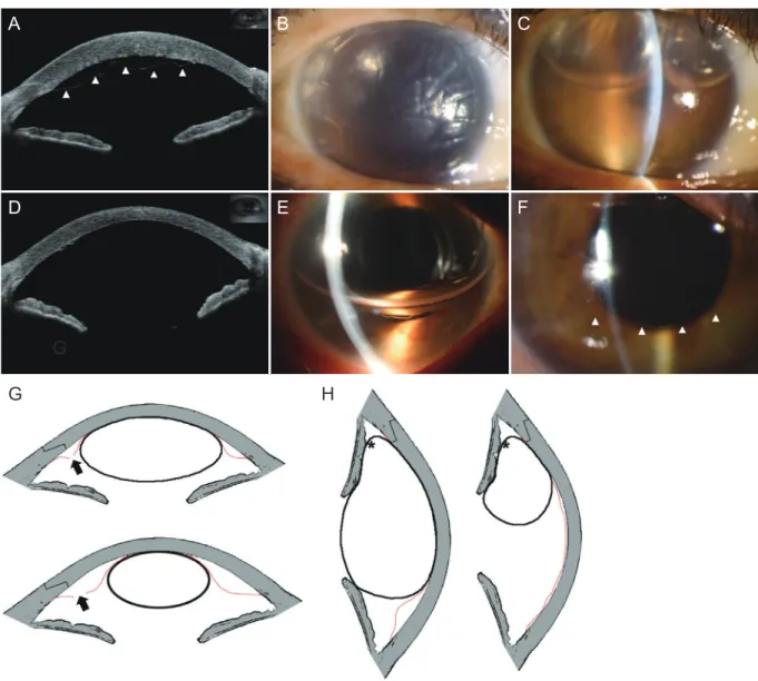
Fig. 1. A comparison of supine and lateral decubitus position after gas tamponade. (A) Anterior optical coherence tomography revealed total Descemet’s membrane detachment (DMD) (arrowheads). (B,C) Slit lamp photographs depicted extensive DMD, and DMD remained with gas bubbles. (D,E) DMD recovered dramatically the day after the left-decubitus position was adopted. (F) Only a small amount of DMD (arrowhead) was detected. (G) In the supine position, the bubble in the anterior chamber could not seal off the incision site and fluids could flow into the incision site (arrows). (H) In a decubitus position, the bubble sealed off the incision site more effectively, even when the bubble became smaller (stellates). The red demarcation line represents Descemet’s membrane.
A
D
G
B
E
C
F
G H
175
Conflict of Interest
No potential conflict of interest relevant to this article was reported.
References
1. Kansal S, Sugar J. Consecutive Descemet membrane de- tachment after successive phacoemulsification. Cornea 2001;20:670-1.
2. Kim T, Sorenson A. Bilateral Descemet membrane detach- ments. Arch Ophthalmol 2000;118:1302-3.
3. Fang JP, Amesur KB, Baratz KH. Preexisting endothelial abnormalities in bilateral postoperative Descemet mem- brane detachment. Arch Ophthalmol 2003;121:903-4.