Intraocular Lens Power Estimation in Combined Phacoemulsification and Pars Plana Vitrectomy in Eyes
with Epiretinal Membranes: A Case-Control Study
Min Kim,
1Hyoung Eun Kim,
1Dong Hyun Lee,
1Hyoung Jun Koh,
1Sung Chul Lee,
2and Sung Soo Kim
21Department of Ophthalmology, Yonsei University College of Medicine, Gangnam Severance Hospital, Seoul;
2Institute of Vision Research, Department of Ophthalmology, Severance Eye and ENT Hospital, Yonsei University College of Medicine, Seoul, Korea.
Received: June 17, 2014 Revised: July 30, 2014 Accepted: August 13, 2014
Corresponding author: Dr. Sung Soo Kim, Institute of Vision Research,
Department of Ophthalmology, Severance Eye and ENT Hospital, Yonsei University College of Medicine, 50-1 Yonsei-ro, Seodaemun-gu, Seoul 120-752, Korea.
Tel: 82-2-2019-3440, Fax: 82-2-3463-1049 E-mail: [email protected]
∙ The authors have no financial conflicts of interest.
© Copyright:
Yonsei University College of Medicine 2015 This is an Open Access article distributed under the terms of the Creative Commons Attribution Non- Commercial License (http://creativecommons.org/
licenses/by-nc/3.0) which permits unrestricted non- commercial use, distribution, and reproduction in any medium, provided the original work is properly cited.
Purpose: To evaluate the accuracy of postoperative refractive outcomes of com- bined phacovitrectomy for epiretinal membrane (ERM) in comparison to cataract surgery alone. Materials and Methods: Thirty-nine eyes that underwent com- bined phacovitrectomy with intraocular lens (IOL) implantation for cataract and ERM (combined surgery group) and 39 eyes that received phacoemulsification for cataract (control group) were analyzed, retrospectively. The predicted preoperative refractive aim was compared with the results of postoperative refraction. Results:
In the combined surgery group, refractive prediction error by A-scan and IOLMas- ter were -0.305±0.717 diopters (D) and -0.356±0.639 D, respectively, compared to 0.215±0.541 and 0.077±0.529 in the control group, showing significantly more myopic change compared to the control group (p=0.001 and p=0.002, respective- ly). Within each group, there was no statistically significant difference in refractive prediction error between A-scan and IOLMaster (all p>0.05). IOL power calcula- tion using adjusted A-scan measurement of axial length based on the macular thickness of the normal contralateral eye still resulted in significant postoperative refractive error (all p<0.05). Postoperative refraction calculated with adjusted axial length based on actual postoperative central foveal thickness change showed the closest value to the actual postoperative achieved refraction (p=0.599). Conclu- sion: Combined phacovitrectomy for ERM resulted in significantly more myopic shift of postoperative refraction, compared to the cataract surgery alone, for both A-scan and IOLMaster. To improve the accuracy of IOL power estimation in eyes with cataract and ERM, sequential surgery for ERM and cataract may need to be considered.
Key Words: Cataract surgery with vitrectomy, epiretinal membranes, myopic shift, phacovitrectomy, IOL power calculation
INTRODUCTION
Pars plana vitrectomy (PPV) combined with cataract surgery are usually per- formed in eyes with vitreoretinal disease and coexisting cataract, particularly when
Institutional Review Boards at the Gangnam Severance Hos- pital. The study was performed in accordance with the tenets of the Declaration of Helsinki and all federal laws.
Surgery
Extracapsular cataract extraction with phacoemulsification (2.8-mm clear corneal incision at the superior limbus) and posterior capsular IOL implantation with an acrylic foldable IOL were performed in all patients. The preoperative IOL power was targeted for emmetropia. The combined surgery group comprised patients who had undergone phacoemulsi- fication and PPV due to an ERM without other retinal pa- thology influencing the macula, and cataract surgery was performed before vitrectomy. Vitreoretinal procedures in- cluded a 23-gauge PPV and removal of epiretinal mem- branes. Indocyanine green-assisted ILM peeling was per- formed in all cases. No gas was used in any eyes during the surgery. The sclerotomy sites were left without any sutures.
All patients were treated by a single vitreoretinal surgeon (SSK).
Preoperative and postoperative examinations
Clinical examinations were performed preoperatively and at 3 months postoperatively. The examinations included manifest refraction, autokeratometry (K) using a ref-kera- tometer (RK-3; CANON Inc., Kanagawa, Japan), measure- ment of anterior chamber depth (ACD), AL using an A- scan and IOLMaster, and central foveal thickness (CFT) by SD-OCT (Cirrus HD-OCT, Carl Zeiss Meditec, Dublin, CA, USA). A-scan biometry was obtained using the appla- nation method. Postoperative refractive prediction error was calculated by subtracting the spherical equivalent (SE) of the actual refraction from the SE of the predicted refraction.
A-scan ultrasonography measured the distance from cor- nea to the ILM, while IOLMaster measured the distance to the RPE. CFT by SD-OCT was defined as the distance be- tween the ILM and the RPE at the fovea.
Statistical analysis
Chi-squared test and independent Student t-tests were per- formed to assess differences in patient demographic data between groups. Independent t-test was used to assess dif- ferences in postoperative refractive prediction errors between groups. Paired t-test was used to evaluate differences in re- fractive prediction error between A-scan and IOLMaster within each group. Statistical analysis was conducted using SPSS software (18.0, SPSS Inc., Chicago, IL, USA). In all a cataract prevents adequate visualization of the retina. It is
a safe and effective way to treat vitreoretinal pathology and cataracts simultaneously, and its functional outcomes are re- ported to be comparable to those of sequential surgery.1-3 In order to achieve a favorable postoperative visual outcome, accurate preoperative intraocular lens (IOL) power calcula- tion is crucial, and precise measurement of axial length (AL) is the most important element in obtaining accurate estimations of the IOL calculation.4,5 However, some stud- ies have reported that the refractive results after combined phacovitrectomy show a postoperative myopic shift, com- pared to predicted refraction obtained by either A-scan ul- trasonography or optical biometry using IOLMaster.6-12 Nevertheless, only a few studies have compared postopera- tive refractive prediction error by A-scan and IOLMaster si- multaneously, and no studies have used spectral domain OCT (SD-OCT) to measure the distance from the internal limiting membrane (ILM) to the retinal pigment epithelium (RPE) in patients with epiretinal membrane (ERM) in asso- ciation with biometry.
In this study, we evaluated whether performing PPV with removal of the ERM combined with cataract surgery af- fects postoperative refractive prediction error with both A- scan and IOLMaster in comparison cataract surgery alone, and we also attempted to outline factors influencing refrac- tive outcomes. Furthermore, we evaluated whether adjusted A-scan measured AL method based on macular thickness of the normal contralateral eye could minimize refractive prediction error.
MATERIALS AND METHODS
Patients
This retrospective case control study was performed in 39 eyes of 39 patients who underwent combined phacoemulsifi- cation and vitrectomy for idiopathic epiretinal membranes (combined surgery group) and 39 eyes of 39 patients who re- ceived phacoemulsification for cataract only (control group) at Gangnam Severance Hospital from July 2008 to January 2010. Patients with diabetes, with any retinal vascular disor- der, or with macular degeneration were excluded. A-scan ul- trasonography (UD-6000, TOMEY, Nagoya, Japan) and IOLMaster (Carl Zeiss Meditec, CA, USA) were used simul- taneously for preoperative AL measurement and IOL calcu- lation using the SRK/T formulae. Informed consent was ob- tained from all patients, and the study was approved by the
opters (D) and -0.356±0.639 D, respectively, compared to 0.215±0.541 and 0.077±0.529 in the control group, respec- tively, revealing significantly more myopic change in the combined surgery group (p=0.001 and p=0.002, respective- ly) (Table 2). However, within each group, there was no sta- tistically significant difference in refractive prediction error between A-scan and IOLMaster measurements (all p>0.05).
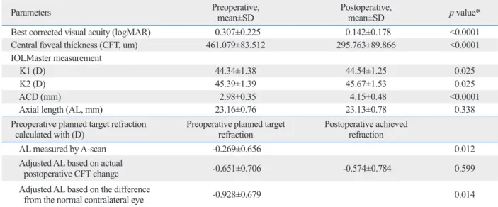
Table 3 shows postoperative changes of visual acuity, mac- ular thickness, and refraction with IOL calculation by A-scan before and after combined surgery. The mean planned target refraction was -0.269±0.656 D, while the mean actual post- operative refractive outcome was -0.574±0.784 (p=0.012), showing a significant myopic shift. When the preoperative planned target refraction was recalculated with an adjusted AL obtained by using the CFT value of the healthy contralat- cases, a p-value less than 0.05 was considered statistically
significant.
RESULTS
Baseline patient demographic data are shown in Table 1.
Seventy-eight eyes of 78 patients were enrolled in the study.
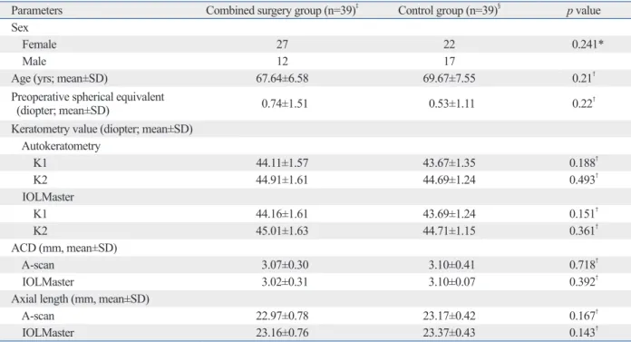
There was no significant difference between the combined surgery group and the control group regarding gender, age, preoperative spherical equivalents, keratometric value, ACD, and axial length. The axial length measured by IOLMaster was longer than that of the A-scan in both groups (p<0.0001) (Table 1). In the combined surgery group, refractive predic- tion error by A-scan and IOLMaster were -0.305±0.717 di- Table 1. Patient Demographic Data
Parameters Combined surgery group (n=39)‡ Control group (n=39)§ p value
Sex
Female 27 22 0.241*
Male 12 17
Age (yrs; mean±SD) 67.64±6.58 69.67±7.55 0.21†
Preoperative spherical equivalent
(diopter; mean±SD) 0.74±1.51 0.53±1.11 0.22†
Keratometry value (diopter; mean±SD) Autokeratometry
K1 44.11±1.57 43.67±1.35 0.188†
K2 44.91±1.61 44.69±1.24 0.493†
IOLMaster
K1 44.16±1.61 43.69±1.24 0.151†
K2 45.01±1.63 44.71±1.15 0.361†
ACD (mm, mean±SD)
A-scan 3.07±0.30 3.10±0.41 0.718†
IOLMaster 3.02±0.31 3.10±0.07 0.392†
Axial length (mm, mean±SD)
A-scan 22.97±0.78 23.17±0.42 0.167†
IOLMaster 23.16±0.76 23.37±0.43 0.143†
IOL, intraocular lens; ACD, anterior chamber depth.
*Chi-squared test was used for statistical analysis.
†Independent t-test was used for statistical analysis.
‡Combined surgery group=pars plana vitrectomy, removal of epiretinal membrane, and phacoemulsification and intraocular lens implantation.
§Control group=phacoemulsification and intraocular lens implantation only.
Table 2. Comparison of Refractive Prediction Error between the Combined Surgery Group and the Control Group Refractive prediction error (D)‡ Combined surgery group (n=39) Control group (n=39) p value*
A-scan -0.305±0.717 0.215±0.541 0.001
IOLMaster -0.356±0.639 0.077±0.529 0.002
p value† 0.616 0.073
IOL, intraocular lens; D, diopters.
*Independent t-test was used for statistical analysis.
†Paired t-test was used for statistical analysis.
‡Refractive prediction error=actual postoperative refraction–predicted refraction.
icant refractive prediction error; meanwhile, postoperative refraction calculated with adjusted AL based on actual post- operative CFT change showed the closest value to the actu- al postoperative achieved refraction.
Combined phacovitrectomy has become a common proce- dure as a result of recent progress in cataract and vitrectomy surgery. However, in combined phacovitrectomy, postopera- tive refractive errors are thought to arise from concomitant macular pathology, such as macular thickening due to epireti- nal membrane. Kovács, et al.6 suggested that myopic shift results from underestimation of the axial length due to a thicker macula using A-scan ultrasonography. In contrast, Manvikar, et al.13 reported that there is no tendency toward a myopic shift in IOL power estimation using IOLMaster, while Falkner-Radler, et al.12 reported that there was myopic refractive change after combined phacovitrectomy despite IOL power calculation with IOLMaster. These studies sug- gested thicker retina preoperatively or anterior displacement of the IOL by the complete gas fill achieved after phacovit- rectomy as potential mechanisms of myopic shift.6-9,11,12
A-scan ultrasound measures the distance between the an- terior surface of the cornea and the ILM, whereas the IOL- Master measures the distance between the anterior corneal surface and the RPE and then calibrates to match the mea- surement of the A-scan. Verhulst and Vrijghem14 reported that the mean difference in axial length between ultrasound and optical biometry was 0.2 mm, and IOLMaster resulted in measurement of a longer axial length. These findings were consistent with our results in that the axial length mea- eral eye (preoperative CFT‒normal contralateral eye CFT),
the target refraction of the implanted IOL was -0.928±0.679 D, still showing a statistically significant difference from the achieved refraction (mean refractive prediction error of 0.311±0.687 D, p=0.014). The estimated IOL power was re- calculated with an adjusted AL by adding the amount of ac- tual postoperative CFT change (preoperative CFT‒postoper- ative CFT using SD-OCT) to AL measured by A-scan, and the planned target refraction became -0.651±0.706 D, show- ing no statistically significant difference between the planned and achieved refractions (p=0.599).
In the combined surgery group, there was a statistically significant increase of both K values and ACD measured by IOLMaster, postoperatively (p=0.025 and p<0.0001, respec- tively). However, AL measured with IOLMaster did not show any significant difference between preoperative and postoperative values (p=0.338) (Table 3).
During the follow-up period, there were no cases with sig- nificant postoperative complications, such as cystoid macu- lar edema, endophthalmitis, or hypotony.
DISCUSSION
Our study showed that the combined phacovitrectomy results in a significantly more myopic shift of postoperative refrac- tion, compared to that of the control group using both A-scan and IOLMaster. Adjusted A-scan measured AL method based on the healthy contralateral eye still resulted in signif-
Table 3. Postoperative Changes of Visual Acuity, Macular Thickness, and Refraction with IOL Calculation by A-Scan and Kera- tometry (K) Value, Anterior Chamber Depth (ACD), and Axial Length Measured by IOLMaster in the Combined Surgery Group
Parameters Preoperative,
mean±SD Postoperative,
mean±SD p value*
Best corrected visual acuity (logMAR) 0.307±0.225 0.142±0.178 <0.0001
Central foveal thickness (CFT, um) 461.079±83.512 295.763±89.866 <0.0001
IOLMaster measurement
K1 (D) 44.34±1.38 44.54±1.25 0.025
K2 (D) 45.39±1.39 45.67±1.53 0.025
ACD (mm) 2.98±0.35 4.15±0.48 <0.0001
Axial length (AL, mm) 23.16±0.76 23.13±0.78 0.338
Preoperative planned target refraction
calculated with (D) Preoperative planned target
refraction Postoperative achieved refraction
AL measured by A-scan -0.269±0.656 0.012
Adjusted AL based on actual
postoperative CFT change -0.651±0.706 -0.574±0.784 0.599
Adjusted AL based on the difference
from the normal contralateral eye -0.928±0.679 0.014
IOL, intraocular lens; D, diopters.
*Paired t-test was used for statistical analysis.
my, and a new generation of formulas may need to be de- veloped in the future.19
In some IOL implantation cases with macular diseases, a double peak was observed by IOLMaster in axial length measurements, with the posterior peak of a double peak representing RPE.20 The report suggested that the anterior peak of the double peak may represent reflection from the surface of the retina, such as an ERM, an inner limiting membrane, or a posterior vitreous membrane. We speculat- ed that this may also be a possible cause of severe myopic shift; however, in our study, there was only one patient in the combined surgery group who showed a double peak in the IOLMaster signal, and the anterior peak of the double peak was initially used to measure axial length. The refrac- tive prediction error caused by this measurement of axial length was -1.6 D. When the measurement was corrected to use the posterior peak of the double peak, the refractive prediction error decreased to -0.8 D.
Since the use of small gauge sutureless incisions is known to reduce surgically induced astigmatism and phacovitrecto- my was found to cause no significant postoperative astig- matic change,19,21 we can assume that performing sutureless vitrectomy combined with phacoemulsification would not cause significant astigmatic change.
There have been many attempts to reduce postoperative refractive prediction error after performing combined cata- ract surgery and PPV. A study conducted by Patel, et al.9 sug- gested that slight residual hyperopia could compensate for the myopic overcorrection. Kovács, et al.6 attempted to deter- mine the adjusted planned ametropia by substituting the full axial length and the implanted IOL power into the SRK/T formula (12 eyes studied). This full axial length was deter- mined in each case by adding the difference between the normal value of macular thickness and results of the OCT scans to the AL of the A-scan. Similarly, Sun and Choi22 proposed a correlation between postoperative refraction and the calculated predicted refraction using an adjusted AL with macular thickness change based on the contralateral eye (23 eyes studied). To apply this calculation prospectively to pa- tients with increased macular thickness planning to undergo combined phacovitrectomy, they subtracted the macular thickness of the contralateral normal eye from the macular thickness of the affected eye, and then added this number to the A-scan measured AL. Although these various attempts to compensate for the myopic overcorrection using adjusted A- scan measured AL have proven partially effective in com- pensating for myopic shift, macular thickness in many pa- sured by IOLMaster was longer than that of A-scan in both
groups (p<0.0001) (Table 1). Thus, we hypothesized that an error in measurement of axial length due to the ERM could cause myopic shift in IOL calculation with A-scan. This is consistent with a previous finding of a postoperative myo- pic shift due to an error in measuring axial length in com- bined surgery.6
IOLMaster measures axial length to the RPE and thus should not be affected by retinal thickening due to the pres- ence of ERM. However, our results showed that there was a similar degree of refractive prediction error obtained when using IOLMaster, compared to A-scan. Studies of IOL po- sitioning reported that during the first postoperative week, IOL moves slightly forward, which is neutralized by a slight backward movement within 3 months.15 Falkner-Radler, et al.12 reported that, compared with patients in the cataract surgery group, patients in the combined surgery group had a mean postoperative myopic shift in refractive error and deep- er ACD in IOL calculation with IOLMaster. The combined surgery group in our study also showed increased ACD postoperatively (p<0.0001) (Table 3).
A few studies have suggested that myopic shift might be the result of a postoperative increase in axial length caused by sclera thinning or stretching in or around the sclerotomy sites after vitrectomy.8,12 In our study, there was no signifi- cant change in axial length measured by IOLMaster after combined surgery (p=0.338) (Table 3). Therefore, it would be difficult to consider postoperative increases in axial length as a potential cause for a myopic shift following phacoemulsification and PPV in IOL power estimation with IOLMaster. Falkner-Radler, et al.12 speculated that the myo- pic shift by the IOLMaster calculation might result from a significantly higher postoperative K2 value than the base- line K2 value in the combined surgery group. This result was in close agreement with those of our study (Table 3);
therefore, it is possible that a change in K value after sur- gery may also contribute to the postoperative refractive pre- diction error upon use of IOLMaster. There were also stud- ies reporting that after PPV, vitreous gel is replaced by the aqueous after PPV, and refractive index decreases slightly, thereby resulting in a myopic shift.16-18 Theoretically, the myopic shift in vitrectomized eyes could be up to -0.5 D.17 Considering that the mean myopic change was -0.36 D in the IOL power calculation with IOLMaster, we speculate that vitrectomy itself could also play a role in causing myo- pic change after phacovitrectomy. Currently, there are no formulas available for vitrectomized eyes or phacovitrecto-
ACKNOWLEDGEMENTS
This study was supported by a faculty research grant of Yonsei University College of Medicine (Seoul, Korea) for 2012 (3-2012-0137).
REFERENCES
1. Demetriades AM, Gottsch JD, Thomsen R, Azab A, Stark WJ, Campochiaro PA, et al. Combined phacoemulsification, intraocular lens implantation, and vitrectomy for eyes with coexisting cataract and vitreoretinal pathology. Am J Ophthalmol 2003;135:291-6.
2. Hwang JU, Yoon YH, Kim DS, Kim JG. Combined phacoemulsi- fication, foldable intraocular lens implantation, and 25-gauge transconjunctival sutureless vitrectomy. J Cataract Refract Surg 2006;32:727-31.
3. Heiligenhaus A, Holtkamp A, Koch J, Schilling H, Bornfeld N, Lösche CC, et al. Combined phacoemulsification and pars plana vitrectomy: clear corneal versus scleral incisions: prospective ran- domized multicenter study. J Cataract Refract Surg 2003;29:1106- 12.
4. McEwan JR, Massengill RK, Friedel SD. Effect of keratometer and axial length measurement errors on primary implant power calculations. J Cataract Refract Surg 1990;16:61-70.
5. Olsen T. Sources of error in intraocular lens power calculation. J Cataract Refract Surg 1992;18:125-9.
6. Kovács I, Ferencz M, Nemes J, Somfai G, Salacz G, Récsán Z.
Intraocular lens power calculation for combined cataract surgery, vitrectomy and peeling of epiretinal membranes for macular oede- ma. Acta Ophthalmol Scand 2007;85:88-91.
7. Suzuki Y, Sakuraba T, Mizutani H, Matsuhashi H, Nakazawa M.
Postoperative refractive error after simultaneous vitrectomy and cataract surgery. Ophthalmic Surg Lasers 2000;31:271-5.
8. Jeoung JW, Chung H, Yu HG. Factors influencing refractive out- comes after combined phacoemulsification and pars plana vitrec- tomy: results of a prospective study. J Cataract Refract Surg 2007;
33:108-14.
9. Patel D, Rahman R, Kumarasamy M. Accuracy of intraocular lens power estimation in eyes having phacovitrectomy for macular holes. J Cataract Refract Surg 2007;33:1760-2.
10. Hotta K, Sugitani A. Refractive changes in silicone oil-filled pseu- dophakic eyes. Retina 2005;25:167-70.
11. Iwase T, Sugiyama K. Investigation of the stability of one-piece acrylic intraocular lenses in cataract surgery and in combined vit- rectomy surgery. Br J Ophthalmol 2006;90:1519-23.
12. Falkner-Radler CI, Benesch T, Binder S. Accuracy of preoperative biometry in vitrectomy combined with cataract surgery for pa- tients with epiretinal membranes and macular holes: results of a prospective controlled clinical trial. J Cataract Refract Surg 2008;
34:1754-60.
13. Manvikar SR, Allen D, Steel DH. Optical biometry in combined phacovitrectomy. J Cataract Refract Surg 2009;35:64-9.
14. Verhulst E, Vrijghem JC. Accuracy of intraocular lens power cal- culations using the Zeiss IOL master. A prospective study. Bull Soc Belge Ophtalmol 2001:61-5.
tients does not decrease completely to a normal value after surgery, and each patient shows a different postoperative CFT change. For this reason, our analysis showed persistent postoperative refractive prediction error even when calculat- ed with the adjusted AL, based on the difference in macular thickness from the healthy contralateral eye (p=0.014) (Ta- ble 3). For the most accurate assessment of CFT and AL with which to maximize postoperative refractive outcomes, it might prove to be better to perform vitrectomy first and then carry out cataract surgery based on the IOL calculation obtained after vitrectomy, in a sequential manner.
Many attempts to correct for myopic shift after combined phacovitrectomy have been based on IOL calculation using A-scan measurements of AL. As the cause of myopic shift in IOL power estimation using IOLMaster seemed to be multifactorial and unpredictable, there are limitations in at- tempts to compensate for the postoperative refractive pre- diction error by IOLMaster. Sequence of surgery, as well as time between surgeries, may need to be taken into consider- ation to improve refractive outcomes in cases under consid- eration for combined phacovitrectomy.19
There are a few limitations to this study. It is a retrospec- tive case control study, and a prospective study with more patients would be needed in the future. Also, further studies comparing the refractive outcomes of combined phacovit- rectomy versus sequential surgery are warranted in the fu- ture to optimize refractive outcomes. The use of the SRK/T formula rather than the Holladay and Hoffer Q formulas may also have contributed to refractive prediction error in addition to axial length.
In conclusion, combined phacovitrectomy resulted in sig- nificantly more myopic shift of postoperative refraction, com- pared to that of the control group, on both A-scan and IOL- Master. Myopic shift in IOL calculations with A-scan may be attributable to an underestimation of axial length due to ERM, while myopic shift in IOL power estimation using IOLMaster may be attributed to a combination of changes in K values, ACD, and refractive index due to removal of vitreous gel. Adjusted A-scan measured AL method based on a healthy contralateral eye still resulted in significant re- fractive prediction error, while postoperative refraction cal- culated with an adjusted AL based on actual postoperative CFT change showed the closest value to the actual postop- erative achieved refraction. Therefore, to improve the accu- racy of IOL power estimation in eyes with cataract and ERM, sequential surgery for ERM and cataract may need to be considered.
19. Hamoudi H, La Cour M. Refractive changes after vitrectomy and phacovitrectomy for macular hole and epiretinal membrane. J Cataract Refract Surg 2013;39:942-7.
20. Kojima T, Tamaoki A, Yoshida N, Kaga T, Suto C, Ichikawa K.
Evaluation of axial length measurement of the eye using partial coherence interferometry and ultrasound in cases of macular dis- ease. Ophthalmology 2010;117:1750-4.
21. Yuen CY, Cheung BT, Tsang CW, Lam RF, Baig NB, Lam DS.
Surgically induced astigmatism in phacoemulsification, pars plana vitrectomy, and combined phacoemulsification and vitrectomy: a comparative study. Eye (Lond) 2009;23:576-80.
22. Sun HJ, Choi KS. Improving intraocular lens power prediction in combined phacoemulsification and vitrectomy in eyes with macu- lar oedema. Acta Ophthalmol 2011;89:575-8.
15. Petternel V, Menapace R, Findl O, Kiss B, Wirtitsch M, Rainer G, et al. Effect of optic edge design and haptic angulation on postop- erative intraocular lens position change. J Cataract Refract Surg 2004;30:52-7.
16. Gao Q, Chen X, Ge J, Liu Y, Jiang Z, Lin Z, et al. Refractive shifts in four selected artificial vitreous substitutes based on Gullstrand- Emsley and Liou-Brennan schematic eyes. Invest Ophthalmol Vis Sci 2009;50:3529-34.
17. Mehdizadeh M, Nowroozzadeh MH. Postoperative induced myo- pia in patients with combined vitrectomy and cataract surgery. J Cataract Refract Surg 2009;35:798-9.
18. Byrne S, Ng J, Hildreth A, Danjoux JP, Steel DH. Refractive change following pseudophakic vitrectomy. BMC Ophthalmol 2008;8:19.