Clinical Study
Nd:YAG Capsulotomy after Phacoemulsification in
Vitrectomized Eyes: Effects of Pars Plana Vitrectomy on Posterior Capsule Opacification
Jong Hwa Jun, Kwang Soo Kim, and Sung Dong Chang
Department of Ophthalmology, School of Medicine, Dongsan Medical Center, Keimyung University, 56 Dalseong-ro, Jung-gu, Daegu 704-701, Republic of Korea
Correspondence should be addressed to Jong Hwa Jun; [email protected]
Received 8 December 2013; Revised 18 March 2014; Accepted 19 March 2014; Published 27 April 2014 Academic Editor: Tamer A. Macky
Copyright © 2014 Jong Hwa Jun et al. This is an open access article distributed under the Creative Commons Attribution License, which permits unrestricted use, distribution, and reproduction in any medium, provided the original work is properly cited.
To compare the progression of posterior capsule opacification (PCO) in patients who required Nd:YAG laser capsulotomy following either combined cataract surgery with pars plana vitrectomy (PPV; C-CV), sequential cataract surgery after PPV (S-CV), or cataract surgery alone (CA). The medical records of 321 patients (408 eyes) who underwent Nd:YAG capsulotomy were retrospectively evaluated. The CA group had a significantly longer time interval from cataract surgery to capsulotomy than that of both the CV group (𝑃 = 0.006) and the S-CV (𝑃 = 0.013) and C-CV (𝑃 = 0.042) subgroups when age-matched comparisons were used.
CV patients who implanted a hydrophobic acrylic IOL had shorter time intervals than those of CA patients (𝑃 = 0.028). CV patients had larger hazard of earlier capsulotomy than CA patients (hazard ratio (HR) = 1.337; 95% confidence interval (CI) 1.100–
1.625; 𝑃 = 0.004). C-CV and S-CV patients both had larger hazard than CA patients in earlier capsulotomy (HR = 1.304; 95%
CI = 1.007–1.688; 𝑃 = 0.044, HR = 1.361; 95% CI = 1.084–1.709; 𝑃 = 0.008, resp.). PCO progresses more rapidly in patients undergoing combined or sequential cataract surgery and PPV than in patients undergoing CA.
1. Introduction
Cataract formation and progression are common postop- erative complications after pars plana vitrectomy (PPV) for a variety of vitreoretinal diseases, including epiretinal membrane and macular hole in phakic patients [1, 2]. Patient age [1, 3], lens opacification before PPV [4], and intravitreal tamponade [5–7] are major risk factors related to cataract progression after PPV. Recently, for cases of vitreoretinal disease combined with moderate to severe lens opacification, combined cataract surgery with PPV has become a routine procedure. The inclusion of cataract extraction leads to better visualization of vitreoretinal structures and earlier visual rehabilitation after PPV can be achieved. However, difficul- ties with capsulorhexis, higher postoperative inflammation, intraoperative miosis, and corneal edema with Descemet’s folds are disadvantages when combining cataract and vit- reoretinal surgery [8, 9]. Moreover, in combined surgeries for epiretinal membrane, increased postoperative macular
thickness and recurrence of epiretinal membrane have been reported [10].
Posterior capsule opacification (PCO) is the most com- mon complication after cataract surgery [11]. Further, when cataract surgery is combined with PPV, either sequentially or simultaneously, PCO is also the most common factor threatening vision [12]. In previous reports, the rate of PCO after phacovitrectomy has ranged from 10.3% to 51.1% and from 8% to 51% in sequential surgery [12–18]. Particularly, in eyes with vitreoretinal disease, postvitrectromy PCO interferes with the ability to diagnose retinal pathology.
Previously, several studies have reported outcomes of pha- coemulsification related to PPV; however, the PCO rate was only reported in limited sample sizes and for relatively short follow-up periods. Moreover, it is unknown whether the cataract surgery performed either with or after PPV could influence the interval from cataract surgery to neodymium- doped: yttrium aluminum garnet (Nd:YAG) capsulotomy due to vision threatening PCO. Hence, the aim of this paper
Volume 2014, Article ID 840958, 8 pages
http://dx.doi.org/10.1155/2014/840958
was to retrospectively evaluate the records of a large sample of patients over a 14-year period who underwent Nd:YAG capsulotomy for clinically significant PCO with an emphasis on comparing the results of PCO after combined PPV and cataract surgery, sequential PPV and cataract surgery, and cataract surgery alone (CA).
2. Materials and Methods
Between January 2000 and December 2012, the medical records of 321 patients (408 eyes) who underwent Nd:YAG laser capsulotomies at the Department of Ophthalmology, Dongsan Medical Center, Keimyung University, Daegu, Korea, were retrospectively evaluated. Patients were divided into two primary groups to investigate whether vitreous removal could influence the progression of PCO; there were 212 eyes from 150 patients in the CA group and 196 eyes from 171 patients in the cataract surgery either with or after PPV group (CV group). 62 patients of CA and 25 patients of CV were enrolled in both eyes. For evaluating capable correlation of outcomes on eyes from same patients, we adopted the point biserial correlation coefficients method. The coefficient of correlation was very low, so we included both eyes of the same patients in this study (data not shown). The CV group was subdivided into two secondary groups: 80 eyes from 71 patients in the combined cataract surgery with PPV group (C-CV group) and 116 eyes from 100 patients in the sequential cataract surgery after PPV group (S-CV group). Patients with a history of uveitis, primary aphakia after cataract extraction, glaucoma filtration surgery, repeated vitrectomy after cataract surgery, intraoperative posterior capsule rupture, combined extracapsular cataract extraction with PPV, and extracapsular fixation of the intraocular (IOL) were excluded. Furthermore, cases who were lost to follow-up for more than 1 year were also excluded to avoid overestimation of the interval to capsulotomy.
Data collection included age at cataract surgery, gender, history of diabetes mellitus (DM), and laterality of surgery.
The patient demographics are shown in Table 1. The key parameters of interest were the time interval from cataract surgery to Nd:YAG laser capsulotomy and the IOL materials.
For investigating the effects of vitreous on PCO, comparisons were performed between the CV and CA groups. The C-CV and S-CV groups were also compared to test the hypothesis that postoperative inflammation could influence PCO pro- gression. The IOL material subgroups were evaluated for the IOL edge effect and vitreous decompression theory after PPV [17].
2.1. Surgical Techniques. All surgeries were performed by one experienced surgeons (KKS) using retrobulbar anesthesia.
From 2000 to 2010, conventional 20-gauge, 3-port PPVs were used (Premiere DP 3672 200, Storz, USA, and Millennium Phaco Vitrectomy A/P CX3173, Bausch Lomb, USA) and after October 2011, 23-gauge, 3-port PPVs were used (Millennium Phaco Vitrectomy A/P CX3173). Cataract surgeries were per- formed with Ten Thousand 10,000 Phacoemulsifier/Aspirator (Alcon Laboratories, USA) from 2000 to 2005 and Millen- nium Phaco Vitrectomy A/P CX3173 (Bausch Lomb, USA)
from 2005 to 2013. Combined cataract and PPV surgeries were performed with continuous curvilinear capsulotomy followed by phacoemulsification and an irrigation/aspiration procedure through the scleral tunnel incision. The PPV was performed without implantation of an IOL. After the PPV, the IOL was implanted in the capsular bag. Routinely, cataract surgeries were performed under retrobulbar anesthesia using a standard clear corneal temporal incision. In a small proportion of cataract surgeries, a scleral tunnel incision method was used. After incision, a continuous curvilinear capsulorhexis was performed, followed by hydrodissection and hydrodelineation. The lens nucleus was removed by phacoemulsification, and the cortical fibers were irrigated and aspirated. The IOL was implanted in the capsular bag and stromal hydration was used for sealing the incision.
2.2. Statistical Analyses. The mean age and incidence of diabetes were significantly different between the CA and CV groups (Table 1). As age and diabetes are major risks for progression of PCO, an analysis of covariance (ANCOVA) test was used to allow for age-matched comparisons between groups. In addition, Cox proportional hazards models were used to assess the relative risks of age and diabetes. Fisher’s exact test, the independent 𝑡-test, and one-way analysis of variance (ANOVA) were used to compare the parameters of interest. Mann-Whitney 𝑈 tests and Kruskal-Wallis tests were used to compare IOL subgroups as the data distribution was not normally distributed due to the low sample sizes of the subgroups. The comparison of parameters for the hydrophobic acrylic IOL subgroup for both the surgical method group and the diabetes subgroup within CV was made with independent 𝑡-tests as the data was normally distributed. Partial correlation analysis was used to assess the relationship between the time interval of the PPV and cataract surgery and the subsequent time interval to cap- sulotomy. For evaluating Nd:YAG capsulotomy free survival after cataract surgery, a Cox proportional hazards model was employed. A 𝑃 value less than 0.05 was considered to indicate a statistically significant difference.
3. Results and Discussion
3.1. Results. Of 321 patients, there were 122 men and 199
women. The mean age at cataract surgery was 61.2 ± 0.5
years (range: 31 to 98 years). The mean age of the CA, CV,
S-CV, and C-CV groups was 65.1 ± 10.5 years (range: 31 to
98 years), 57.1 ± 9.0 years (range: 38 to 81 years), 57.2 ± 9.6
years (range: 38 to 81 years), and 57.0 ± 8.5 years (range: 40
to 78 years), respectively. The mean age at cataract surgery
of the CV group was significantly younger than that of the
CA group (𝑃 < 0.001). There was no difference in the mean
age at cataract surgery between the C-CV and S-CV groups
(𝑃 = 0.914). There were also no differences in gender (𝑃 =
0.093) and laterality proportion (𝑃 = 0.747). There was a
higher proportion of DM patients in the CV group (57.7%)
than in the CA group (20.3%) (𝑃 < 0.001). The S-CV group
had a higher proportion of DM patients than the C-CV group
(73.8% versus 46.6%) (𝑃 = 0.000). All comparative statistics
are shown in Table 1.
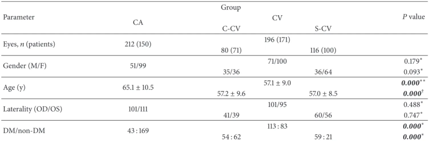
Table 1: Patient demographics.
Parameter
Group
𝑃 value
CA CV
C-CV S-CV
Eyes, 𝑛 (patients) 212 (150) 196 (171)
80 (71) 116 (100)
Gender (M/F) 51/99 71/100 0.179 ∗
35/36 36/64 0.093 ∗
Age (y) 65.1 ± 10.5 57.1 ± 9.0 0.000 ∗∗
57.2 ± 9.6 57.0 ± 8.5 0.000 †
Laterality (OD/OS) 101/111 101/95 0.488 ∗
41/39 60/56 0.747 ∗
DM/non-DM 43 : 169 113 : 83 0.000 ∗
54 : 62 59 : 21 0.000 ∗
Note. CA: cataract surgery alone; CV: cataract surgery with or after pars plana vitrectomy (PPV); C-CV: combined cataract surgery with PPV; S-CV: sequential cataract surgery after PPV; DM: diabetes mellitus.
∗ Fisher’s exact test.
∗∗ Independent 𝑡-test.
† One-way ANOVA test.
Table 2: Age-matched comparison of the mean time interval from cataract surgery to Nd:YAG capsulotomy.
Parameter Group
𝑃 value
CA CV
Mean time interval (M) 36.3 ± 26.4 29.1 ± 23.1 0.006 ∗
CA C-CV S-CV
Mean time interval (M) 36.3 ± 26.4 29.5 ± 24.4 28.8 ± 22.2 0.023 ∗
∗ ANCOVA test.
Table 3: Time interval differences from cataract surgery to Nd:YAG capsulotomy within subgroups.
Group 𝑃 value
Matching of subgroup
CA C-CV 0.042 ∗
CA S-CV 0.013 ∗
C-CV S-CV 0.321 ∗
∗ Contrast test after ANCOVA test.
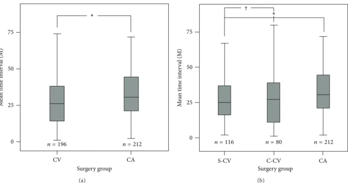
The mean time interval from cataract surgery to Nd:YAG capsulotomy was significantly longer in the CA group than in the CV group (36.3 ± 26.4 versus 29.1 ± 23.1 months;
𝑃 = 0.006). There was also a significant difference in mean time intervals from cataract surgery to Nd:YAG capsulotomy between the three groups when patients were matched for age: 36.3 ± 26.4 months (CA), 29.5 ± 24.4 months (C-CV), and 28.8 ± 22.2 months (S-CV; 𝑃 = 0.023; Table 2). Nd:YAG capsulotomy was performed later in the CA group than in the C-CV group (𝑃 = 0.013) and the S-CV group (𝑃 = 0.042;
Table 3; Figure 1). In vitrectomized eyes, regardless of the surgical sequence, the clinically significant PCO meant that the mean Nd:YAG capsulotomy free survival time was less than that in nonvitrectomized CA eyes.
Considering the IOL materials of silicone, hydrophobic acrylic, hydrophilic acrylic, and polymethyl methacrylate (PMMA), there were no differences in Nd:YAG capsulotomy free survival times between material types for the CV group
(𝑃 = 0.838). The IOL subgroup for CA eyes showed no difference in Nd:YAG capsulotomy free survival times between material types (P = 0.066), but, in the hydrophobic acrylic IOL subgroup, there was a significant difference in the Nd:YAG capsulotomy free survival time between the CA and CV groups. The CA subgroup in which eyes implanted the hydrophobic acrylic IOL showed a longer Nd:YAG capsulo- tomy free survival time (𝑃 = 0.028; Table 4; Figure 2).
There were no differences in Nd:YAG capsulotomy free survival times between DM and non-DM patients in either the CA group or the C-CV and S-CV subgroups. Nd:YAG capsulotomy free survival time was not correlated with diabetes history (Table 5). There was a weak yet significant correlation between the time intervals between PPV and cataract surgeries in the S-CV group and the Nd:YAG capsulotomy free survival time (𝑟 = 0.248, 𝑃 = 0.009;
Figure 3).
The survival curves for each group are shown in Figure 4.
The duration of Nd:YAG capsulotomy free survival after cataract surgery showed that the CA group had longer survival than the CV group (𝑃 = 0.003). Surgical sequence was significantly related to the timing of capsulotomy (𝑃 = 0.013), but age, DM, laterality of surgery, and gender were not (CV versus CA: 𝑃 = 0.973, 0.875, 0.103, and 0.774; S-CV and C-CV versus CA: 𝑃 = 0.974, 0.855, 0.105, and 0.757, resp.).
CV patients had larger hazard of earlier Nd:YAG capsulotomy
than CA’s by Cox proportional hazard model (hazard ratio
(HR) = 1.337, 95% CI 1.100–1.625, 𝑃 = 0.004). Both C-CV and
75
50
25
0
M ea n time in te rv al (M)
CV CA
Surgery group
n = 196 n = 212
∗
(a)
75
50
25
0
M ea n time in te rv al (M)
CA Surgery group
∗
S-CV C-CV
†
n = 116 n = 80 n = 212
(b)
Figure 1: Comparisons of mean time interval from cataract surgery to Nd:YAG capsulotomy in each group (gray bars). The CV group had a shorter time interval than the CA group ((a), 𝑃 = 0.006). The S-CV and C-CV groups had shorter time intervals than the CA group, ((b), 𝑃 = 0.013 and 0.043, resp.). There was no difference between the S-CV and C-CV groups (𝑃 = 0.321). The asterisk indicates a statistically significant difference in the mean time interval. A cross indicates no difference between S-CV and C-CV groups.
100
SI HO HI PMMA
IOL subgroup 75
50
25
0
M ea n time in te rv al (M)
∗
†
n = 66
n = 19 n = 132 n = 99
(a)
SI HO HI PMMA
IOL subgroup 75
50
25
0
M ea n time in te rv al (M)
∗
†
n = 16
n = 19 n = 11 n = 17
(b)
Figure 2: Comparisons of mean time intervals from cataract surgery to Nd:YAG capsulotomy within IOL subgroups. (a) Means comparison of IOL subgroup in the CV group, and (b) means the comparison of the CA group. Within the CV and CA groups, the IOL subgroup showed no difference in the mean time interval to Nd:YAG capsulotomy (𝑃 = 0.838 and 0.066, resp.). However, the hydrophobic acrylic IOL subgroup within the CV group showed a significantly earlier time to Nd:YAG capsulotomy than that of the CA group (𝑃 = 0.028). SI, HO, HI, and PMMA refer to silicone, hydrophobic acrylic, hydrophilic acrylic, and polymethyl methacrylate IOL materials, respectively. The asterisk indicates a statistical difference of the mean time interval to capsulotomy between the hydrophobic acrylic IOL subgroups of CV and CA.
The cross indicates no difference in the mean time interval of capsulotomy within the IOL subgroup.
Table 4: Comparison of the mean time interval within the IOL subgroup in cataract surgery with or after vitrectomy and cataract surgery alone.
Subgroup 𝑃 value
Silicone Hydrophobic acrylic Hydrophilic acrylic PMMA
Mean time interval (M)
CV 30.7 ± 29.3 29.1 ± 21.6 25.8 ± 19.9 27.1 ± 20.7 0.838 ∗
CA 36.4 ± 25.3 35.5 ± 22.1 22.6 ± 8.7 32.8 ± 15.9 0.066 ∗
𝑃 value 0.084 ∗∗ 0.028 † 0.909 ∗∗ 0.378 ∗∗
Note. IOL: intraocular lens; PMMA: polymethyl methacrylate.
∗ Kruskal-Wallis test.
∗∗ Mann-Whitney 𝑈 test.
† Independent 𝑡-test.
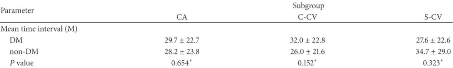
Table 5: Comparisons of the mean time interval from cataract surgery to Nd:YAG capsulotomy within the PPV subgroup.
Parameter Subgroup
CA C-CV S-CV
Mean time interval (M)
DM 29.7 ± 22.7 32.0 ± 22.8 27.6 ± 22.6
non-DM 28.2 ± 23.8 26.0 ± 21.6 34.7 ± 29.0
𝑃 value 0.654 ∗ 0.152 ∗ 0.323 ∗
∗ Independent 𝑡-test.
100
100 80
80 60
60 40
40 20
20 0
0
T ime in te rv al o f ca psulo to m y (M)
Time interval of sequence (M)
Figure 3: Age-matched partial correlation between the time interval between the PPV and cataract surgeries and the time interval between the cataract surgery and capsulotomy (𝑃 = 0.009, 𝑟 = 0.248). A longer time interval between PPV and cataract surgeries was associated with a longer time interval from cataract surgery to Nd:YAG capsulotomy.
S-CV patients had larger hazard than CA patients in earlier Nd:YAG capsulotomy, respectively (HR = 1.304, 95% CI = 1.007–1.688; 𝑃 = 0.044; HR = 1.361, 95% CI = 1.084–1.709, 𝑃 = 0.008, resp.). There was no difference in hazard between the C-CV and the S-CV groups (𝑃 = 0.767).
3.2. Discussion. Several previous studies have investigated factors associated with PCO. From biochemical investiga-
tions, various growth factors, extracellular matrices, inte- grins, and matrix metalloproteinases are related to lens epithelial cell (LEC) proliferation, migration, and transd- ifferentiation. Postoperative inflammation in the anterior chamber due to surgical trauma, the IOL design, and surgical technique contribute to PCO formation with progression in cataract surgery [11, 19]. Recently, several studies reported that PCO may occur after cataract surgery performed with or after PPV and causative factors were hypothesized. However, the effects of vitreous body and vitrectomy on PCO progres- sion after cataract surgery are still unknown.
We retrospectively evaluated Nd:YAG capsulotomy free survival times in patients over an extended period, whereas previous studies only focused on the PCO value and rate of laser capsulotomy after sequential or combined surgery.
However, the postoperative PCO free duration is also impor- tant for patients and ophthalmologists who need to plan post- PPV PCO and vitreoretinal pathology treatments, such as laser photocoagulation or intravitreal injections. The long- term analysis of PCO data should enable an understanding of factors that relate to its progression after cataract surgery with or after PPV.
Toda et al. [17] reported more extensive PCO formation after combined cataract surgery with vitrectomy than CA.
In their study, the combined surgery group showed a higher PCO value than that of the CA group in both DM and non- DM patients. They suggested that elevated cytokines caused by postoperative inflammation accelerated the LEC prolifer- ation via autocrine and/or paracrine signaling [17, 20–23]. In addition, Iwase et al. [24] showed a lower PCO rate using 23-gauge phacovitrectomy than when 20-gauge phacovitrec- tomy was used. They assumed that this was because the 23- gauge phacovitrectomy lowered postoperative inflammation.
However, in their study, although there was no difference in
the rate of capsulotomy between 23-gauge phacovitrectomy
1.0 0.8 0.6 0.4 0.2 0.0
1.0 0.8 0.6 0.4 0.2 Cu m s u rv iv al 0.0
0
0
24
24
12 36
48 72 96 120 144
Time interval (M) (a)
1.0 0.8 0.6 0.4 0.2 0.0 1.0
0.8 0.6 0.4 0.2 0.0
Cu m s u rv iv al
0 12 24 36
0 24 48 72 96 120 144
Time interval (M) (b)
5 4 3 2 1
C u m haza rd
0 0
12 24 36 48 60 72 84 96 108 120 Time interval (M)
Cataract surgery alone Cataract surgery with/after PPV
(c)
5 4 3 2 1
C u m haza rd
0
0 12 24 36 48 60 72 84 96 108 120 Time interval (M)
Cataract surgery alone
Combined cataract surgery with PPV Sequential cataract surgery after PPV
(d)
Figure 4: Cox proportional hazards model. Surgical option was identified as a risk factor that influenced the time interval from cataract surgery to capsulotomy. The CV group had a higher risk of a shorter Nd:YAG capsulotomy free survival time than the CA group ((c), hazard ratio (HR) = 1.337; 95% confidence interval (CI) 1.100–1.625; 𝑃 = 0.003). Combined surgery and sequential surgery were associated with an increased risk of a shorter Nd:YAG capsulotomy free survival time ((d), HR = 1.304; 95% CI; 1.007–1.688; 𝑃 = 0.44, HR = 1.361; 95% CI = 1.084–1.709; 𝑃 = 0.008, resp.). The CA group showed a longer Nd:YAG capsulotomy free survival than the CV group (a) and both the C-CV and S-CV groups (b).
and cataract only surgery, 23-gauge phacovitrectomy showed a higher PCO value than the cataract surgery only group.
Rahman et al. assumed that this discrepancy was due to the effect of posterior vitreous pressure on PCO. Furthermore, sequential cataract surgery after vitrectomy has been shown to lead to a higher incidence of PCO than combined surgery
[18]. In our study, Nd:YAG capsulotomy free survival times
showed no difference in both surgical option groups by
both statistical comparison and survival plot. Nevertheless,
in sequential surgery, the time interval from PPV to cataract
surgery had a weak but highly significant correlation with
the time interval from cataract surgery to capsulotomy. It
is hypothesized that postoperative inflammation may be related to PCO progression; hence, as the interval from PPV to cataract surgery increases, the post-PPV inflammation decreases.
Theoretically, loss of compression of the vitreous body after PPV may cause early PCO formation in sequential and combined surgeries. Nishi et al. [25] suggested that capsular bend formation is the mainstay of edge effect in square edge IOL, but, according to the loss of compression theory, elimination of the vitreous body causes decompression of the posterior capsule on the optic edge. This phenomenon incurs a loss of acute angle formation in the capsular bag- optic apparatus and more rapid LEC proliferation may occur.
In our study, the timing of Nd:YAG capsulotomy after cataract surgery was not dependent on the IOL material in vitrectomized eyes. The only significant difference in Nd:YAG capsulotomy free survival times between vitrectomized and nonvitrectomized eyes was when a hydrophobic acrylic IOL was used. The optic edge technology was applied in hydrophobic acrylic IOL; this indicated that a short Nd:YAG capsulotomy free survival time may be due to early PCO formation mainly caused by a loss of vitreous compression.
It is also possible that there is a change of vitreous circulation after vitrectomy. In the nonvitrectomized state, vitreous circulation of oxygen is limited by the vitreous body, which maintains a relatively low oxygenation state around the lens. Elevated oxygen tension after vitrectomy induces a relatively higher concentration of oxygen distribution near the lens [26, 27]. Therefore, after cataract surgery with or following PPV, relatively higher oxygen tension may be induced near the LECs and more rapid LEC proliferation may occur. As is known from neovascular glaucoma in proliferative diabetic retinopathy, cataract surgery would induce a higher concentration of vitreoretinal factors such as vascular endothelial growth factor near the LEC remnants.
This promotes the survival of LECs in a hypoxic state [28]
and leads to increased early migration and proliferation in the posterior capsule.
Our study was retrospective in nature, and there were numerous improvements in surgical materials and tech- niques over the 14-year review period, leading to potential differences in outcomes between the early and late study periods. Furthermore, in present study, there were a thousand or more cases with past history of laser capsulotomy; we decided to include selective cases that could identify at least one-year follow-up observation in medical records. However, with these exclusions, selection bias could occur because the patients who were attended to our Ophthalmology Department would have specific problems like diabetes, con- comitant vitreoretinal disease, and other medical disorders.
Particularly, patients with vitreoretinal disease would have chronicity of vision problems and would be enthusiastic to regular follow-up observations. Actually, the majority of exclusion was lost to follow-up; their distributions were higher in simple cataract surgery patients. However, our study provides some comprehensive indicators of the patho- physiology of PCO after cataract surgery with or after PPV. It should also be noted that the ages and proportion of patients with DM were different in each group. As age and diabetes
may be related to the rate of PCO after phacovitrectomy or cataract surgery only [17, 29], these intergroup differences may have introduced bias into our study. However, the use of age matching reduced the potential influence of this bias and age and diabetes were not identified as risk factors, which has also been noted in a previous study [18]. Furthermore, we retrospectively evaluated the surgical outcomes during longer period, so evolution of surgical techniques and surgical instruments, especially intraocular lenses, could be potential bias of this study. This point would be the limitations of this retrospective study.
4. Conclusions
We found that the progressions of PCO are more rapid in patients who underwent combined or sequential cataract surgery and vitrectomy than in patients undergoing CA.
For cataract surgery with or after vitrectomy, the formation of postoperative PCO must be considered and appropriate patient counseling and follow-up management should be provided.
Conflict of Interests
The authors declare that there is no conflict of interests regar- ding the publication of this paper.
References
[1] G. M. Cherfan, R. G. Michels, S. De Bustros, C. Enger, and B. M. Glaser, “Nuclear sclerotic cataract after vitrectomy for idiopathic epiretinal membranes causing macular pucker,”
American Journal of Ophthalmology, vol. 111, no. 4, pp. 434–438, 1991.
[2] J. T. Thompson, “The role of patient age and intraocular gas use in cataract progression after vitrectomy for macular holes and epiretinal membranes,” American Journal of Ophthalmology, vol. 137, no. 2, pp. 250–257, 2004.
[3] N. S. Melberg and M. A. Thomas, “Nuclear sclerotic cataract after vitrectomy in patients younger than 50 years of age,”
Ophthalmology, vol. 102, no. 10, pp. 1466–1471, 1995.
[4] S. De Bustros, J. T. Thompson, R. G. Michels, C. Enger, T. A.
Rice, and B. M. Glaser, “Nuclear sclerosis after vitrectomy for idiopathic epiretinal membranes,” American Journal of Ophthal- mology, vol. 105, no. 2, pp. 160–164, 1988.
[5] K. H. Lucke, M. H. Foerster, and H. Laqua, “Long-term results of vitrectomy and silicone oil in 500 cases of complicated retinal detachments,” American Journal of Ophthalmology, vol. 104, no.
6, pp. 624–633, 1987.
[6] J. L. Federman and H. D. Schubert, “Complications associated with the use of silicone oil in 150 eyes after retina-vitreous sur- gery,” Ophthalmology, vol. 95, no. 7, pp. 870–876, 1988.
[7] B. A. Blodi and S. A. Paluska, “Cataract after vitrectomy in young patients,” Ophthalmology, vol. 104, no. 7, pp. 1092–1095, 1997.
[8] S. B. Koenig, W. F. Mieler, D. P. Han, and G. W. Abrams, “Com-
bined phacoemulsification, pars plana vitrectomy, and posterior
chamber intraocular lens insertion,” Archives of Ophthalmology,
vol. 110, no. 8, pp. 1101–1104, 1992.
[9] K. Scharwey, S. Pavlovic, and K. W. Jacobi, “Combined clear cor- neal phacoemulsification, vitreoretinal surgery, and intraocular lens implantation,” Journal of Cataract and Refractive Surgery, vol. 25, no. 5, pp. 693–698, 1999.
[10] G. Yiu, K. V. Marra, S. Wagley et al., “Surgical outcomes after epiretinal membrane peeling combined with cataract surgery,”
British Journal of Ophthalmology, vol. 97, no. 9, Article ID 303189, pp. 1197–1201, 2013.
[11] N. Awasthi, S. Guo, and B. J. Wagner, “Posterior capsular opaci- fication: a problem reduced but not yet eradicated,” Archives of Ophthalmology, vol. 127, no. 4, pp. 555–562, 2009.
[12] M. A. Chang, M. K. Parides, S. Chang, and R. E. Braunstein,
“Outcome of phacoemulsification after pars plana vitrectomy,”
Ophthalmology, vol. 109, no. 5, pp. 948–954, 2002.
[13] S. M. Pinter, “Phacoemulsification in eyes with past pars plana vitrectomy: case-control study,” Journal of Cataract and Refrac- tive Surgery, vol. 25, no. 4, pp. 556–561, 1999.
[14] R. Ling, P. Simcock, J. McCoombes, and S. Shaw, “Presbyopic phacovitrectomy,” British Journal of Ophthalmology, vol. 87, no.
11, pp. 1333–1335, 2003.
[15] Y. Mochizuki, T. Kubota, Y. Hata et al., “Surgical results of combined pars plana vitrectomy, phacoemulsification, and intr- aocular lens implantation for various vitreoretinal diseases,”
European Journal of Ophthalmology, vol. 16, no. 2, pp. 279–286, 2006.
[16] Z. Szij´art´o, B. Haszonits, Z. Bir´o, and B. Kov´acs, “Phacoemul- sification on previously vitrectomized eyes: results of a 10-year period,” European Journal of Ophthalmology, vol. 17, no. 4, pp.
601–604, 2007.
[17] J. Toda, S. Kato, T. Oshika, and G. Sugita, “Posterior capsule opa- cification after combined cataract surgery and vitrectomy,” Jou- rnal of Cataract and Refractive Surgery, vol. 33, no. 1, pp. 104–107, 2007.
[18] J. H. Roh, H. J. Sohn, D. Y. Lee, K. H. Shyn, and D. H. Nam,
“Comparison of posterior capsular opacification between a combined procedure and a sequential procedure of pars plana vitrectomy and cataract surgery,” Ophthalmologica, vol. 224, no.
1, pp. 42–46, 2009.
[19] I. M. Wormstone, L. Wang, and C. S. C. Liu, “Posterior capsule opacification,” Experimental Eye Research, vol. 88, no. 2, pp. 257–
269, 2009.
[20] G. Ariki and N. Ogino, “Postoperative anterior chamber inflam- mation after posterior chamber intraocular lens implantation concurrent with pars plana vitrectomy and lensectomy,” Journal of Japanese Ophthalmological Society, vol. 96, no. 10, pp. 1300–
1305, 1992.
[21] O. Nishi and K. Nishi, “Intraocular lens encapsulation by shri- nkage of the capsulorhexis opening,” Journal of Cataract and Refractive Surgery, vol. 19, no. 4, pp. 544–545, 1993.
[22] N. Tachi, M. Kondo, H. Uchida, and N. Ogino, “Anterior cha- mber inflammation after vitrectomy in posterior vitreous mem- brane syndrome and phacoemulsification and intraocular lens implantation,” Journal of Japanese Ophthalmological Society, vol.
99, no. 3, pp. 329–335, 1995.
[23] O. Nishi, “Effects of the cytokines on the proliferation of and collagen synthesis by human cataract lens epithelial cells,” Bri- tish Journal of Ophthalmology, vol. 80, no. 1, pp. 63–68, 1996.
[24] T. Iwase, B. C. Oveson, and Y. Nishi, “Posterior capsule opacifi- cation following 20- and 23-gauge phacovitrectomy (posterior capsule opacification following phacovitrectomy),” Eye, vol. 26, pp. 1459–1464, 2012.
[25] O. Nishi, K. Nishi, C. Mano, M. Ichihara, and T. Honda, “The inhibition of lens epithelial cell migration by a discontinuous capsular bend created by a band-shaped circular loop or a capsule-bending ring,” Ophthalmic Surgery and Lasers, vol. 29, no. 2, pp. 119–125, 1998.
[26] I. A. Barbazetto, J. Liang, S. Chang, L. Zheng, A. Spector, and J.
P. Dillon, “Oxygen tension in the rabbit lens and vitreous before and after vitrectomy,” Experimental Eye Research, vol. 78, no. 5, pp. 917–924, 2004.
[27] N. M. Holekamp, Y.-B. Shui, and D. C. Beebe, “Vitrectomy sur- gery increases oxygen exposure to the lens: a possible mech- anism for nuclear cataract formation,” American Journal of Ophthalmology, vol. 139, no. 2, pp. 302–310, 2005.
[28] S. Neelam, M. M. Brooks, and P. R. Cammarata, “Lenticular cyt- oprotection. Part 1: the role of hypoxia inducible factors-1𝛼 and -2𝛼 and vascular endothelial growth factor in lens epithelial cell survival in hypoxia,” Molecular Vision, vol. 19, pp. 1–15, 2013.
[29] Y. Ebihara, S. Kato, T. Oshika, M. Yoshizaki, and G. Sugita, “Pos-
terior capsule opacification after cataract surgery in patients
with diabetes mellitus,” Journal of Cataract and Refractive
Surgery, vol. 32, no. 7, pp. 1184–1187, 2006.
Submit your manuscripts at http://www.hindawi.com
Stem Cells International
Hindawi Publishing Corporation
http://www.hindawi.com Volume 2014
Hindawi Publishing Corporation
http://www.hindawi.com Volume 2014
INFLAMMATION
Hindawi Publishing Corporation
http://www.hindawi.com Volume 2014
Behavioural Neurology
Endocrinology International Journal of
Hindawi Publishing Corporation
http://www.hindawi.com Volume 2014
Hindawi Publishing Corporation
http://www.hindawi.com Volume 2014
Disease Markers
Hindawi Publishing Corporation
http://www.hindawi.com Volume 2014
BioMed
Research International
Oncology Journal of
Hindawi Publishing Corporation
http://www.hindawi.com Volume 2014
Hindawi Publishing Corporation
http://www.hindawi.com Volume 2014
Oxidative Medicine and Cellular Longevity
Hindawi Publishing Corporation
http://www.hindawi.com Volume 2014
PPAR Research The Scientific World Journal
Hindawi Publishing Corporation
http://www.hindawi.com Volume 2014
Immunology Research
Hindawi Publishing Corporation
http://www.hindawi.com Volume 2014
Journal of
Obesity Journal of
Hindawi Publishing Corporation
http://www.hindawi.com Volume 2014
Hindawi Publishing Corporation
http://www.hindawi.com Volume 2014
Computational and Mathematical Methods in Medicine
Ophthalmology Journal of
Hindawi Publishing Corporation
http://www.hindawi.com Volume 2014
Diabetes Research Journal of
Hindawi Publishing Corporation
http://www.hindawi.com Volume 2014
Hindawi Publishing Corporation
http://www.hindawi.com Volume 2014
Research and Treatment
AIDS
Hindawi Publishing Corporation
http://www.hindawi.com Volume 2014
Gastroenterology Research and Practice
Hindawi Publishing Corporation
http://www.hindawi.com Volume 2014
Parkinson’s Disease
Evidence-Based Complementary and Alternative Medicine
Volume 2014 Hindawi Publishing Corporation
http://www.hindawi.com