© 2014 The Korean Ophthalmological Society
This is an Open Access article distributed under the terms of the Creative Commons Attribution Non-Commercial License (http://creativecommons.org/licenses /by-nc/3.0/) which permits unrestricted non-commercial use, distribution, and reproduction in any medium, provided the original work is properly cited.
Original Article
Comparison of Surgically-induced Astigmatism after Combined Phacoemulsification and 23-Gauge Vitrectomy:
2.2-mm vs. 2.75-mm Cataract Surgery
Yong-Kyu Kim1*, Yong Woo Kim2*, Se Joon Woo1, Kyu Hyung Park1
1Department of Ophthalmology, Seoul National University Bundang Hospital, Seoul National University College of Medicine, Seongnam, Korea
2Department of Ophthalmology, Seoul National University Hospital, Seoul National University College of Medicine, Seoul, Korea
Purpose: The 2.2-mm microincision cataract surgery and small-gauge vitrectomy system is known to result in less surgically-induced astigmatism (SIA) in comparison to conventional surgical methods. We compared the amounts of SIA after combined phacoemulsification and 23-gauge transconjunctival sutureless vitrectomy (23G-TSV) using the 2.2-mm microincision and 2.75-mm standard incision methods.
Methods: We studied 59 patients (61 eyes) who underwent combined phacoemulsification and 23G-TSV from November 2008 to September 2012. Twenty-eight patients (28 eyes) underwent 2.2-mm microincision coaxial phacoemulsification, and 31 patients (33 eyes) underwent 2.75-mm standard incision phacoemulsification. SIA was evaluated using Naeser’s polar method with the simulated keratometric values obtained from corneal topog- raphy. Preoperative and 1-week and 1-month postoperative KP (Naeser’s polar value along the specific axis) and ΔKP values were compared between the 2.2-mm microincision and 2.75-mm standard incision groups.
Results: One week after surgery, both groups exhibited similar amounts of SIA (-ΔKP[120], 0.40 ± 0.41 vs. 0.51
± 0.56 diopters [D]; p = 0.390). One month after surgery, however, the amount of SIA was significantly smaller in the 2.2-mm microincision group as compared to the 2.75-mm standard incision group (-ΔKP[120], 0.31 ± 0.54 vs. 0.56 ± 0.42 D; p = 0.045).
Conclusions: In combined phacoemulsification with 23G-TSV, 2.2-mm microincision coaxial phacoemulsifica- tion induces less SIA than does 2.75-mm standard coaxial phacoemulsification.
Key Words: Microincision, Phacoemulsification, Surgically-induced astigmatism, Transconjunctival sutureless vitrectomy
Owing to the development of new surgical tools and techniques, numerous ophthalmic surgeries can now be
performed non-invasively. New small-gauge instruments are continually developed, and most retinal surgeries are now performed by transconjunctival sutureless vitrectomy (TSV) [1-3]. Cataract surgeries through small corneal inci- sions are also increasing in popularity [4,5]. Small-gauge TSV (23- or 25-gauge) requires a smaller incision and therefore involves less surgically-induced astigmatism (SIA) than does its 20-gauge counterpart [5-12]. Microinci- sion coaxial cataract surgery requires a smaller incision than
Received: March 4, 2013 Accepted: May 28, 2013
Corresponding Author: Se Joon Woo, MD. Department of Ophthalmolo- gy, Seoul National University Bundang Hospital, #82 Gumi-ro 173beon- gil, Bundang-gu, Seongnam 463-707, Korea. Tel: 82-31-787-7377, Fax: 82- 31-787-4057, E-mail: [email protected]
*These authors contributed equally to this work.
that required for standard coaxial cataract surgery, but does not carry the steep learning curve of the bimanual micro- incision technique [4,13]. However, it remains unknown whether microincision coaxial cataract surgery combined with vitrectomy will produce less SIA than does the standard coaxial cataract surgery combination. In this study, we com- pared 2.2-mm microincision coaxial cataract surgery and 2.75-mm standard coaxial cataract surgery, both in combi- nation with 23-gauge transconjunctival sutureless vitrecto- my (23G-TSV), with respect to the severity of SIA.
Materials and Methods
Patients
This retrospective study comprised 59 patients (61 eyes) who underwent combined phacoemulsification and 23G-TSV from November 2008 to September 2012 at the retina and vitreous disease referral center of Seoul Nation- al University Bundang Hospital. All operations were per- formed by a single surgeon (SJW). Twenty-eight patients (28 eyes) underwent 2.2-mm microincision coaxial phacoemul- sification, and 31 patients (33 eyes) underwent 2.75-mm standard incision phacoemulsification. This study adhered to the tenets of the Declaration of Helsinki and was ap- proved by the institutional review board of Seoul National University Bundang Hospital. Those patients with a histo- ry of corneal trauma or corneal disease (dystrophies, de- generations, infections, etc.) that might influence corneal topography were excluded from the study. Patients who underwent additional scleral buckling or cryotherapy and those who were not available for serial corneal topography were also excluded. Two patients, one from each group, re- quired intraoperative sclerotomy site sutures for persistent leakage after cannula removal. No intraoperative or post- operative complications occurred. The patient demograph- ic characteristics, preoperative diagnoses, and operative procedures are summarized in Table 1.
Surgical procedure
Preoperatively, the pupil was dilated with a topical com- bination of 0.5% tropicamide and 0.5% phenylephrine hy- drochloride (Mydrin-P; Santen Pharmaceutical, Osaka, Japan). All surgeries were performed under local anesthesia
using a sub-Tenon injection of 2 mL of 2% lidocaine. A clear corneal incision was made at 11 o’clock. For the 2.2- mm microincision surgery, the initial incision was made with a 2.2-mm double-blade corneal knife. A 2.75-mm dou- ble-blade corneal knife was used for the 2.75-mm standard incision surgery. An additional puncture for insertion of the second instrument was made using a super-sharp blade at the 2 o’clock position of the limbus. Sodium hyaluronate 1.0% (Healon; Abbott Laboratories, Abbott Park, IL, USA) was used to stabilize the anterior chamber and to protect the corneal endothelium during the surgery. A 5.5-mm con- tinuous curvilinear capsulorhexis was performed using a 25-gauge needle and capsulorhexis forceps. Hydrodissec- tion and hydrodelineation were performed with balanced salt solution. Phacoemulsification was performed using the Intrepid Infiniti system (Alcon Laboratories, Fort Worth, TX, USA). After phacoemulsification, the remnant cortical lens material was removed with a coaxial irrigation/aspira- tion tip. An acrylic foldable intraocular lens (IOL), either an Akreos MI60 (Bausch & Lomb, Rochester, NY, USA) or an AcrySof SN60WF (Alcon Laboratories), whichever was available at the time of the operation, was inserted into the capsular bag. The Akreos MI60 was inserted into the cap- sular bag using a Viscoject injector and Viscoglide car- tridge system (Medicel AG, Widnau, Switzerland), while the AcrySof SN60WF was inserted into the capsular bag with a Monarch injector and C-cartridge system (Alcon Laboratories). Before vitrectomy, the corneal incision was sutured temporarily with a single stitch of 10-0 nylon to prevent eyeball collapse during the vitrectomy. Upon con- clusion of the surgery, the 10-0 nylon sutures were removed according to standard clinical practice. No case required suturing of the corneal incision due to wound leakage.
Upon completion of the cataract surgery, 23G-TSV was performed using the Accurus vitrectomy system (Alcon Laboratories). A 23-gauge stiletto blade (45° angle; DORC, Zuidland, The Netherlands) was inserted at a 15° to 30°
angle through the conjunctiva, sclera, and pars plana, 3.5 mm from the corneoscleral limbus, at the superotemporal, superonasal, and inferotemporal quadrants, respectively. A microcannula (DORC) was then inserted through the con- junctival incision and into the scleral tunnel using a spe- cially designed blunt inserter. At the end of the operation, the cannulae were withdrawn from their scleral tunnels, and the conjunctiva was pushed laterally with a cotton wool applicator to seal the puncture site. If there was per-
sistent leakage from the wound, the sclerotomy site was sutured with 8-0 vicryl. Postoperative treatment consisted of 0.5% levofloxacin (Cravit, Santen Pharmaceutical) and fluorometholone acetate 0.01% (Flarex, Alcon Laborato- ries) eye drops 4 times a day for 4 weeks.
Topographic and astigmatic evaluation
Corneal topography was performed using the Orbscan II system (Bausch & Lomb) before surgery and 1 week and 1 month after surgery. In this study, we evaluated astigma- tism using the polar method proposed by Naeser [14], Naeser and Hjortdal [15], and Naeser et al. [16]. Any pair of polar values separated by an arc of 45° (e.g., KP[90] and KP[135], KP[120] and KP[165], etc.) indicates the presence of net astigmatism [17]. Because the main corneal incision was made at the 11 o’clock position (120° from the horizon-
tal baseline), we evaluated astigmatism using KP(120) and KP(165) as follows:
In this setting, a positive ΔKP(120) indicates a steepen- ing of the meridian in which the incision was made, or Table 1. Patient demographic information, preoperative diagnoses, and operative procedures
2.2-mm microincision group 2.75-mm standard incision group p-value* Demographics
No. of eyes (patients) 28 (28) 33 (31) -
Male : female 16 : 12 15 : 18 0.363
Age 63.6 ± 8.8 64.6 ± 7.7 0.616
Right : left 14 : 14 14 : 19 0.554
Preoperative diagnosis (%) 0.155
RRD, retinal tear 2 (7.1) 2 (6.1)
Macular hole 1 (3.6) 8 (24.2)
Idiopathic ERM 13 (46.4) 13 (39.4)
PDR TRD, VH 7 (25.0) 9 (27.3)
RVO VH 1 (3.6) 1 (3.0)
AMD SRH, VH 3 (10.7) 0
Asteroid hyalosis 1 (3.6) 0
Operative procedures (%) 0.183
PE & PCL 28 (100) 33 (100)
Membrane peeling, removal 17 (60.7) 26 (78.8)
Endolaser 11 (39.3) 12 (36.4)
Intravitreal gas (SF6) injection 1 (3.6) 10 (30.3)
Intravitreal bevacizumab injection 1 (3.6) 3 (9.1)
Intravitreal SO injection 0 2 (6.1)
Sutured sclerotomy wound (%) 1 (3.6) 1 (3.0) >0.999
RRD = rhegmatogenous retinal detachment; ERM = epiretinal membrane; PDR = proliferative diabetic retinopathy; TRD = tractional retinal detachment; VH = vitreous hemorrhage; RVO = retinal vein occlusion; AMD = age-related macular degeneration; SRH = subretinal hemorrhage; PE & PCL = phacoemulsification and posterior chamber lens insertion; SF6 = sulfur hexafluoride; SO = silicone oil.
*p-value from Student’s t-test, chi-square test, or Fisher’s exact test.
KP(120) = M{sin2(α - 30) - cos2(α - 30)}
KP(165) = M{sin2(α - 75) - cos2(α - 75)}
SIA expressed as KP(120) = ΔKP(120) = KP(120)postop - KP(120)preop
SIA expressed as KP(165) = ΔKP(165) = KP(165)postop - KP(165)preop
KP = Naeser’s polar value along the specifi c axis
M = magnitude of the net astigmatism (diopters, D) taken from simulated keratometry (Sim K) values obtained from corneal topography
α = direction of the steepest meridian (degrees) SIA = surgically-induced astigmatism
with-the-rule change. A negative ΔKP(120) indicates flat- tening of the meridian in which the incision was made, or against-the-rule change. A positive ΔKP(165) indicates in- duced anticlockwise torque, while a negative ΔKP(165) in- dicates induced clockwise torque [16].
Statistical analysis
SPSS ver. 15.0 (SPSS Inc., Chicago, IL, USA) was used for all statistical analyses. Preoperative and postoperative Sim K astigmatism, KP(120) and KP(165), were compared using repeated-measures ANOVA. The ΔKP values were compared between the 2.2-mm microincision group and the
KP (120)
p < 0.001
0.4 0.0
0.2 -0.4
-0.2
Preoperative 1 wk 1 mon
p = 0.006
p = 0.260 p < 0.001
0.0 -0.4
-0.2 -0.8
-0.6
Preoperative 1 wk 1 mon
p < 0.001
p = 0.527
KP (120)
p < 0.001
0.4 0.0
0.2 -0.4
-0.2
Preoperative 1 wk 1 mon
p = 0.006
p = 0.260 p < 0.001
0.0 -0.4
-0.2 -0.8
-0.6
Preoperative 1 wk 1 mon
p < 0.001
p = 0.527
Fig. 1. Serial changes in KP(120) in the 2.2-mm microincision group (A, n = 28) and the 2.75-mm standard incision group (B, n = 33). A p-values was calculated using the paired t-test.
A B
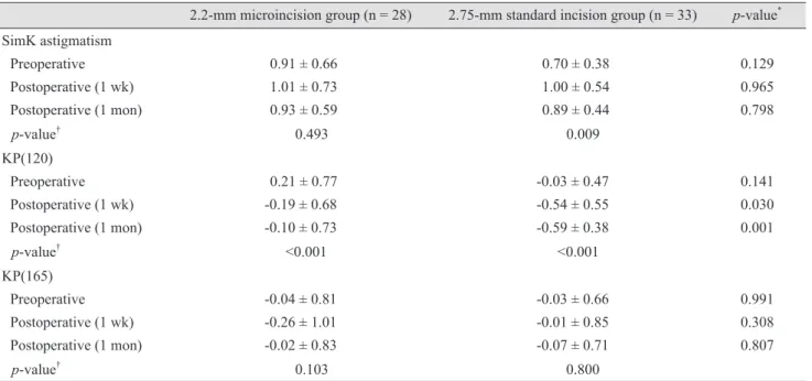
Table 2. Serial changes in Sim K astigmatism and KP values in the 2.2-mm microincision and 2.75-mm standard incision groups 2.2-mm microincision group (n = 28) 2.75-mm standard incision group (n = 33) p-value* SimK astigmatism
Preoperative Postoperative (1 wk) Postoperative (1 mon)
0.91 ± 0.66 1.01 ± 0.73 0.93 ± 0.59
0.70 ± 0.38 1.00 ± 0.54 0.89 ± 0.44
0.129 0.965 0.798
p-value† 0.493 0.009
KP(120) Preoperative Postoperative (1 wk) Postoperative (1 mon)
0.21 ± 0.77 -0.19 ± 0.68 -0.10 ± 0.73
-0.03 ± 0.47 -0.54 ± 0.55 -0.59 ± 0.38
0.141 0.030 0.001
p-value† <0.001 <0.001
KP(165) Preoperative Postoperative (1 wk) Postoperative (1 mon)
-0.04 ± 0.81 -0.26 ± 1.01 -0.02 ± 0.83
-0.03 ± 0.66 -0.01 ± 0.85 -0.07 ± 0.71
0.991 0.308 0.807
p-value† 0.103 0.800
Sim K = simulated keratometry.
*Student’s t-test; †Repeated-measures ANOVA.
2.75-mm standard incision group using Student’s t-test. A p-value less than 0.05 was considered statistically significant.
Results
The 2.2-mm microincision group showed no significant serial changes related to Sim K astigmatism (p = 0.493), while the 2.75-mm standard incision group did show sig- nificant changes ( p = 0.009) (Table 2). However, both groups showed significant changes (p < 0.001 for both) when we evaluated serial astigmatic changes using KP(120) (Table 2 and Fig. 1). Both groups showed similar negative changes in KP(120) 1 week after surgery (ΔKP[120], -0.40
± 0.41 for 2.2 mm vs. -0.51 ± 0.56 for 2.75 mm; p = 0.390) (Table 3 and Fig. 2). However, 1 month after surgery, the amount of SIA had decreased in the 2.2-mm microincision
group (ΔKP[120] = -0.31 ± 0.54), but had not changed in the 2.75-mm standard incision group (ΔKP[120] = -0.56 ± 0.42). The SIA values 1 month after surgery differed sig- nificantly between the two groups (p = 0.045) (Table 3 and Fig. 2). There was no significant induced torque along the surgical meridian (ΔKP[165]) in either group (Table 3).
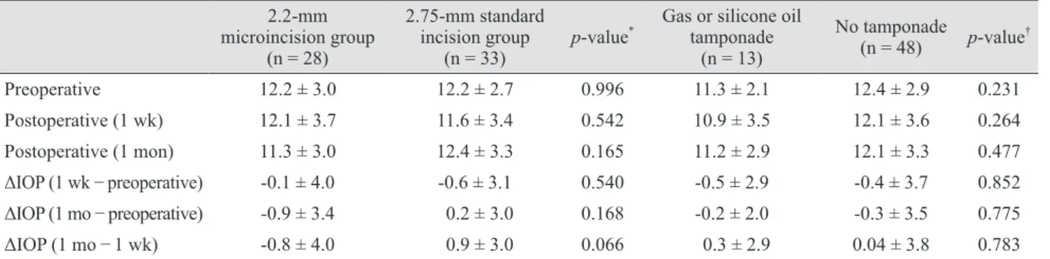
There were no significant differences in postoperative intraocular pressure (IOP) when the 2.2-mm microincision group was compared to the 2.75-mm standard incision group, or when the intraocular tamponade group was com- pared to the non-tamponade group (Table 4).
Discussion
In this study, we compared SIA between 2.2-mm-micro- incision and 2.75-mm standard incision coaxial phacoemul- sification combined with 23G-TSV. Stabilization of the cor- neal curvature and astigmatic and refractive changes after cataract surgery have been reported to be complete by two weeks after surgery when the incision size is <3.0 mm [18,19]. We evaluated astigmatic changes one week and one month after surgery to identify early postoperative astig- matic changes and serial changes in corneal astigmatism.
Both groups showed similar negative changes in KP(120) one week after surgery, with no significant difference in ΔKP(120) between the groups at this time. This means that both groups showed flattening of the cornea along the inci- sion site to a degree that did not differ significantly between the groups. However, after one month, the difference in SIA expressed as ΔKP(120) between groups increased sig- nificantly. The measurements obtained one-month postop- eratively therefore suggest that the 2.2-mm microincision surgery induced less SIA than the 2.75-mm standard incision surgery.
ΔKP(120)
p = 0.390
0.0 -0.4
-0.2 -0.8
-0.6
1 wk 1 mon
p = 0.045
2.2 mm 2.75 mm
Fig. 2. Comparison of surgically-induced astigmatism (represent- ed as ΔKP[120]) between the 2.2-mm microincision group and the 2.75-mm standard incision group. A p-values was calculated using Student’s t-test.
Table 3. Comparison of surgically-induced astigmatism in the 2.2-mm microincision group vs. the 2.75-mm standard incision group
2.2-mm microincision group (n = 28) 2.75-mm standard incision group (n = 33) p-value* ΔKP(120)
Postoperative (1 wk) Postoperative (1 mon)
-0.40 ± 0.41 -0.31 ± 0.54
-0.51 ± 0.56 -0.56 ± 0.42
0.390 0.045 ΔKP(165)
Postoperative (1 wk) Postoperative (1 mon)
-0.22 ± 0.55 0.01 ± 0.48
0.02 ± 0.65 -0.04 ± 0.55
0.126 0.705
*Student’s t-test.
The KP(165) value did not change significantly in either group, which means that no significant torque was induced.
Although we studied cases with combined cataract and vit- rectomy surgery, our result is comparable to those of previ- ous studies on the effect of incision size on SIA after cata- ract surgery. We summarized the results of such previous studies in Table 5. Most of the studies measured astigmatic changes 1 to 3 months after surgery and showed a trend of decreasing SIA as the size of the corneal incision decreased.
Furthermore, SIA as measured 1 to 3 months after surgery differed significantly between patients treated with 2.2-mm versus ≥2.75-mm incisions. In our study, the SIA one month after surgery was significantly smaller in the 2.2-mm mi- croincision group as compared to the 2.75-mm standard in- cision group. Therefore, the advantage of small-incision cataract surgery in terms of reducing SIA remains valid for
the combination cataract and 23G-TSV surgery.
We speculate that the combined vitrectomy surgery did not greatly influence the overall SIA patterns associated with corneal incision size due to the use of small-gauge sclerotomies without sutures. In previous studies compar- ing SIA between 23-gauge and 20-gauge vitrectomy com- bined with 2.2-mm microincision cataract surgery, the 20-gauge group showed significantly larger SIA at all post- operative times from 1 week through 12 weeks [20]. Al- though scleral incisions and sutures have less influence on corneal astigmatism in comparison to corneal incisions, vitrectomy alone induced astigmatism of approximately 3.0 D with a maximum change of almost 9.0 D during the first postoperative week [21]. This means that we cannot guar- antee results similar to those of our present study in the setting of 20-gauge vitrectomy.
Table 5. Summary of previous studies comparing surgically-induced astigmatism after cataract surgeries with different incision sizes
Researcher Methods* Results†
Kim et al. (2011) [12] 1.8-mm vs. 2.2-mm vs. 2.75-mm 2 mon
1.8-mm (0.21 D) ≈ 2.2-mm (0.29 D) < 2.75-mm (0.44 D) Can et al. (2010) [13] 1.2 to 1.4-mm (biaxial) vs. 2.2-mm
vs. 2.8-mm
90 day
1.2 to 1.4-mm (biaxial) (0.13 D) < 2.2-mm (0.24 D) < 2.8-mm (0.46 D) Wang et al. (2009) [9] 2.2-mm vs. 2.6-mm vs. 3.0-mm 30 day
2.2-mm (0.5 D) ≈ 2.6-mm (0.6 D) < 3.0-mm (0.9 D) 90 days
2.2-mm (0.4 D) ≈ 2.6-mm (0.5 D) < 3.0-mm (0.6 D) Masket et al. (2009) [10] 2.2-mm vs. 3.0-mm
intra-patient control
6 wk
2.2-mm (0.35 D) < 3.0-mm (0.67 D) D =diopter.
*Corneal incision sizes are shown here; those without parentheses were all performed using the coaxial technique; †The symbol ≈ denotes a statistically insignificant difference and < denotes a statistically significant difference. The numbers in the parentheses are mean surgically-induced astigmatism represented in D.
Table 4. Comparison of preoperative and postoperative IOP changes (mmHg) 2.2-mm
microincision group (n = 28)
2.75-mm standard incision group
(n = 33) p-value* Gas or silicone oil tamponade
(n = 13)
No tamponade
(n = 48) p-value†
Preoperative 12.2 ± 3.0 12.2 ± 2.7 0.996 11.3 ± 2.1 12.4 ± 2.9 0.231
Postoperative (1 wk) 12.1 ± 3.7 11.6 ± 3.4 0.542 10.9 ± 3.5 12.1 ± 3.6 0.264
Postoperative (1 mon) 11.3 ± 3.0 12.4 ± 3.3 0.165 11.2 ± 2.9 12.1 ± 3.3 0.477
ΔIOP (1 wk − preoperative) -0.1 ± 4.0 -0.6 ± 3.1 0.540 -0.5 ± 2.9 -0.4 ± 3.7 0.852
ΔIOP (1 mo − preoperative) -0.9 ± 3.4 0.2 ± 3.0 0.168 -0.2 ± 2.0 -0.3 ± 3.5 0.775
ΔIOP (1 mo − 1 wk) -0.8 ± 4.0 0.9 ± 3.0 0.066 0.3 ± 2.9 0.04 ± 3.8 0.783
IOP = intraocular pressure.
*Student’s t-test;†Mann-Whitney U-test.
Our study was limited by its small size, retrospective de- sign, and short follow-up periods, as well as our inability to match the two groups fully with respect to their preop- erative diagnoses and operative procedures. The numbers of patients who underwent intravitreal gas injection dif- fered significantly. However, we used sulfur hexafluoride (SF6) gas, which is retained in the eye for only a short peri- od (less than two weeks). Among patients who underwent microincision cataract surgery and 23G-TSV, SIA one week after surgery was significantly larger in the gas tam- ponade group than in the non-gas tamponade group. How- ever, this difference was diminished and statistically insig- nificant four weeks after surgery [20]. There were also no significant differences between the gas tamponade group and the non-gas tamponade group in the magnitude of IOP changes after surgery (Table 4). We therefore believe that the gas tamponade procedure did not affect our results sig- nificantly. Furthermore, the follow-up period of one month was relatively short. Although we found significantly less SIA in the 2.2-mm microincision group as compared to the 2.75-mm standard incision group after one month, the de- gree of astigmatism varied significantly from that observed preoperatively. In the 2.2-mm microincision group, SIA decreased at 1 month as compared to the SIA at 1 week, but this difference was not significant. Further study is needed to demonstrate the slow progression of any astigmatic changes in patients who have undergone 2.2-mm microin- cision surgery.
SIA was even less pronounced after biaxial microinci- sion surgery, which can be performed using an even small- er incision (1.2 to 1.4 mm) [13]. This implies that further reduction of the incision size would further decrease SIA.
However, biaxial microincision surgery has some disad- vantages, including a longer surgery, a steep learning curve, and poor corneal wound integrity [13,22]. Microincision coaxial phacoemulsification has advantages over other methods in this regard.
In conclusion, 2.2-mm microincision coaxial cataract surgery produced less SIA than did 2.75-mm standard co- axial cataract surgery in cases of combined phacoemulsifi- cation and 23G-TSV surgery. The combination of 23G-TSV and microincision cataract surgery, two less invasive sur- geries that require smaller incisions, improved patients’
chances of successful visual rehabilitation.
Conflict of Interest
No potential conflict of interest relevant to this article was reported.
Acknowledgements
This study was supported in part by a grant from the Korea Health Technology R&D Project, Ministry of Health and Welfare, Republic of Korea (grant no. A111161).
References
1. Lakhanpal RR, Humayun MS, de Juan E Jr, et al. Outcomes of 140 consecutive cases of 25-gauge transconjunctival sur- gery for posterior segment disease. Ophthalmology 2005;
112:817-24.
2. Eckardt C. Transconjunctival sutureless 23-gauge vitrecto- my. Retina 2005;25:208-11.
3. Fujii GY, De Juan E Jr, Humayun MS, et al. Initial experi- ence using the transconjunctival sutureless vitrectomy sys- tem for vitreoretinal surgery. Ophthalmology 2002;109:
1814-20.
4. Vasavada V, Vasavada V, Raj SM, Vasavada AR. Intraoper- ative performance and postoperative outcomes of microco- axial phacoemulsification. Observational study. J Cataract Refract Surg 2007;33:1019-24.
5. Alio J, Rodriguez-Prats JL, Galal A, Ramzy M. Outcomes of microincision cataract surgery versus coaxial phacoemul- sification. Ophthalmology 2005;112:1997-2003.
6. Okamoto F, Okamoto C, Sakata N, et al. Changes in corne- al topography after 25-gauge transconjunctival sutureless vitrectomy versus after 20-gauge standard vitrectomy.
Ophthalmology 2007;114:2138-41.
7. Yanyali A, Celik E, Horozoglu F, Nohutcu AF. Corneal topographic changes after transconjunctival (25-gauge) su- tureless vitrectomy. Am J Ophthalmol 2005;140:939-41.
8. Kim YK, Hyon JY, Woo SJ, et al. Surgically induced astig- matism after 23-gauge transconjunctival sutureless vitrec- tomy. Eye (Lond) 2010;24:799-804.
9. Wang J, Zhang EK, Fan WY, et al. The effect of micro-in- cision and small-incision coaxial phaco-emulsification on corneal astigmatism. Clin Experiment Ophthalmol 2009;37:
664-9.
10. Masket S, Wang L, Belani S. Induced astigmatism with 2.2- and 3.0-mm coaxial phacoemulsification incisions. J Re- fract Surg 2009;25:21-4.
11. Moon SC, Mohamed T, Fine IH. Comparison of surgically induced astigmatisms after clear corneal incisions of dif- ferent sizes. Korean J Ophthalmol 2007;21:1-5.
12. Kim EC, Byun YS, Kim MS. Microincision versus small-incision coaxial cataract surgery using different power modes for hard nuclear cataract. J Cataract Refract Surg 2011;37:1799-805.
13. Can I, Takmaz T, Yildiz Y, et al. Coaxial, microcoaxial, and biaxial microincision cataract surgery: prospective comparative study. J Cataract Refract Surg 2010;36:740-6.
14. Naeser K. Conversion of keratometer readings to polar val- ues. J Cataract Refract Surg 1990;16:741-5.
15. Naeser K, Hjortdal JO. Bivariate analysis of surgically in- duced regular astigmatism. Mathematical analysis and graphical display. Ophthalmic Physiol Opt 1999;19:50-61.
16. Naeser K, Knudsen EB, Hansen MK. Bivariate polar value analysis of surgically induced astigmatism. J Refract Surg
2002;18:72-8.
17. Naeser K, Behrens JK. Correlation between polar values and vector analysis. J Cataract Refract Surg 1997;23:76-81.
18. Masket S, Tennen DG. Astigmatic stabilization of 3.0 mm temporal clear corneal cataract incisions. J Cataract Re- fract Surg 1996;22:1451-5.
19. Oshika T, Tsuboi S. Astigmatic and refractive stabilization after cataract surgery. Ophthalmic Surg 1995;26:309-15.
20. Park DH, Shin JP, Kim SY. Surgically induced astigmatism in combined phacoemulsification and vitrectomy; 23-gauge transconjunctival sutureless vitrectomy versus 20-gauge standard vitrectomy. Graefes Arch Clin Exp Ophthalmol 2009;247:1331-7.
21. Wirbelauer C, Hoerauf H, Roider J, Laqua H. Corneal shape changes after pars plana vitrectomy. Graefes Arch Clin Exp Ophthalmol 1998;236:822-8.
22. Berdahl JP, DeStafeno JJ, Kim T. Corneal wound architec- ture and integrity after phacoemulsification evaluation of coaxial, microincision coaxial, and microincision bimanual techniques. J Cataract Refract Surg 2007;33:510-5.
![Fig. 2. Comparison of surgically-induced astigmatism (represent- (represent-ed as ΔKP[120]) between the 2.2-mm microincision group and the 2.75-mm standard incision group](https://thumb-ap.123doks.com/thumbv2/123dokinfo/5464626.658153/5.892.80.819.156.308/comparison-surgically-astigmatism-represent-represent-microincision-standard-incision.webp)