ISSN 0378-6471 (Print)⋅ISSN 2092-9374 (Online)
http://dx.doi.org/10.3341/jkos.2015.56.5.709
Original Article
유리체절제술과 백내장 병합수술 중 시행한 후낭절제술이 굴절력에 미치는 영향
Postoperative Refractive Errors after Posterior Capsulectomy during Combined Vitrectomy and Cataract Surgery
김정훈⋅한상범⋅이승준⋅김무상
Jung Hoon Kim, MD, Sang Beom Han, MD, Seung Jun Lee, MD, Moo Sang Kim, MD
강원대학교 의학전문대학원 안과학교실
Department of Ophthalmology, Kangwon National University School of Medicine, Chuncheon, Korea
Purpose: To evaluate the difference between the predicted and actual postoperative refractions after combined vitrectomy and cataract surgeries with a posterior capsulectomy.
Methods: We performed a retrospective study of 33 eyes of 33 patients who underwent combined vitrectomy and cataract sur- geries in our hospital between March 2013 and May 2014. The patients were divided into 2 groups: those who underwent a pos- terior capsulectomy (group A) and those who underwent combined vitrectomy and cataract surgeries (group B). The actual re- fractive errors were analyzed 3 months after surgery using spherical equivalent. We compared the results between the predicted and actual refractive errors in the two groups.
Results: Group A consisted of 25 eyes of 25 patients and group B of 8 eyes of 8 patients. In group A, the average difference be- tween predicted and actual postoperative refractive errors was -0.16 ± 0.38 D (p = 0.083). The predicted refraction was -0.33 ± 0.46 D and actual refraction was -0.49 ± 0.55 D in group A. There was no statistically significant difference between the predicted refraction and actual refractive errors in group A (p = 0.083). In group B, the average difference between predicted and actual postoperative refractive errors was 0.27 ± 0.29 D (p = 0.078). Additionally, the predicted refraction was -0.49 ± 0.77 D and actual refraction was -0.22 ± 0.59 D. The difference between the predicted refraction and actual refractive errors in group B was not statistically significant (p = 0.078).
Conclusions: When a posterior capsulectomy is performed during combined vitrectomy and cataract surgery, no significant dif- ference in refractive errors between the predicted refraction and actual refractive errors was observed 3 months after surgery.
Compared with combined vitrectomy and cataract surgeries only, a small amount of myopic refractive change tended to occur 3 months after surgery.
J Korean Ophthalmol Soc 2015;56(5):709-714
Key Words: Cataract surgery, Intraocular lens, Posterior capsulectomy, Refraction, Vitrectomy
■Received: 2014. 9. 12. ■ Revised: 2014. 11. 19.
■Accepted: 2015. 4. 20.
■Address reprint requests to Moo Sang Kim, MD
Department of Ophthalmology, Kangwon National University Hospital, #156 Baengnyeong-ro, Chuncheon 200-722, Korea Tel: 82-33-258-2014, Fax: 82-33-258-2191
E-mail: [email protected]
ⓒ2015 The Korean Ophthalmological Society
This is an Open Access article distributed under the terms of the Creative Commons Attribution Non-Commercial License (http://creativecommons.org/licenses/by-nc/3.0/) which permits unrestricted non-commercial use, distribution, and reproduction in any medium, provided the original work is properly cited.
유리체절제술은 최근의 비약적인 수술 기구 및 술기의 발전으로 그 빈도가 점차 증가하고 있다. 유리체절제술은 그 자체로서 백내장을 발생시키거나 진행시키는 것으로 알 려졌는데 이로 인하여 성공적인 유리체절제술 시행 이후에 백내장 수술이 추가적으로 요구되기도 한다.1,2 유리체절제 술을 시행 받은 눈은 술 후 해부학적 변화로 인하여 백내장 수술 중 후낭 파열 및 수정체 소대 손상과 같은 합병증이
증가되어 백내장 수술에 어려움이 있는 것으로 알려져 있 다.3,4 이러한 이유들로 유리체망막질환에서 유리체절제술 과 백내장을 동시에 수술하는 경우가 늘고 있다.
한편 후발백내장은 인공수정체 삽입술의 흔한 합병증으로 알려져 있고 Neodymium:yttrium-aluminum-garne (Nd:YAG) 레이저 후낭절개술은 후발백내장에 대한 비교적 안전한 치 료로서 치료 시행 시에 시력향상, 대비감도의 증가를 보이 는 것으로 보고되고 있다.5-7 이러한 합병증을 사전에 예방 하기 위한 방법으로 술자에 따라 유리체절제술과 백내장 병 합수술 중에 유리체절제기를 이용하여 후낭절제술(posterior capsulectomy)을 시행하기도 한다.8-10
최근 환자들의 시기능에 대한 기대치가 높아지면서 백내 장 수술에서 정확한 굴절력의 예측이 성공적인 수술을 위 한 중요한 요인으로 여겨지고 있다. Nd:YAG 레이저 후낭 절개술은 전방에서의 인공수정체 위치 변화 및 굴절력 변 화를 일으킬 수 있는 것으로 보고된 바 있으나11-13 아직까 지 국내에서 유리체절제술과 백내장 병합수술 중 시행한 후낭절제술이 일으키는 술 후 굴절력 변화에 대해서 연구 된 바는 없었다. 저자들은 유리체절제기를 이용한 후낭절 제술이 술 전의 안축장을 이용한 인공수정체 도수계산에 따른 예측 굴절력과 술 후의 실제 굴절력 간의 차이에 미치 는 영향에 대하여 알아보았다.
대상과 방법
저자들은 2013년 3월부터 2014년 6월까지 본원에서 망 막질환과 백내장으로 유리체절제술과 백내장 병합수술을 시행 받는 환자들을 대상으로 하였고 수술 중 유리체절제 기를 이용하여 후낭절제술을 시행한 환자를 A군, 후낭절제 술을 시행하지 않은 환자를 B군으로 분류하고 후향적으로 의무기록을 분석하였다. 병합수술은 초음파수정체유화술을 시행한 후 유리체절제술을 시행하였다. 백내장 수술은 이 측투명각막절개를 통하여 진행하였고 수정체초음파유화술 을 완료한 뒤에 후낭에 인공수정체를 삽입하였으며 절개창 은 10-0 nylon으로 봉합하였다. 유리체절제술은 고식적인 23게이지 표준 3포트 평면부 유리체절제술을 시행하였으며 술 중 유리체절제기를 이용하여 후낭의 중심부에 유리체절 제기를 위치시킨 후 직경 약 4 mm 정도의 크기로 후낭절제 술을 시행하였다. 대상군은 유리체절제술과 백내장 병합수 술 이후 3개월 이상 경과 관찰이 가능했던 경우만 포함시켰 으며, 인공수정체는 6.0 mm 크기의 광학부 직경을 가진 1 piece 아크릴재질의 Clare® (Cristalnes Indystrie, Lannion, France)와 Biovue4 PAL® (Aaren Scientific, Ontario, CA, USA)을 삽입한 경우만 포함하였으며 유리체절제술 중 안
내충전물을 사용한 경우는 연구 대상에서 제외하였다. 기 대 술 후 굴절도는 SRK-T 공식을 통하여 계산하였고, 안축 장의 측정은 IOL master® (Carl Zeiss, Jena, Germany)를 이용 하여 측정하였다. 술 후 시력 및 굴절도는 수술 후 3개월 뒤 외래 방문 시를 기준으로 하였고 굴절 검사는 Autorefractor (KW-2000, Kowa, Nagoya, Japan)를 사용하였다. 수술 후 3개 월째의 실제 굴절력과 SRK-T 공식을 이용한 예측 굴절력의 차이를 계산하였으며 이때 예측 굴절력은 IOL master®에 내장되어 있는 SRK-T 공식을 이용하였고, IOL Master에 특 화된 A 상수 값(118.5 for Clare®, 118.4 for Biovue4 PAL®) 을 사용하였으며 실제 굴절력은 구면렌즈대응치(spherical equivalent)로 계산하였다. 안축장의 길이는 공식 간의 차이 가 크지 않고 예상굴절력이 대체로 정확한 22.0 mm에서 25.0 mm를 대상으로 하였고 각막곡률도는 41.0 diopter (D) 에서 47.0D 사이의 환자를 대상으로 하였다. 통계분석은 SPSS 통계 분석 프로그램(version 12.0, SPSS Inc., Chicago, IL, USA)을 사용하여 두 군의 수술 전 예측 굴절값과 수술 후 실제 굴절값의 차이 비교 시에는 Mann-Whitney 검정을, 각 군의 수술 전과 후의 굴절값 차이는 Wilcoxon 부호순위 검정을 시행하였으며, p값이 0.05 미만인 경우에 통계적으 로 유의하다고 판단하였다.
결 과
유리체절제술과 백내장 병합수술을 시행하고 유리체절 제기를 사용하여 후낭절제술을 시행한 환자는 25명 25안으 로 남자가 15명 여자가 10명이었다. 수술 중에 수정체 후낭 절제술과 관련하여 발생한 합병증은 없었다. A군에 속하는 25명의 평균나이는 66.28 ± 10.43세였고, 평균 각막곡률도 는 44.45 ± 1.19D, 평균 안축장의 길이는 23.47 ± 0.81 mm, 수술 전 평균 최대교정시력(logMAR)은 0.46 ± 0.33이었다.
후낭절제술 없이 유리체절제술과 백내장 병합수술을 시 행한 환자는 8명 8안으로 남자가 7명 여자가 1명이었다. B 군에 속하는 8명 환자들의 평균나이는 59.50 ± 19.49세였으 며 평균 각막곡률도는 43.71 ± 1.27D, 평균 안축장의 길이 는 24.03 ± 1.41 mm, 수술 전 평균 최대교정시력(logMAR) 은 0.48 ± 0.32였다(Table 1). 두 그룹 간 나이와 평균 각막 곡률도, 안축장의 길이 및 수술 전 평균 최대교정시력에서 통계적으로 유의한 차이는 없었다(p=0.584, 0.172, 0.284 and 0.832, respectively).
수술 후 평균 최대교정시력(logMAR)은 A군은 0.15 ± 0.11이었고, B군은 0.14 ± 0.14였으며 두 군 간의 통계적으 로 유의한 차이는 없었다(Table 2, p=0.701).
수술 전 예측 굴절력과 수술 후 3개월에 측정한 실제 굴
Table 1. Baseline characteristics of phacovitrectomy with posterior capsulectomy group (group A) and without posterior capsu-
lectomy group (group B)Characteristics Group A Group B p-value
Number (patients/eyes) 25/25 8/8 -
Age (years) 66.28 ± 10.43 59.50 ± 19.49 0.584*
Sex (male/female) 15/10 7/1 -
Axial length (mm) 23.47 ± 0.81 24.03 ± 1.41 0.284*
Preoperative keratometry (diopter) 44.45 ± 1.19 43.71 ± 1.27 0.172*
Preoperative BCVA (log MAR) 0.46 ± 0.33 0.48 ± 0.32 0.832*
Values are presented as mean ± SD unless otherwise indicated.
BCVA = best-corrected visual acuity.
*Mann-Whitney U test.
Table 2. Preoperative best-corrected visual acuity (BCVA) and postoperative BCVA in phacovitrectomy with posterior capsu-
lectomy group (group A) and without posterior capsulectomy group (group B) at 3 months after the operationGroup A (n = 25) Group B (n = 8) p-value
Preoperative BCVA (log MAR) 0.46 ± 0.33 0.48 ± 0.32 0.832*
Postoperative BCVA (log MAR) 0.15 ± 0.11 0.14 ± 0.14 0.701*
Values are presented as mean ± SD.
*Mann-Whitney U test.
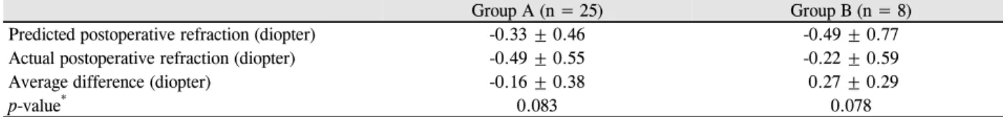
Table 3. Predicted postoperative refraction and actual postoperative refraction in phacovitrectomy with posterior capsulectomy
group (group A) and without posterior capsulectomy group (group B) at 3 months after the operationGroup A (n = 25) Group B (n = 8)
Predicted postoperative refraction (diopter) -0.33 ± 0.46 -0.49 ± 0.77
Actual postoperative refraction (diopter) -0.49 ± 0.55 -0.22 ± 0.59
Average difference (diopter) -0.16 ± 0.38 0.27 ± 0.29
p-value* 0.083 0.078
Values are presented as mean ± SD.
*Wilcoxon signed rank test.
절력의 차이를 각 군별로 비교한 결과를 보면, A군에서 수 술 전 예측 굴절력은 -0.33 ± 0.46D였고 수술 후 측정한 실 제 굴절력은 -0.49 ± 0.55D로 통계적으로 유의한 차이를 보 이지 않았다(Table 3, p=0.083). 평균적으로 -0.16 ± 0.38D 의 차이를 보였으며 총 25안 중 15안에서는 SRK-T로 예상 하였던 도수보다 -0.1D에서 -0.8D까지 근시 이행을 보였고 8안에서는 +0.1D에서 +0.7D까지 원시성 변화를 보였으며 2안은 예측 굴절력과 실제 굴절력이 일치하였다. B군에서 수술 전 예측 굴절력은 -0.49 ± 0.77D, 수술 후 측정한 실제 굴절력은 -0.22 ± 0.59D였고 평균적으로 0.27 ± 0.29D의 차 이를 보였으며 통계적으로 유의한 차이를 보이지 않았다 (Table 3, p=0.078).
고 찰
최근에 망막유리체질환이 합병된 백내장 환자들에서 유 리체절제술과 백내장 병합수술을 시행하는 경우가 점차 늘 고 있다. 유리체절제술의 합병증으로 백내장이 발생하는 경우는 수술의 방법 및 술자에 따라 다르게 보고되고 있는
데, 한 연구에서는 당뇨합병증으로 인한 유리체절제술 후 백내장의 발생을 80%까지 보고한 바 있다.14 특히 수정체 핵경화 백내장이 있는 노인은 대부분의 경우에서 백내장 진행이 심해져 백내장 수술을 시행하게 되는 경우가 많다. 따라서 이러한 병합수술은 추가적인 재수술의 가능성을 줄 여 환자에게 삶의 질 증대와 경제적 비용의 감소를 가져올 수 있다.
한편 유리체절제술의 시행은 이후에 백내장 수술 시행 시에 술기의 난이도를 높이고, 술 중 합병증의 가능성을 높 이는 것으로 알려졌다.15-17 이는 유리체절제술 후에 발생하 는 안구의 해부학적 변화에 기인하는데, 동공 산동이 충분 히 되지 않고, 수정체 후낭 혼탁 및 수정체 후 유착을 동반 할 수 있으며, 안구 내 유리체가 제거된 안구의 경우에는 백내장 수술 중 관류와 흡입시의 불균형이 쉽게 발생하고 전방 깊이의 급격한 변화를 초래하게 되어 후낭 파열, 수정 체 소대 손상과 같은 합병증 발생을 증가시키게 된다.3,4 유 리체절제술과 백내장 병합수술의 시행은 이러한 합병증의 발생을 피할 수 있고 백내장이 합병된 경우 선명한 주변부 망막 관찰이 가능해져 유리체절제술을 좀 더 용이하게 할
수도 있다.
후낭 혼탁은 백내장 수술 후 시력저하를 일으키는 대표 적인 합병증으로 10-50%까지 발생한다고 보고되었다.18,19 후낭 혼탁은 Nd:YAG 레이저를 이용하여 비교적 안전하고 간단하게 치료가 가능한데, 안압상승, 앞유리체혼탁, 낭포 황반부종, 망막박리, 안내염등의 합병증을 발생시키기도 한
다.20-24 유리체절제술과 백내장 병합수술을 시행 중에 후낭
절제술의 시행은 이러한 합병증을 미연에 방지할 수 있는 것으로 보인다. Park and Lee25는 유리체절제술과 백내장 병합수술 중 후낭절제술을 시행한 26안을 후향적으로 분석 하여 2안에서 시력에 영향을 주지 않는 1 mm 정도의 가벼 운 인공수정체 중심이탈 외에는 다른 합병증이 발생하지 않았으며 술 중 후낭절제술이 비교적 간단하고 안전하며 후낭 혼탁 예방에 효과적이었다고 하였다. 본 연구에서도 병합수술을 시행 중에 후낭절제술을 시행하였던 25안에서 유리체절제기를 이용하여 비교적 간단한 방법으로 제거할 수 있었고 후낭절제술과 관련된 합병증 없이 성공적으로 수술을 완료하였다.
수정체의 후낭은 해부학적으로 안구의 전방과 후방을 분 리하는 기능을 한다. 이러한 후낭의 기능은 방수와 유리체 사이에서 프로스타글란딘이나 혈관형성물질 같은 저분자 혹은 고분자물질들이 쉽게 통과할 수 없도록 하는 장벽 역 할을 한다.26 후낭절개술, 후낭절제술등으로 후낭이 제거되 어 전방과 후방 사이의 장벽으로서의 기능을 상실한 경우 에는 안구내의 염증매개물질과 혈관내피세포성장인자들이 이동하는 통로로 작용할 수 있으며, 특히 신생혈관형성을 유발하는 당뇨망막병증이나 망막정맥폐쇄증등의 허혈성망 막 질환에서는 신생혈관내피세포성장인자가 전방각으로 이동함으로써 신생혈관 녹내장이 발생할 가능성이 존재한
다.27,28 따라서 허혈성망막질환을 가진 환자에서는 병합 수
술 중 후낭절제술의 시행이 신중하게 고려되어야 할 것으 로 생각한다.
본 연구에서 유리체절제술과 백내장 병합수술을 받은 환 자에서 후낭절제술을 함께 시행하였을 때 굴절력의 변화를 보면 수술 전 예측 굴절력은 -0.33 ± 0.46D였고 수술 후 측 정한 실제 굴절력은 -0.49 ± 0.55D로 통계적으로 유의한 차 이를 보이지 않았다(Table 3, p=0.083). 임상적으로 중요한 것은 수술 중 시행한 후낭절제술이 수술 후에 굴절력을 변 화시키는 정도가 안경도수 혹은 인공수정체도수를 변경할 정도로 큰지 알아보는 것인데 환자들의 평균적인 변화를 보면 예측 굴절력보다 -0.16 ± 0.38D의 근시이행을 보여 예 측 굴절력과 미미한 차이를 보였다. 한편 통계적으로 유의 하진 않으나 후낭절제술을 시행하지 않은 경우에서 수술 전 예측굴절력과 수술 후의 굴절력 간의 평균 차이 0.27 ±
0.29D와 비교하여 약간의 근시성 변화를 보이는 경향이 나 타났다.
전방 내에서 인공수정체의 위치변화는 백내장 수술 이후 굴절오차를 발생시킬 수 있는 원인이 된다. 후낭은 전방과 후방을 나누는 장벽으로서 기능을 하는데, 사체안을 대상 으로 하였던 Yang et al29의 보고에 의하면 후낭은 3.76 ± 0.83 g의 장력을 견딘다고 알려져 있다. 병합수술 중의 후 낭절제술은 후낭의 개구부 형성으로 인하여 장벽의 기능을 상실하게 되며, 인공수정체는 후방으로부터의 압력(vitreous pressure)의 직접적인 영향에 놓이게 된다. 이는 특히 안내 충전물로 가스를 주입한 경우 더 큰 영향을 받을 것으로 보 이며, Suzuki et al30은 유리체절제술에서 안내가스충전물이 인공수정체의 전방이동을 일으켜 근시 이행을 나타낼 수 있다고 설명하였다. Nd:YAG 레이저 후낭절개술을 이용한 후낭혼탁의 치료 이후 인공수정체의 위치변화 및 굴절력변 화에 대해서는 상반된 연구들이 존재한다. Thornval and Naeser11는 52명의 인공수정체안을 대상으로 하여 후낭절 개술 이후 전방 깊이는 4.06 mm에서 4.07 mm로 증가하였 고, 굴절값은 0.30D에서 0.24D로 감소하였으나 유의한 변 화는 보이지 않았다고 하였다. Findl et al12은 32안을 대상 으로 하여 연구를 진행하였고 시술 이후 약간의 원시성 변 화와 인공수정체의 후방이동을 보였으나 유의한 차이는 없 었다고 보고하였다. 53안을 대상으로 하였던 Hu et al13, 22 안을 대상으로 하였던 Paik et al31은 전방 깊이의 감소를 보 였으나 통계학적으로 유의하지는 않았다고 하였다. 상기 연구들은 공통적으로 후낭절개 이후 인공수정체의 위치변 화와 굴절력을 설명하고 있는데, 후낭혼탁의 발생 및 Nd:YAG 레이저 후낭절개술의 시행시기는 백내장 수술 후 비교적 장기간의 시간이 지난 이후로 인공수정체가 이미 수정체낭 내에서 안정적인 위치를 가졌을 것이라고 생각된 다. 반면, 유리체절제술과 백내장 병합 수술 중에 후낭절개 술을 시행하는 경우는 인공수정체가 수정체낭 내에서 이동 성을 가지고 있는 시점이어서 후낭 개구부를 통한 후방의 압력에 비교적 민감할 것으로 판단된다. 이렇게 유발되는 전방으로의 인공수정체 위치 변화는 본 연구의 결과처럼 수술 후 근시성 변화에 영향을 미칠 것으로 생각한다.
다만 본 연구에서는 전방의 깊이 측정이나 인공수정체의 위치 변화 측정에 대한 분석이 이루어지지 않아 유리체절 제술과 백내장 병합수술에서 후낭절제술이 약한 근시성 이 행을 보이는 경향이 후낭절개에 의한 후방 압력에서 기인 하는지 아니면 다른 원인이 기여하는지 보다 정확한 원인 을 밝힐 수 없었다. 또한 추적관찰 기간이 3개월로 비교적 짧아 시간이 지난 뒤 인공수정체의 위치 변화에서 기인한 보다 장기적인 실제 굴절력까지 연구 대상으로 포함하지 못
하였다는 한계점이 있다. 추후 Pentacam® (Oculus, Wetzlar, Germany) 등을 활용한 전방에서의 인공수정체의 정밀한 위치 측정이 이러한 연구에 도움이 될 것으로 보인다.
결론적으로, 유리체절제술과 백내장 병합수술 시 후낭절 제술을 시행한 경우에서, 수술 후 측정되는 굴절력은 SRK-T 공식을 통해 예상한 인공수정체 도수 계산과 비교 하여 유의한 차이가 없었다. 다만 병합수술만 시행하였던 경우와 비교하여 약한 정도의 근시 이행이 나타나는 경향 을 보였다. 이러한 결과는 병합수술 시에 후낭절제술을 시 행하려는 환자에서 수술 후 보다 정확한 굴절상태를 예측 하는 데 도움이 될 것으로 보여지며 추후 더 많은 환자들을 대상으로 장기간의 연구가 필요할 것으로 생각한다.
REFERENCES
1) Heimann H, Bartz-Schmidt KU, Bornfeld N, et al. Scleral buckling versus primary vitrectomy in rhegmatogenous retinal detachment:
a prospective randomized multicenter clinical study. Ophthalmology 2007;114:2142-54.
2) Han NS, Lee SB, Kim YB, Jo YJ. Results of triple surgery: cataract extraction, intraocular lens implantation and vitrectomy for retinal detachment. J Korean Ophthalmol Soc 2004;45:2041-6.
3) Misra A, Burton RL. Incidence of intraoperative complications during phacoemulsification in vitrectomized and nonvitrectomized eyes: prospective study. J Cataract Refract Surg 2005;31:1011-4.
4) Biró Z, Kovacs B. Results of cataract surgery in previously vitrec- tomized eyes. J Cataract Refract Surg 2002;28:1003-6.
5) Tan JC, Spalton DJ, Arden GB. The effect of neodymium: YAG capsulotomy on contrast sensitivity and the evaluation of methods for its assessment. Ophthalmology 1999;106:703-9.
6) Magno BV, Datiles MB, Lasa MS, et al. Evaluation of visual func- tion following neodymium:YAG laser posterior capsulotomy.
Ophthalmology 1997;104:1287-93.
7) Weiblinger RP. Review of the clinical literature on the use of the Nd:YAG laser for posterior capsulotomy. J Cataract Refract Surg 1986;12:162-70.
8) Gimbel HV, Neuhann T. Development, advantages, and methods of the continuous circular capsulorhexis technique. J Cataract Refract Surg 1990;16:31-7.
9) Gimbel HV. Posterior continuous curvilinear capsulorhexis and optic capture of the intraocular lens to prevent secondary opacifica- tion in pediatric cataract surgery. J Cataract Refract Surg 1997;23 Suppl 1:652-6.
10) Galand A, van Cauwenberge F, Moosavi J. Posterior capsulorhexis in adult eyes with intact and clear capsules. J Cataract Refract Surg 1996;22:458-61.
11) Thornval P, Naeser K. Refraction and anterior chamber depth be- fore and after neodymium: YAG laser treatment for posterior cap- sule opacification in pseudophakic eyes: a prospective study. J Cataract Refract Surg 1995;21:457-60.
12) Findl O, Drexler W, Menapace R, et al. Changes in intraocular lens
position after neodymium: YAG capsulotomy. J Cataract Refract Surg 1999;25:659-62.
13) Hu CY, Woung LC, Wang MC, Jian JH. Influence of laser posterior capsulotomy on anterior chamber depth, refraction, and intraocular pressure. J Cataract Refract Surg 2000;26:1183-9.
14) Blankenship GW. Stability of pars plana vitrectomy results for dia- betic retinopathy complications. A comparison of five-year and six-month postvitrectomy findings. Arch Ophthalmol 1981;99:1009-12.
15) Kim JW, Yang JW, Jee DH. Stability of four-haptic intraocular lens in combined phacoemulsification and vitrectomy. J Korean Ophthalmol Soc 2010;51:829-34.
16) Hutton WL, Pesicka GA, Fuller DG. Cataract extraction in the dia- betic eye after vitrectomy. Am J Ophthalmol 1987;104:1-4.
17) Kim EY, Ahn JH, Lew HM, Yang HS. Effect of vitrectomy on IOL calculation for cataract surgery : study of vitrectomized eyes. J Korean Ophthalmol Soc 2008;49:1759-64.
18) Kappelhof JP, Vrensen GF. The pathology of after-cataract. A minireview. Acta Ophthalmol Suppl 1992;13-24.
19) McDonnell PJ, Zarbin MA, Green WR. Posterior capsule opacifi- cation in pseudophakic eyes. Ophthalmology 1983;90:1548-53.
20) Steinert RF, Puliafito CA, Kumar SR, et al. Cystoid macular ede- ma, retinal detachment, and glaucoma after Nd:YAG laser posteri- or capsulotomy. Am J Ophthalmol 1991;112:373-80.
21) Stark WJ, Worthen D, Holladay JT, Murray G. Neodymium: YAG lasers. An FDA report. Ophthalmology 1985;92:209-12.
22) Bath PE, Fankhauser F. Long-term results of Nd:YAG laser poste- rior capsulotomy with the Swiss laser. J Cataract Refract Surg 1986;12:150-3.
23) Kumagai K, Ogino N, Shinjo U, et al. Vitreous opacification after neodymium:YAG posterior capsulotomy. J Cataract Refract Surg 1999;25:981-4.
24) Lewis H, Singer TR, Hanscom TA, Straatsma BR. A prospective study of cystoid macular edema after neodymium: YAG laser pos- terior capsulotomy. Ophthalmology 1987;94:478-82.
25) Park SE, Lee SJ. Mechanized posterior capsulectomy during com- bined vitrectomy and cataract surgery. J Korean Ophthalmol Soc 2007;48:1335-40.
26) Ohrloff C, Schalnus R, Rothe R, Spitznas M. Role of the posterior capsule in the aqueous-vitreous barrier in aphakic and pseudo- phakic eyes. J Cataract Refract Surg 1990;16:198-201.
27) Weinreb RN, Wasserstrom JP, Parker W. Neovascular glaucoma following neodymium-YAG laser posterior capsulotomy. Arch Ophthalmol 1986;104:730-1.
28) Poliner LS, Christianson DJ, Escoffery RF, et al. Neovascular glau- coma after intracapsular and extracapsular cataract extraction in di- abetic patients. Am J Ophthalmol 1985;100:637-43.
29) Yang X, Zou L, Binrong M, et al. Tensile strength of lens capsules in eye-bank eyes. J Cataract Refract Surg 1998;24:543-6.
30) Suzuki Y, Sakuraba T, Mizutani H, et al. Postoperative refractive error after simultaneous vitrectomy and cataract surgery.
Ophthalmic Surg Lasers 2000;31:271-5.
31) Paik JS, Ku HC, Lee YC, Kim HS. The change in ACD and re- fraction after Nd:YAG laser posterior capsulotomy. J Korean Ophthalmol Soc 2006;47:905-12.
= 국문초록 =
유리체절제술과 백내장 병합수술 중 시행한 후낭절제술이 굴절력에 미치는 영향
목적: 유리체절제술과 백내장 병합수술 중 후낭절제술을 시행한 경우에서 수술 전 예상 굴절력과 수술 후 실제 굴절력 간의 차이를 알아보았다.
대상과 방법: 2013년 3월부터 2014년 5월까지 본원에서 유리체절제술과 백내장 병합수술을 받은 환자들 중 수술 후 3개월까지 추적관 찰이 가능했던 33명 33안을 대상으로 수술 중 후낭절제술을 시행했던 환자들을 A군, 시행하지 않았던 환자들을 B군으로 나누어 두 군의 수술 전 예상 굴절력과 수술 후 실제 굴절력을 후향적으로 분석하였다. 실제 굴절력은 수술 후 3개월에 측정한 구면렌즈대응치 로 평가하였다.
결과: 유리체절제술과 백내장 병합수술 시 인공수정체 삽입술 후 후낭절제술을 시행했던 A군은 25명 25안이었고, 후낭절제술을 시행 하지 않았던 B군은 8명 8안이었다. 수술 후 예상 굴절력과 실제 굴절력 간의 차이를 보면 A군에서 수술 전 인공수정체 예상 굴절력이 -0.33 ± 0.46D였고 실제 굴절력은 -0.49 ± 0.55D로 평균적으로 -0.16 ± 0.38D (p=0.083)의 차이를 보였으나 통계적으로 유의하 지 않았으며, B군에서 예상 굴절력은 -0.49 ± 0.77D였고 실제 굴절력은 -0.22 ± 0.59D로 평균 차이는 0.27 ± 0.29D (p=0.078)였고 통계적으로 유의한 차이를 보이지 않았다.
결론: 유리체절제술과 백내장 병합수술에서 후낭절제술을 시행한 경우, 수술 후 3개월에 측정한 실제 굴절력은 예측하였던 굴절력과 통계적으로 유의한 차이를 보이지 않았다. 다만 후낭절제술을 시행하지 않은 경우보다 약간의 근시 이행을 나타내는 경향을 보였다.
<대한안과학회지 2015;56(5):709-714>