Synthesis of Mn-doped Zn
2SiO
4phosphor particles by solid-state method at relatively low temperature
and their photoluminescence characteristics
Jin-Hwa, Lee
1, Seung-Ok, Choi
2and Dong-Kyu, Lee
1*1Department of Industrial Chemical Engineering, College of Engineering, Chungbuk National University
2Dong Yang Oil & Fat Co., Ltd.,
상대적으로 낮은 온도에서의 고상법에 의한 망간이 도핑된 Zn
2SiO
4형광체 입자의 제조 및 형광특성
이진화
1, 최성옥
2, 이동규
1*1충북대학교 공과대학 공업화학과
2(주)동양유지
Abstract Mn-doped Zn2SiO4 phosphor particles having submicrometer sizes were synthesized by a solid-state reaction method using methyl hydrogen polysiloxane-treated ZnO, fumed SiO2 and various Mn sources. The crystallization and photoluminescent properties of the phosphor particles were investigated by X-ray diffraction(XRD), scanning electron microscope(SEM), and by their photoluminescence(PL) spectra. Due to the effect of the dispersion and coherence of the methyl hydrogen polysiloxane-treated ZnO, the Mn-doped Zn2SiO4
particles were successfully obtained by a solid state method at 1000℃, and the maximum PL intensity of the prepared particles under vacuum ultra violet(VUV) excitation occurred at a Mn concentration of 0.02mol and a sintering temperature of 1000℃.
요 약 Methyl hydrogen polysiloxne으로 처리한 ZnO, fumed SiO2와 다양한 망간 전구체를 이용하여 서브마이크로미 터 크기를 갖는 망간이 도핑된 Zn2SiO4 형광체 입자를 고상법으로 제조하였다. 결정화와 광발광 특성은 XRD, SEM, PL스펙트라를 이용하여 분석하였다. 고상법으로 제조한 망간 도핑된 Zn2SiO4는 methyl hydrogen polysiloxne 처리한 ZnO의 분산과 응집 때문에 1000℃에서 성공적으로 얻어졌고, 진공자외선 여기하에서 제조된 입자의 최대 PL강도는 0.02mol Mn, 1000℃에서 확인되었다.
Key Words : Solid-State Reaction, Phosphor, Photoluminescence, Zn2SiO4
*Corresponding Author : Lee, Dong-Kyu([email protected])
Received October 20, 2009 Revised December 24, 2009 Accepted January 20, 2010
1. Introduction
Inorganic phosphor materials have been widely used in modern lighting and display parts, such as fluorescent lamps, cathode-ray tubes, field emission displays and plasma display panels[1]. In particular, the luminescent properties of inorganic phosphors have been widely
investigated for commercial use in flat panel displays(FPDs) in the recent years[2,3]. It is highly desirable to develop novel low-voltage phosphors for next generation field emission displays(FEDs), that have a high efficiency and good chemical stability under electron-beam bombardment in a high vacuum system[4,5]. Phosphor particles must have a small
diameter and narrow size distribution. Moreover, they must be non-aggregated, and have a spherical morphology for good luminescent characteristics. Mn-doped Zn2SiO4 is mainly used as a green-emitting phosphor material in plasma display panels because of its good luminescence characteristics and chemical stability under vacuum ultraviolet (VUV) excitation[6]. Green-emitting Mn-doped Zn2SiO4 is a well-known phosphor for its high luminescent efficiency and chemical stability. The emission of Zn2SiO4:Mn at 524nm is attributed to a d-level spin-forbidden transition for Mn2+.
Up to the present time, the commercial Mn-doped Zn2SiO4 green phosphors have been synthesized mainly with a solid-state reaction method[7]. In the solid-state reaction method, a high reaction temperature, a long heating time, and a milling process are required. Due to this combination of requirements, new synthesis methods such as r. f. magnetron sputtering[8], sol-gel[9-12], hydrothermal[13,14], and an ultrasonic spray pyrolysis method[6] have been studied.
The objectives of the present study are to synthesize green-light-emitting phosphor particles with a submicron size at relatively low temperature using a solid-state reaction method, and to characterize their photoluminescent(PL) properties. The effect of different Mn sources and sintering temperatures on the PL characteristics of Mn-doped Zn2SiO4 phosphor particles is also investigated.
2. Experimental
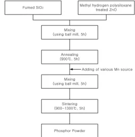
Mn-doped Zn2SiO4 green phosphors were synthesized with a solid-state method. Fumed SiO2 and methyl hydrogen polysiloxane-treated ZnO powders were mixed in a ball mill and sintered at 900℃ for 5h in an air atmosphere to prepare Zn2SiO4 particles as a host material. After being reground, a respective Mn source(MnCO3, MnO2) was added and mixed homogeneously in the ball mill to prepare Mn-doped Zn2SiO4 phosphor[15]. This mixture were sintered at 100 0℃, 1100℃, 1200℃, and 1300℃ for 5h in an air atmosphere. The preparation procedure with the solid-state reaction method is shown in Fig. 1.
In all cases, the phase and crystallinity of the prepared
phosphor particles were characterized using X-ray diffractometer(XRD, Sintag Model XDS 2000) with CuK α radiation. The particle morphology was determined in accordance with a scanning electron microscopy(SEM, Hitachi S-2500C). The photoluminescence characteristics of prepared particles were measured by a vacuum ultraviolet photoluminescence spectro- meter(VUV PL, Milton Roy 3000 Array) using a Kr lamp.
Fumed SiO2 Methyl hydrogen polysiloxane treated ZnO
Mixing (using ball mill, 5h)
Annealing (900℃, 5h)
Mixing (using ball mill, 5h)
Adding of various Mn source
Sintering (900-1300℃, 5h)
Phosphor Powder
[Fig. 1] Synthesis procedure of the Mn-doped Zn2SiO4
phosphor particles using the solid-state reaction method.
3. Results and discussion
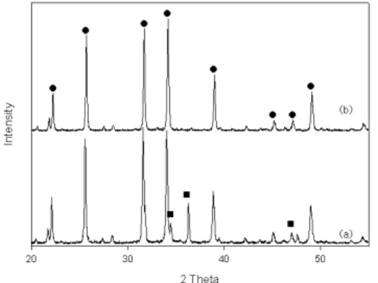
Fig. 2 shows the XRD patterns of Zn2SiO4 annealed at 900℃ with pure ZnO and methyl hydrogen polysiloxane-treated ZnO. When the pure ZnO was used as a Zn source, the peak of the ZnO was detected, but when the methyl hydrogen polysiloxane-treated ZnO was used as a Zn source, only the pure Zn2SiO4's peak was detected. Due to the effect of the dispersion and coherence of the methyl hydrogen polysiloxane-treated ZnO, only a relatively low sintering temperature was needed for the Zn2SiO4 crystal synthesis. Fig. 3 shows the XRD patterns of the Mn-doped Zn2SiO4 phosphor particles synthesized by the solid-state reaction method when sintered at 1000℃, 1100℃, 1200℃, and 1300℃
using the Mn sources of MnCO3 and MnO2. All of the XRD analyses showed a typical Zn2SiO4 crystal structure
with a 1000℃ sintering temperature. From the XRD analysis, the characteristic peaks of the dopants were not observed.
[Fig. 2] XRD patterns of the Zn2SiO2 host material annealed at 900℃(●:Zn2SiO4,■:ZnO).
(a)pure ZnO, (b)methyl hydrogen polysiloxane-treated ZnO
(a) (b)
[Fig. 3] XRD patterns of the Zn2SiO4:Mn phosphor prepared with the solid-state reaction method at various sintering temperatures.
(a) MnCO3 as the Mn source, (b)MnO2 as the Mn source
Fig. 4 and 5 are the SEM micrographs of the Mn-doped Zn2SiO4 phosphor particles prepared with different MnCO3 concentrations and different sintering temperatures. In Fig. 4, as the MnCO3 concentration increased from 0.01 to 0.04mol, the particle size increased from 0.5 to 1.2㎛. These as-prepared particles were sintered at 1000℃ for 5h. The results show that smaller particles were synthesized at a lower Mn concentration, and more agglomerated particles were synthesized at a higher Mn concentration. In Fig. 5, as the sintering
temperature increased from 1000℃ to 1300℃, the particle size increased from 0.5 to 20㎛.
(a) (b)
(c) (d)
[Fig. 4] SEM micrographs of the Mn-doped Zn2SiO4 phosphor particles prepared using MnCO3 as the Mn source at different Mn concentrations.
(a) 0.01mol, (b)0.02mol, (c)0.03mol, (d)0.04mol
(a) (b)
(c) (d)
[Fig. 5] SEM micrographs of the Mn-doped Zn2SiO4
phosphor particles prepared using MnCO3 as the Mn source at different sintering temperatures.
(a)1000℃, (b)1100℃, (c)1200℃, (d)1300℃
Fig. 6 and 7 show the SEM micrographs of the Mn-doped Zn2SiO4 phosphors particle prepared with different MnO2 concentrations and different sintering temperatures. In Fig. 6, as the MnO2 concentration increased from 0.01 to 0.04mol, the particle size increased from 0.4 to 1.5㎛. These as-prepared particles were
sintered at 1000℃ for 5h. The results show that smaller particles were synthesized at a lower Mn concentration, and more agglomerated particles were synthesized at a higher Mn concentration. In Fig. 7, as the sintering temperature increased from 1000℃ to 1300℃, the particle size increased from 0.4 to 15㎛.
Green light was emitted from these phosphor particles under VUV irradiation. Fig. 8 shows the dependence of Mn activator(MnCO3, MnO2) concentrations on the PL intensity for the Mn-doped Zn2SiO4 phosphor particles sintered at 1000℃. It was found that there were distinct differences in the PL intensity of the Mn-doped Zn2SiO4 phosphor particles at Mn concentrations ranging from 0.01mol to 0.03mol. The maximum PL intensity of a phosphor particle was shown at a Mn concentration of 0.02mol, and it decreased when the Mn concentration increased due to the concentration quenching effect[16].
(a) (b)
(c) (d)
[Fig. 6] SEM micrographs of the Mn-doped Zn2SiO4
phosphor particles prepared using MnO2 as theMn source at different Mn concentrations.
(a)0.01mol, (b)0.02mol, (c)0.03mol, (d)0.04mol
(a) (b)
(c) (d)
[Fig. 7] SEM micrographs of the Mn-doped Zn2SiO4
phosphor particles prepared using MnO2 as the Mn source at different sintering temperatures.
(a)1000℃, (b)1100℃, (c)1200℃, (d)1300℃
nm
350 400 450 500 550 600 650 700 750 0.0
0.2 0.4 0.6 0.8
1.0 C o m m e r c i a l
Mn/Zn= 0.050 Mn/Zn= 0.075 Mn/Zn= 0.025
nm
350 400 450 500 550 600 650 700 750 0.0
0.2 0.4 0.6 0.8
1.0 Mn/Zn= 0.050C o m m e r c i a l Mn/Zn= 0.025 Mn/Zn= 0.075
(a) (b)
[Fig. 8] PL spectra of the Mn-doped Zn2SiO4 particles prepared from different Mn concentrations.
(a)MnCO3 as the Mn source, (b)MnO2 as the Mn source
nm
350 400 450 500 550 600 650 700 750 0.0
0.2 0.4 0.6 0.8 1.0
1000 oC C o m m e r c i a l 900 oC
1100 oC 1200 oC
1300 oC
nm
350 400 450 500 550 600 650 700 750 0.0
0.2 0.4 0.6 0.8 1.0
1300 oC C o m m e r c i a l
1200 oC 1100 oC 900 oC 1000 oC
(a) (b)
[Fig. 9] PL spectra of the Mn-doped Zn2SiO4 particles prepared from different sintering temperatures.
(a)MnCO3 as the Mn source, (b)MnO2 as the Mn source
The intensity of green luminescence depends highly on the sintering temperature in a range of 900℃-1300℃.
With an increasing sintering temperature, the intensity of the green emission decreases and reaches a maximum at a 1000℃ sintering temperature. Fig. 9 shows these results.
In addition, the PL intensity of using MnO2 as a Mn source was higher than that using MnCO3 as a Mn source.
This result may be attributed to the effects of the particle size and shape of the phosphor particles.
4. Conclusion
Mn-doped Zn2SiO4 phosphor particles by a solid-state method were prepared at a relatively low temperature using methyl hydrogen polysiloxane-treated ZnO, fumed SiO2 and various Mn sources. The photoluminescent and crystalline properties of the particles were investigated as a function of the Mn source, sintering temperature and the Mn concentration. XRD results indicated that the Mn-doped Zn2SiO4 particles were successfully obtained by a solid-state method at a temperature of 1000℃, and that the phosphor particle sintered at 1200℃ for 5hr. had the highest crystallinity of Zn2SiO4. The phosphor particles prepared at 1000℃ were comparatively uniform in size at 0.5-1.0㎛. With these, at a high temperature of 1300℃, large irregular particles were obtained. Due to the effect of the dispersion and coherence of the methyl hydrogen polysiloxane-treated ZnO, Mn-doped Zn2SiO4 phosphor particles were produced at lower temperatures compared to a conventional solid-state reaction method. The PL intensity decreased as the temperature increased with in the range of 1000℃ to 1300℃, and the optimal doping concentration of Mn was 0.02mol using MnCO3 and MnO2 as a Mn source. The optimal sintering temperature was 1000℃.
References
[1] H. C. Lu, H. K. Chen, T. Y. Tseng, W. L. Kuo. M. S.
Alam, and B.M. Cheng, "Photolumine-scence of phosphors for PDP with VUV excitation", Journal of electron spectroscopy and related phenomena, Vol. 144, pp. 983-985, 2005.
[2] A. A. Talin, K. A. Dean, and J. E. Jaskie, "Field emission displays: a critical review", Solid-state electronics, Vol. 45 No. 6, 963-976, 2001.
[3] H. Chander, D. Haranath, V. Shanker, and P. Sharma,
"Synthesis of nanocrystals of long persisting phosphor by modified combustion technique", Journal of crystal growth, Vol. 271 No. 1/2 , pp.307-312, 2004.
[4] X. Yu, and Y. Wang, "Synthesis and VUV spectral properties of nanoscaled Zn2SiO4:Mn2+ green phosphor", Journal of Physics and Chemistry of Solids, Vol. 70 No.
8, pp. 1146-1149, 2009.
[5] W. B. Im, J.H. Kang, D. C. Lee, S. Lee, D. Y. Jeon, Y. C. Kang, and K. Y. Jung, "Origin of PL intensity increase of CaMgSi2O6:Eu2+ phosphor after baking process for PDPs application", Solid state communications, Vol. 133 No. 3, pp. 197-201, 2005.
[6] C. H. Lee, Y. C. Kang, K. Y. Jung, and J. G. Choi,
"Nano-sized Y2O3:Eu phosphor particles prepared by spray pyrolysis", Materials science & engineering B, Solid-state materials for advanced technology, Vol. 117 No. 2, pp.210-215, 2005.
[7] E. S. Park, T. H. Cho, and H. J. Chang, "Luminescent properties of Zn2SiO4 phosphors doped with Mn2+", Journal of advanced science, Vol. 11 No. 3, pp.205-210, 1999.
[8] S. M. Chung, S. H. Han, Y. J. Kim,"Charac-terization of compositional variation and lumine-scence of ZnGa2O4:Mn thin film phosphor", Materials letters, Vol.
59 No. 7, pp.786-789, 2005.
[9] L. Reynaud, C. Brouca-Cabarrecq, A. Mosset, H.
Ahamdane, "A new solution route to silicates. Part 3:
Aqueous sol-gel synthesis of willemite and potassium antimony silicate", Materials research bulletin, Vol. 31 No. 9, pp.1133-1139, 1996.
[10] R. Selomulya, S. Ski, K. Pita, C. H. Kam, Q. Y.
Zhang, and S. Buddhudu, "Luminescence properties of Zn2SiO4:Mn2+ thin-films by a sol–gel process", Materials Science and Enginee-ring B, Vol. 100 No. 2, pp.136-141, 2003.
[11] L. Zhou, J. and Shi, M. Gong, "Red phosphor SrY2O4:Eu3+ synthesized by the sol-gel method", Journal of luminescence, Vol. 113 No. 3/4, pp.285-290, 2005.
[12] M. Oikawa and S. Fujihara, "Sol-gel prepara-tion and luminescent properties of CeO2:Ln(Ln=Eu3+ and Sm3+) thin films", Journal of the European Ceramic Society, Vol. 25 No. 12, pp.2921-2924, 2005.
[13] S. W. Lu, T. Copeland, B. I. Lee, W. Tong, B. K.
Wagner, W. Park, and F. Zhang, "Synthesis and luminescent properties of Mn2+ doped Zn2SiO4
phosphors by a hydrothermal method", Journal of Physics and Chemistry of Solids, Vol. 62 No. 4, pp.
777-781, 2001.
[14] Y. B. Khollam, S. B. Deshpande, P. K. Khanna, P. A.
Joy, H. S. Potdar, "Microwave-accelerated hydrothermal synthesis of blue white phosphor: Sr2CeO4", Materials letters, Vol. 58 No. 20, pp.2521-2524, 2004.
[15] T. H. Cho, and H. J. Chang, "Preparation and characterizations of Zn2SiO4:Mn green phosphors", Ceramics international, Vol. 29 No. 6, pp.611-618, 2003.
Dong-kyu Lee
[Regular member]• Feb. 1981 : Chungbuk National Univ., Dept. of Chem. Eng., MS
• Feb. 1989 : Chungbuk National Univ., Dept. of Chem. Eng., PhD
• Mar. 1984 ∼ current : Chung- buk National Univ., Dept. of Ind. Eng. Chemistry, Professor
<Research Interests>
Phosphor, Ultrasonic Spray Pyrolysis Method, Sol-gel method, Pearlescent Pigment
Jin-Hwa Lee
[Regular member]• Feb. 1999 : Chungbuk National Univ., Dept. of Ind. Eng. Chemi- stry MS
• Feb. 2006 : Chungbuk National Univ., Dept. of Chem. Eng., PhD
<Research Interests>
Phosphor, Ultrasonic Spray Pyrolysis Method, Sol-gel method, Pearlescent Pigment
Seung-Ok Choi
[Regular member]• Aug. 1994 : Chungbuk National Univ., Dept. of Ind. Eng. Chemi- stry MS
• Aug. 1999 : Chungbuk National Univ., Dept. of Chem. Eng., PhD
<Research Interests>
Surfactant, Organic Materials, Phosphor, NMR analysis