Pathophysiological Regulation of Vascular Smooth Muscle Cells by Prostaglandin F
2α-dependent Activation of Phospholipase C-β3
Ki Ung Kang1†, Jun Young Oh1†, Yun Ha Lee1†, Hye Sun Lee2, Seo Yeon Jin2 and Sun Sik Bae2*
1Pre-Medical School, Pusan National University School of Medicine
2Gene and Cell Therapy Center for Vessel-Associated Disease, Department of Pharmacology, Pusan National University of School of Medicine, Yangsan 50612, Korea
Received August 3, 2018 /Revised October 16, 2018 /Accepted October 17, 2018
Atherosclerosis is an obstructive vessel disease mainly caused by chronic arterial inflammation to which the proliferation and migration of vascular smooth muscle cells (VSMCs) is the main patho- logical response. In the present study, the primary responsible inflammatory cytokine and its signaling pathway was investigated. The proliferation and migration of VSMCs was significantly enhanced by the prostaglandin F2α (PGF2α), while neither was affected by tumor necrosis factor α. Prostacyclin I2
was seen to enhance the proliferation of VSMCs while simultaneously suppressing their migration.
Both prostaglandin D2 and prostaglandin E2 significantly enhanced the migration of VSMCs, however, proliferation was not affected by either of them. The proliferation and migration of VSMCs stimulated by PGF2α progressed in a dose-dependent manner; the EC50 value of both proliferation and migration was 0.1 μM. VSMCs highly expressed the phospholipase isoform C-β3 (PLC-β3) while others such as PLC-β1, PLC-β2, and PLC-β4 were not expressed. Inhibition of the PLCs by U73122 completely blocked the PGF2α-induced migration of VSMCs, and, in addition, silencing PLC-β3 significantly di- minished the PGF2α-induced proliferation and migration of VSMCs. Given these results, we suggest that PGF2α plays a crucial role in the proliferation and migration of VSMCs, and activation of PLC-β3 could be involved in their PGF2α-dependent migration.
Key words : Atherosclerosis, migration, proliferation, prostaglandin, VSMC
†Authors contributed equally.
*Corresponding author
*Tel : +82-51-510-8065, Fax : +82-51-510-8068
*E-mail : [email protected]
This is an Open-Access article distributed under the terms of the Creative Commons Attribution Non-Commercial License (http://creativecommons.org/licenses/by-nc/3.0) which permits unrestricted non-commercial use, distribution, and reproduction in any medium, provided the original work is properly cited.
Journal of Life Science 2018 Vol. 28. No. 12. 1516~1522 DOI : https://doi.org/10.5352/JLS.2018.28.12.1516
Introduction
Atherosclerosis is a chronic inflammatory disease which is a major leading cause of sudden death in worldwide [17].
Protruding of plaques into the arterial lumen or rupture fol- lowed by thrombosis results in clinical complications such as myocardial infarction and stroke [16]. The mechanistic pathway of initiation and progression of atherosclerosis is still unknown but hypertension and metabolic stress such as obesity, diabetes, high low-density lipoprotein (LDL), high cholesterol, and high triacylglycerol levels have been reported to be associated with atherogenesis [19]. It also has been reported that metabolic stress is closely associated with chronic inflammation [13]. Thus, atherosclerosis, metabolic
stress, and chronic inflammation seem to have vicious rela- tion each other. Currently, how the burden of inflammation affect the progression of atherosclerosis remains to be eluci- dated.
Initiation of atherosclerosis is started with adhesion of monocytes to endothelial layer and infiltration into the in- tima layer of arterial walls [17]. Circulating LDL is oxidized and consistently taken up by the monocytes. Meanwhile monocytes differentiate into the macrophage and massive loading of lipid leads to apoptosis of macrophage which is shown as fatty streak in atherosclerotic lesions. The thickness of arterial walls during the progression of atherosclerosis is mainly acquired by vascular smooth muscle cells (VSMCs) [3]. For example, VSMCs in medial layer migrate into intimal layer and rapidly proliferate and secrete extracellular matrix proteins such as collagen and fibronectin. Since the stability of plaque is maintained by these extracellular matrix pro- teins, viability of VSMCs is regarded as crucial target to pro- tect cardiovascular embolism. It is also notable that VSMCs in medial layer do not proliferate, however, unlike cardiac or skeletal muscle cells, VSMCs have phenotypic plasticity [27]. Therefore, contractile phenotype of VSMCs could be
converted into synthetic phenotype. For example, contractile phenotype of VSMCs could be converted into synthetic type VSMCs by tumor necrosis factor and platelet-derived growth factor [1, 11]. Thus, unveiling the responsible factors that modulate phenotypic change, migration, and pro- liferation of VSMCs might be important for the under- standing the pathogenesis of atherosclerosis.
Prostanoid is active lipid compound consisting of prosta- glandins, thromboxane, and prostacyclin that regulates vari- ety of cellular physiologies [24]. It is generated from arach- idonic acid by sequential enzymatic activities. Cyclooxyge- nase (COX) is involved in catalyzing the rate limiting step of prostanoid biosynthesis. Prostaglandin H2 (PGH2) synthe- sized by COX is converted to thromboxane A2 (TXA2), pros- taglandin D2 (PGD2), prostaglandin E2 (PGE2), prostacyclin I2 (PGI2), and prostaglandin F2α (PGF2α) by different enzymes.
Prostanoids regulate variety of cellular pathophysiologies.
For example, PGE2 and PGF2α strongly induce the endo- metrial smooth muscle contraction thereby being used for the termination of pregnancy [5, 21]. Recently, it has been reported that PGD2 is involved in the inflammatory re- sponses of airway smooth muscle cells and the allergic re- sponses [22, 25]. Prostanoids activate five basic types of G protein coupled receptor (GPCR) named type D, E, F, I, and T prostanoid receptor (DP, EP, FP, IP, and TP, respectively) [12]. DP and EP receptors consist of subtypes. For example, there are two subtypes of DP receptors such as DP1 and DP2, and four subtypes of EP receptors such as EP1, EP2, EP3, and EP4. DP1, EP2, EP4 and IP receptors are coupled with Gs protein thereby elevate intracellular cAMP level whereas DP2 and EP3 receptors are coupled with Gi protein thereby reduce intracellular cAMP level. EP1, FP, and TP receptors are coupled with Gq protein thereby elevate the intracellular Ca2+ level through the activation of phospholi- pase C (PLC)-β family of enzyme.
There are three major isoforms of PLC enzymes, i.e., PLC- β, PLC-γ, and PLC-δ [9]. PLC-β is activated by GPCR where- as PLC-γ is mainly activated by growth factors that activate receptor tyrosine kinases. PLC-β has four different subtypes such as PLC-β1, -β2, -β3, and -β4. The expression and func- tion of each subtype of PLC-β show differences in tissues and organs. For example, PLC-β1 and PLC-β4 are mainly expressed in the brain and shows epilepsy and ataxia in mice lacking PLC-β1 and PLC-β4, respectively [15]. Although the role of PLC-β3 in the activation of Stat3 transcriptional factor
in immune cells has been elucidated [14], the role of PLC-β3 in vasculature is largely unknown. In the present study, we have explored the responsible inflammatory cytokines that mediates VSMC proliferation and migration and their mech- anistic pathways.
Materials and Methods
Materials
Dulbecco’s modified eagle medium (DMEM) culture me- dium, fetal bovine serum (FBS), trypsin EDTA, and anti- biotics were purchased from Hyclone Laboratories, Inc.
(Logan, UT, USA). Pan-PLC inhibitor (U73122) was obtained from Merck Millipore (Billerica, MA, USA). Human TNFα was purchased from Koma Biotech (Seoul, Korea). PGD2, PGE2, PGF2α, and PGI2 were obtained from Cayman Chemi- cal (Ann Arbor, MI, USA). Anti-PLC-β1, - β2, - β3, and -β4 were kind gifts from Dr. Pann-Ghill Suh (UNIST, Ulsan, Korea). To obtain brain extract as a control, 3-week old rat brain was isolated and homogenized in the lysis buffer con- taining 20 mM Tris-HCl, pH 7.4, 1% Triton X-100, 5 mM EGTA/EDTA, and 150 mM NaCl. Established vascular smooth muscle cells (A10) were obtained from American Type Cell Culture Inc., and cultured under DMEM medium supplemented with 10% FBS. Cells were lysed with lysis buf- fer as mentioned above and used for control. DAPI and all other high quality reagents were purchased from Sigma- Aldrich unless otherwise indicated.
Preparation of primary vascular smooth muscle cells from rat aorta
VSMCs were isolated from 4-week-old male Sprague- Dawley rats using a tissue explanting method as described previously [29]. Briefly, rats were euthanized by intravenous ketamine (100 mg/kg) injection and perfused with PBS for 5 min. Thoracic aorta was aseptically isolated and the sur- rounding fat and connective tissues were discarded. Vessels were longitudinally cut and the lumen sides were scraped with a razor blade to remove intima. Vessels were cut into 3-5 mm lengths and explanted lumen side down on colla- gen-coated culture dishes. Seven days after explanting, tis- sue fragments were discarded and sprouted VSMCs were collected. All the animal use was permitted by Pusan National University Institutional Animal Care and Use Com- mittee (approval number: PNU-2016-1960).
A B
Fig. 1. Regulation of VSMC proliferation and migration by pros- taglandins. (A) VSMCs were stimulated with the indicat- ed inflammatory cytokines and prostaglandins for 4 days. Cells were fixed and stained with DAPI, and num- ber of cells were counted under the microscopic fields (10×). (B) VSMCs were stimulated with the indicated in- flammatory cytokines and prostaglandins for 3 hr.
Migrated cells were stained with DAPI, and number of cells were counted under the microscopic fields (10×).
Data are means ± S.D. of three independent experiments (n=3). Asterisks indicate statistical significance (p<0.05).
Lentiviral knockdown
For gene silencing, HEK293-FT packaging cells were grown to ~70% confluence in 6-well plates. The cells were triple transfected with 5 μg of pLKO.1 lentiviral construct carrying shRNA of PLC- β3, 1 μg of Δ8.9 expressing Gag and Pol gene of virus, and 1 μg of pVSV-G expressing enve- lope protein of viral particle using the calcium phosphate method. The medium was replaced with a fresh medium at 8 hr post-transfection. Lentiviral supernatants were har- vested at 24 hr post-transfection and passed through 0.45 μm filters. Cell-free viral culture supernatants were used to infect the contractile VSMCs in the presence of 8 μg/ml of polybrene. An additional round of infection was done at 48 hr and 72 hr post-transfection. The infected cells were iso- lated by 10 μg/ml puromycin for 2 days.
Western blot analysis
Cell lysates were subjected to SDS-polyacrylamide gel electrophoresis on 10% polyacrylamide gel under reducing conditions. Proteins were transferred to nitrocellulose mem- branes, which were immunoblotted using indicated primary antibodies and IRDye-conjugated secondary antibodies (Li- COR biosciences). Western blots were developed using Odyssey (Li-COR biosciences).
Measurement of migration and proliferation VSMCs were grown and starved serum for 6 hrs before plating on ChemoTx chamber. Cells were detached with trypsin-EDTA and washed with serum-free DMEM. For mi- gration assay, bottom side of ChemoTx membrane was coat- ed with type I collagen for 30 min and 2×10⁴ serum-starved cells in 100 μl volume were placed on top side of ChemoTx membrane. Migration was induced by placing the cell over-laid ChemoTx membrane on top of serum-free medium either in the presence or absence of inflammatory cytokines for 3 hr. ChemoTx membrane was fixed with 4% paraf- ormaldehyde and non-migrated cells on top side of mem- brane were removed by gently wiping with cotton swab.
Membrane was stained with DAPI and migrated cells were counted under the fluorescent microscope at 20× magnitude (Axiovert 200). For proliferation assay, VSMCs (2×10⁴) were plated on a six-well plate and stimulated with the in- flammatory cytokines for four days. Cells were fixed with 4% paraformaldehyde, and the nuclei were stained with DAPI. Stained cells were captured with a fluorescence mi- croscope at ×20 magnification.
Statistical analysis
Results are expressed as means ± SEM of multiple experi- ments. When comparing two groups, an unpaired Student’s t-test was used to assess differences. P-values less than 0.05 were considered significant and indicated by *.
Results
PGF2α stimulates both proliferation and migration of VSMCs
To identify responsible inflammatory cytokines that in- duce VSMC migration and proliferation, we treated VSMCs with maximum dose of various inflammatory cytokines. As shown in Fig. 1A, stimulation of VSMCs with PGF2α (10 μM) and PGI2 (10 μM) significantly induced proliferation whereas TNFα (50 ng/ml), PGD2, (10 μM) and PGE2 (10 μM) had no effect on the proliferation. As shown in Fig. 1B, stimulation of VSMCs with PGF2α, PGD2, and PGE2 significantly induced migration, however, TNFα had no effect on the migration.
Stimulation of VSMCs with PGI2 markedly suppressed pro- liferation of VSMCs. Therefore, PGF2α significantly induced both proliferation and migration whereas other inflamma- tory cytokines didn’t have stimulatory effects on both pro- liferation and migration.
PGF2α stimulates proliferation and migration of VSMCs in a dose-dependent manner
Since the pathophysiological concentration of inflamma- tory cytokine is important for the biological relevance, we examined the dosage effect of PGF2α on the proliferation and
A B
Fig. 2. Dose-dependent proliferation and migration of VSMCs by PGF2α.VSMCs were stimulated with PGF2α with the indicated doses. Proliferation (A) and migration (B) were measured as described in “Materials and Methods”.
Data are means ± S.D. of three independent experiments (n=3). Asterisks indicate statistical significance (p<0.05).
A B C
Fig. 3. Inhibition of PGF2α-induced migration of VSMCs by block- ing of PLC activity. (A) Rat brain (control), established smooth muscle cells line (A10), and VSMCs lysates were performed Western blot analysis. Each PLC-β isoform was visualized by the indicated antibodies. (B and C) VSMCs were pretreated with U73122 (2 μM) and migra- tion was measured as described in “Materials and Me- thods”. Data are means ± S.D. of three independent ex- periments (n=3). Asterisks indicate statistical significance (p<0.05).
A
B C D
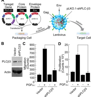
Fig. 4. Attenuation of PGF2α-induced VSMC proliferation and migration by silencing of PLC-β3. (A) Schematic repre- sentation of knockdown strategy using lentivirus. (B) VSMCs were infected by lentivirus carrying shRNA of PLC-β3. Expression of PLC-β3 was verified by Western blot analysis as described in “Materials and Methods”.
After silencing of PLC-β3, PGF2α-induced proliferation (C) and migration (D) of VSMCs were measured as de- scribed in “Materials and Methods”. Data are means ± S.D. of three independent experiments (n=3). Asterisks indicate statistical significance (p<0.05).
migration of VSMCs. As shown in Fig. 2A, PGF2α stimulated VSMC proliferation in a dose-dependent manner. The ap- proximate EC50 was about 0.1 μM and the proliferation was induced at minimum dose of 0.01 μM. In addition, PGF2α
stimulated VSMC migration in a dose-dependent manner and the EC50 (0.1 μM) was similar to that of proliferation.
Therefore, PGF2α significantly enhances the proliferation and migration of VSMCs at the pathophysiological concentration.
Inhibition of PLC attenuates PGF2α-induced VSMC migration
Since PGF2α receptor is coupled with PLC-β family of en- zymes, we next examined the expressions of PLC-β subtypes in VSMCs. As shown in Fig. 3A, PLC-β1, -β2, and -β4 were
expressed in rat brain (control) whereas only PLC-β3 sub- type was expressed in established vascular smooth muscle cells lines (A10) as well as VSMCs. In addition, PLC-β3 was not expressed in the brain. As shown in Figs. 3B and 3C, inhibition of PLC by U73122 (2 μM) significantly blocked PGF2α-induced VSMC migration. Therefore, PLC-β3 is ex- clusively expressed in VSMCs and the inhibition of PLC completely blocks PGF2α-induced VSMC migration.
Silencing of PLC-β3 attenuates PGF2α-induced VSMC proliferation and migration
Since only PLC-β3 was expressed in VSMCs and PGF2α- induced VSMC migration was completely blocked by pan- PLC inhibitor, we next examined the effect of PLC-β3 knock- down on the VSMC proliferation and migration. As shown in Fig. 4A and Fig. 4B, lentiviral expression of short hairpin loop of PLC-β3 (shPLC-β3) significantly reduced the ex- pression of PLC-β3. In addition, knockdown of PLC-β3 sig-
nificantly suppressed the PGF2α-induced proliferation and migration of VSMCs (Fig. 4C, Fig. 4D). Therefore, these re- sults suggest that PLC-β3 plays an essential role in PGF2α
-dependent proliferation and migration of VSMCs.
Discussion
Atherosclerosis is a disease that is associated with com- plex pathophysiological conditions. Especially, chronic in- flammation seems to be an important risk factor during the disease progression [17]. In addition, proliferation and mi- gration of VSMCs is the key pathogenic responses of occlu- sive blood vessel diseases [3]. In the present study, we pro- vide several lines of evidences that link inflammation and VSMC physiologies. First, among the inflammatory prosta- noids that are generated by COX enzyme, PGF2α could in- duce the proliferation and migration of VSMCs. Second, VSMC uniquely expressed PLC-β3 subtypes and other sub- types scarcely expressed. Third, inhibition or silencing of PLC-β3 completely blocked PGF2α-induced proliferation and migration of VSMCs. Therefore, we suggest that PLC-β3 is the major responsible enzyme that medicates occlusive ves- sel disease by chronic inflammation.
It has been known that COX which produces various prostanoids is deeply involved in the progression of athero- sclerosis [7]. For example, COX-2 expression is restricted to atherosclerotic plaque lesions and is not observed in the nor- mal arteries [6]. In addition, COX-2 is highly expressed in symptomatic lesions rather than asymptomatic lesion [8]. It has also been reported that low-dose of aspirin which sup- presses COX enzymatic activity has beneficial effect on the atherothrombosis [20]. Therefore, it is clear that COX and its enzymatic products might be involved in the disease pro- gression of atherosclerosis.
COX produces several key inflammatory cytokines such as PGD2, PGE2, PGI2, and PGF2α [24]. Each of these cytokines has pro-inflammatory or anti-inflammatory properties, and the balance of these cytokines production affects many path- ophysiological processes. For example, PGE2 induces in- flammation whereas the metabolites of PGD2 such as ∆12PGJ2
and 15-deoxy-∆12,14-PGJ2 (15-d-PGJ2) have anti-inflammatory properties [23]. PGI2 has beneficial effect on the cardiovas- cular system, i.e., it enhances vascular dilatation and inhibits blood clotting. PGF2α has detrimental effects on the car- diovascular system by protecting the action of 15-d-PGJ2. Indeed, our results also showed that each of these prosta-
noids differentially affected VSMC proliferation and migra- tion (Fig. 1). It is also notable that only the PGF2α activated VSMCs proliferation and migration. It has been reported that the level of PGF2α is elevated in patients with various arthritis [2]. In line with this, our results also showed that PGF2α was able to induce VSMC proliferation and migration at the pathological concentration (Fig. 2). In addition, dele- tion of PGF2α receptor diminishes inflammation-induced pul- monary fibrosis [18]. Furthermore, ablation of PGF2α receptor results in the low blood pressure and blockade of athero- genesis [28]. Therefore, it is reasonable to suggest that PGF2α
plays an essential role in occlusive vascular disease.
PGF2α activates its cognate FP receptor [12]. The involve- ment of PLC in FP receptor signal transduction has been suggested. For example, pharmacological inhibition of FP re- ceptor modifies calcium mobilization and phosphoinositide turnover [10]. Likewise our results also showed that in- hibition of PLC by pharmacological inhibitor such as U73122 completely blocked PGF2α-induced VSMC migration (Fig. 3).
The involvement between PGF2α and PLC in VSMCs almost never reported. Our results provide novel key findings that PLC-β3 is a crucial downstream regulator of PGF2α receptor signaling pathway. First, VSMCs exclusively expressed PLC- β3 as judged by western blot analysis (Fig. 3A). Other report has shown that all four PLC-β subtypes are expressed in vascular smooth muscle cells isolated from pig aorta [4]. The discrepancy in the expression of each PLC isoform may be due to the species difference. It is also possible that meth- odological differences may lead to the contamination of ad- ventitial fibroblast cells. Second, inhibition of PLC activity downregulated almost all the PGF2α-induced VSMC migra- tion (Fig. 3B, Fig. 3C). Likewise, it has been reported that calcium mobilization and phosphoinositide turnover is cou- pled with PGF2α receptor [10]. It is also known that PGF2α
receptor is coupled with Gq family of G protein which is coupled with PLC-β family [26]. Third, selective silencing of PLC-β3 completely blocked the PGF2α-induced VSMC proliferation and migration (Fig. 4). Since silencing of PLC-β 3 completely blocked PGF2α-induced VSMC proliferation and migration, it is reasonable to suggest that PLC-β3 is the major leading isoform that regulates PGF2α-induced pro- liferation and migration of VSMCs. In these regards, we sug- gest that inflammatory prostanoid such as PGF2α regulates pathophysiological response of VSMCs through the activa- tion of PLC-β3. Targeting PLC-β3-specific pathway would provide beneficial therapeutic strategy for the cardiovascular
disease and others.
Acknowledgement
This work was supported for two years by Pusan National University Research Grant (2017).
References
1. Ali, M. S., Starke, R. M., Jabbour, P. M., Tjoumakaris, S.
I., Gonzalez, L. F., Rosenwasser, R. H., Owens, G. K., Koch, W. J., Greig, N. H. and Dumont, A. S. 2013. TNF-alpha in- duces phenotypic modulation in cerebral vascular smooth muscle cells: implications for cerebral aneurysm pathology.
J. Cereb. Blood Flow Metab. 33, 1564-1573.
2. Basu, S., Whiteman, M., Mattey, D. L. and Halliwell, B. 2001.
Raised levels of F(2)-isoprostanes and prostaglandin F(2al- pha) in different rheumatic diseases. Ann. Rheum. Dis. 60, 627-631.
3. Bennett, M. R., Sinha, S. and Owens, G. K. 2016. Vascular Smooth Muscle Cells in Atherosclerosis. Circ. Res. 118, 692- 702.
4. Blayney, L. M., Gapper, P. W. and Newby, A. C. 1996.
Phospholipase C isoforms in vascular smooth muscle and their regulation by G-proteins. Br. J. Pharmacol. 118, 1003- 1011.
5. Bolognese, R. J. and Corson, S. L. 1975. Interruption of preg- nancy by prostaglandin 15-methyl F2alpha. Fertil. Steril. 26, 695-699.
6. Burleigh, M. E., Babaev, V. R., Yancey, P. G., Major, A. S., McCaleb, J. L., Oates, J. A., Morrow, J. D., Fazio, S. and Linton, M. F. 2005. Cyclooxygenase-2 promotes early athero- sclerotic lesion formation in ApoE-deficient and C57BL/6 mice. J. Mol. Cell. Cardiol. 39, 443-452.
7. Cipollone, F., Cicolini, G. and Bucci, M. 2008. Cyclooxyge- nase and prostaglandin synthases in atherosclerosis: recent insights and future perspectives. Pharmacol. Ther. 118, 161- 180.
8. Cipollone, F., Prontera, C., Pini, B., Marini, M., Fazia, M., De Cesare, D., Iezzi, A., Ucchino, S., Boccoli, G., Saba, V., Chiarelli, F., Cuccurullo, F. and Mezzetti, A. 2001. Overex- pression of functionally coupled cyclooxygenase-2 and pros- taglandin E synthase in symptomatic atherosclerotic plaques as a basis of prostaglandin E(2)-dependent plaque instability.
Circulation 104, 921-927.
9. Gresset, A., Sondek, J. and Harden, T. K. 2012. The phospho- lipase C isozymes and their regulation. Subcell. Biochem. 58, 61-94.
10. Griffin, B. W., Magnino, P. E., Pang, I. H. and Sharif, N.
A. 1998. Pharmacological characterization of an FP prosta- glandin receptor on rat vascular smooth muscle cells (A7r5) coupled to phosphoinositide turnover and intracellular cal- cium mobilization. J. Pharmacol. Exp. Ther. 286, 411-418.
11. Ha, J. M., Yun, S. J., Kim, Y. W., Jin, S. Y., Lee, H. S., Song,
S. H., Shin, H. K. and Bae, S. S. 2015. Platelet-derived growth factor regulates vascular smooth muscle phenotype via mammalian target of rapamycin complex 1. Biochem. Bio- phys. Res. Commun. 464, 57-62.
12. Hohjoh, H., Inazumi, T., Tsuchiya, S. and Sugimoto, Y. 2014.
Prostanoid receptors and acute inflammation in skin. Biochi- mie 107 Pt A, 78-81.
13. Holvoet, P. 2008. Relations between metabolic syndrome, oxidative stress and inflammation and cardiovascular dis- ease. Verh. K. Acad. Geneeskd. Belg. 70, 193-219.
14. Kawakami, T. and Xiao, W. 2013. Phospholipase C-beta in immune cells. Adv. Biol. Regul. 53, 249-257.
15. Kim, D., Jun, K. S., Lee, S. B., Kang, N. G., Min, D. S., Kim, Y. H., Ryu, S. H., Suh, P. G. and Shin, H. S. 1997. Phospholi- pase C isozymes selectively couple to specific neurotrans- mitter receptors. Nature 389, 290-293.
16. Kurkowska-Jastrzebska, I., Karlinski, M. A., Blazejewska- Hyzorek, B., Sarzynska-Dlugosz, I., Filipiak, K. J. and Czlon- kowska, A. 2016. Carotid intima media thickness and blood biomarkers of atherosclerosis in patients after stroke or my- ocardial infarction. Croat. Med. J. 57, 548-557.
17. Libby, P., Ridker, P. M. and Maseri, A. 2002. Inflammation and atherosclerosis. Circulation 105, 1135-1143.
18. Oga, T., Matsuoka, T., Yao, C., Nonomura, K., Kitaoka, S., Sakata, D., Kita, Y., Tanizawa, K., Taguchi, Y., Chin, K., Mishima, M., Shimizu, T. and Narumiya, S. 2009. Prosta- glandin F(2alpha) receptor signaling facilitates bleomycin- induced pulmonary fibrosis independently of transforming growth factor-beta. Nat. Med. 15, 1426-1430.
19. Pansuria, M., Xi, H., Li, L., Yang, X. F. and Wang, H. 2012.
Insulin resistance, metabolic stress, and atherosclerosis.
Front. Biosci. (Schol Ed) 4, 916-931.
20. Patrono, C., Garcia Rodriguez, L. A., Landolfi, R. and Bai- gent, C. 2005. Low-dose aspirin for the prevention of athero- thrombosis. N. Engl. J. Med. 353, 2373-2383.
21. Salazar, H. and Archer, D. F. 1974. Ultrastructural changes of the human corpus luteum of pregnancy induced by pros- taglandin E2 in vitro. Eur. J. Obstet. Gynecol. Reprod. Biol. 4, S19-33.
22. Santini, G., Mores, N., Malerba, M., Mondino, C., Macis, G.
and Montuschi, P. 2016. Investigational prostaglandin D2 receptor antagonists for airway inflammation. Expert. Opin.
Investig. Drugs 25, 639-652.
23. Sasaguri, T. and Miwa, Y. 2004. Prostaglandin J2 family and the cardiovascular system. Curr. Vasc. Pharmacol. 2, 103-114.
24. Smith, W. L., Urade, Y. and Jakobsson, P. J. 2011. Enzymes of the cyclooxygenase pathways of prostanoid biosynthesis.
Chem. Rev. 111, 5821-5865.
25. Ulven, T. and Kostenis, E. 2006. Targeting the prostaglandin D2 receptors DP and CRTH2 for treatment of inflammation.
Curr. Top. Med. Chem. 6, 1427-1444.
26. Watanabe, T., Nakao, A., Emerling, D., Hashimoto, Y., Tsukamoto, K., Horie, Y., Kinoshita, M. and Kurokawa, K.
1994. Prostaglandin-F2-Alpha enhances tyrosine phosphor- ylation and DNA-synthesis through phospholipase-C cou- pled receptor via Ca2+-dependent intracellular pathway in
초록:Prostaglandin F2α 의존적 phospholipase C-β3 활성화에 의한 혈관평활근세포의 병태생리 조절 연구
강기웅1†․오준영1†․이윤한1†․이혜선2․진서연2․배순식2*
(1부산대학교 의과대학 의예과, 2부산대학교 의과대학 약리학교실)
죽상동맥경화는 대동맥의 만성염증에 의해 주로 발병되는 폐쇄동맥질환이다. 혈관평활근세포의 증식 및 이동 은 죽상동맥경화 발병의 주된 병리적 반응이다. 본 연구에서는 죽상동맥경화 발병기전을 유도하는 표적 염증반응
물질의 탐색 및 이들에 의한 신호전달 기전을 연구하였다. 혈관평활근세포의 증식 및 이동은 prostaglandin F2α
(PGF2α)에 의해 의미 있게 증가하였으나 tumor necrosis factor α (TNFα)에 의해서는 증가하지 않았다. Prostacy- clin I2 (PGI2)는 혈관평활근세포의 증식은 촉진시켰으나 이동은 오히려 억제하였다. prostaglandin D2 (PGD2) 및 prostaglandin E2 (PGE2)는 혈관평활근세포의 증식을 촉진시켰으나 이동에는 영향을 미치지 않았다. PGF2α는 용 량 의존적으로 혈관평활근세포의 증식 및 이동을 촉진시켰고 EC50는 약 0.1 μM로 관찰되었다. 혈관평활근세포에 서 phospholipase C-β3 (PLC-β3) 아형의 발현은 매우 높았으나 PLC-β1, PLC-β2, 및 PLC-β4의 발현은 관찰되지
않았다. U73122 처리를 통해 PLC의 활성을 억제하면 PGF2α에 의한 혈관평활근세포의 이동이 억제되었다. 또한
PLC-β3의 발현을 억제하면 PGF2α에 의한 혈관평활근세포의 증식 및 이동이 억제되었다. 이러한 결과들을 바탕으
로 PGF2α 는 혈관평활근세포의 증식 및 이동에 중요한 역할을 수행하고, 여기에는 PLC-β3가 필수적인 역할을 담
당하고 있음을 제안한다.
Nih-3t3 cells. J. Biol. Chem. 269, 17619-17625.
27. Yoshida, T. and Owens, G. K. 2005. Molecular determinants of vascular smooth muscle cell diversity. Circ. Res. 96, 280- 291.
28. Yu, Y., Lucitt, M. B., Stubbe, J., Cheng, Y., Friis, U. G., Hansen, P. B., Jensen, B. L., Smyth, E. M. and FitzGerald, G. A. 2009. Prostaglandin F2alpha elevates blood pressure
and promotes atherosclerosis. Proc. Natl. Acad. Sci. USA. 106, 7985-7990.
29. Yun, S. J., Ha, J. M., Kim, E. K., Kim, Y. W., Jin, S. Y., Lee, D. H., Song, S. H., Kim, C. D., Shin, H. K. and Bae, S. S.
2014. Akt1 isoform modulates phenotypic conversion of vas- cular smooth muscle cells. Biochim. Biophys. Acta 1842, 2184- 2192.