Received: December 2, 2019 Revised: February 21, 2020 Accepted: March 11, 2020
Address for Correspondence: Byung-Koo Yoon, Department of Obstetrics, Gynecology and Women’s Health, Samsung Medical Center, Sungkyunkwan University School of Medicine, 81 Irwon-ro, Gangnam-gu, Seoul 06351, Korea
Tel: 82-2-3410-3519, E-mail: [email protected], ORCID: https://orcid.org/0000-0002-1326-6102
Address for Correspondence: Chi-Dug Kang, Department of Biochemistry, Pusan National University School of Medicine, 49 Busandaehak-ro, Mulgeum-eup, Yangsan 50612, Korea
Tel: 82-51-510-8082, E-mail: [email protected], ORCID: https://orcid.org/0000-0002-7860-5347
ORIGINAL ARTICLE
INTRODUCTION
Coronary heart disease (CHD) arising from athero- sclerosis is the leading cause of death and morbidity worldwide [1]. The pathophysiology of CHD is com- plex and not yet fully understood. The oxidative hy- pothesis is a classical theory on atherosclerosis that oxi-
dative stress is a critical and final common mechanism [2]. Growing evidence indicates that inflammation also influences all stages of atherosclerosis [3].
Apoptosis is a form of programed cell death that oc- curs in multicellular organisms and is involved in vari- ous biologic processes including development, aging, and chemical-induced cell death. Inappropriate apop-
17ββ-Estradiol Inhibits Lysophosphatidylcholine-Induced Apoptosis in Cultured Vascular Smooth Muscle Cells
Byung-Koo Yoon1,2, Young-Hee Kang2, Won-Jong Oh3, Cheong-Rae Roh1, Duk-Kyung Kim4, Chi-Dug Kang5
1Department of Obstetrics, Gynecology and Women’s Health, Samsung Medical Center, Sungkyunkwan University School of Medicine, Seoul, Korea, 2Samsung Biomedical Research Institute, Samsung Medical Center, Seoul, Korea, 3Korea Brain Research Institute, Daegu, Korea, 4Department of Medicine, Samsung Medical Center, Sungkyunkwan University School of Medicine, Seoul, Korea, 5Department of Biochemistry, Pusan National University School of Medicine, Yangsan, Korea
Objectives: Coronary heart disease (CHD) risk increases in women after menopause, but menopausal hormone therapy (MHT) helps prevent CHD if started early after menopause. To explore the mechanism underlying the direct vascular actions of estrogen, the effects of 17β-estradiol (E2) on apoptosis of vascular smooth muscle cells (VSMCs) induced with lysophosphatidylcholine (lysoPC), an active component of oxidized low-density lipoprotein, were investigated in the present study.
Methods: VSMCs were isolated from rat aortas. Apoptosis and protein expression of caspases were assessed using propidium iodide staining and Western blot analysis, respectively. Intracellular formation of reactive oxygen species (ROS) was examined using dichlorofluorescein diacetate, a cell-permeable oxidation-sensitive probe, and quantitated with flow cytometry. Nuclear factor-κB (NF-κB) activation was determined after transfection with a reporter plasmid containing the luciferase reporter gene.
Results: After pre-treatment for 24 hours, 17β-E2 suppressed lysoPC-induced (15 mM) apoptotic cell death in a dose-dependent manner with statistical significance at near physiological concentration. 17β-E2 (10−6 M) also increased protein levels of caspase-9 and -8 precursors and decreased the active form of caspase-3. Western blot analysis using subcellular fractions showed that 17β-E2 decreased mitochondrial Bax levels and concomitantly increased cytosolic Bax expression. Furthermore, intracellular production of ROS and NF- κB-mediated transcriptional activity were reduced with 17β-E2. In addition, estrogen effects on apoptosis were partially blocked by ICI 182,780, a specific estrogen receptor antagonist.
Conclusions: In cultured VSMCs treated with lysoPC, 17β-E2 reduced apoptotic cell death by down-regulating both extrinsic and intrinsic apoptosis pathways, contributing to the preventive action of MHT against CHD.
Key Words: Apoptosis, Atherosclerosis, Estrogens, Myocytes, Smooth muscle
tosis is associated with many human diseases such as neurodegenerative diseases, ischemic damage, autoim- mune disorders, and cancers [4].
Vascular smooth muscle cells (VSMCs) are strongly implicated across all stages of CHD. Migration of VSMCs from the media into the subendothelial region and aberrant proliferation are characteristic findings in atheroma formation. Apoptosis is frequently associated with increased proliferation, which largely accounts for high rates of VSMC turnover [5]. Furthermore, VSMCs are predominant in the fibrous atheroma cap and lesional apoptosis of VSMC triggers plaque progres- sion and its vulnerability to rupture [6]. In addition, the number of VSMCs in the media layer decreases through apoptosis in association with arterial aging [7,8].
Oxidatively modified low-density lipoprotein (LDL) contributes significantly to atherosclerosis by adversely affecting not only the monocytes, macrophages, and vascular endothelial cells (VECs), but also VSMCs [9].
We previously reported that lysophosphatidylcholine (lysoPC), an active component of oxidized LDL, exerts a cytotoxic effect mainly via apoptosis in VSMCs [10].
There is convincing evidence regarding a causal re- lationship between estrogen deficiency and CHD in women. Women with early menopause have an in- creased risk of CHD [11]. In addition, estrogen therapy, if initiated early after menopause, reduces CHD risk [12]. Estrogen has various action mechanisms underly- ing its cardioprotective effects. In addition to the ben- eficial systemic effects on lipid profiles, blood pressure, and glucose metabolism, the direct action of estrogen on arteries may be important [13].
To explore the mechanism underlying direct vascular actions of estrogen in CHD, the effects of 17β-estradiol (E2) on lysoPC-induced apoptosis in cultured VSMCs were investigated in the present study.
MATERIALS AND METHODS
Materials
Sprague–Dawley rats were purchased from Charles River Japan (Hino, Japan). Dulbecco’s modified Eagle’s medium (DMEM) and Ham’s F-12 medium (DMEM/
F-12) without phenol red, fetal bovine serum (FBS), trypsin-ethylenediaminetetraacetic acid (EDTA), and penicillin-streptomycin were obtained from GIBCO BRL (Grand Island, NY, USA). 2’,7’-dichlorofluorescein diacetate (DCF-DA) was purchased from (Calbiochem,
Darmstadt, Germany). LysoPC, 17β-E2, ICI 182,780, and all other chemicals were from Sigma Chemical Co. (St. Louis, MO, USA). Monoclonal antibody for α-smooth muscle actin was obtained from DAKO (Glostrup, Denmark), anti-β-actin antibody was from Sigma Chemical Co., and antibodies against Bax, Bcl- 2, precursors of caspase-9 and -8, active form of cas- pase-3, and cytochrome c oxidase subunit IV were bought from Cell Signaling Technology, Inc. (Beverly, MA, USA). DCF-DA and ICI 182,780 were dissolved in dimethyl sulfoxide (DMSO) and 17β-E2 in ethanol (EtOH). All the other chemicals were resolved in dis- tilled water.
Cell culture
The thoracic aortas from 3-month-old Sprague–Daw- ley rats (160–180 g) were got rid of and the VSMCs were isolated with an enzymatic digestion method as previously described [14]. The VSMCs were cultured in DMEM/F-12 (50 : 50) containing 10% FBS without phenol red and antibiotics. The cells were positively stained for α-smooth muscle actin. Cells in a nearly confluent state were made quiescent by incubation for at least 48 hours in a defined serum-free medium in- cluding insulin (0.5 mM), transferrin (5 mg/mL), and ascorbate (0.2 mM).
This study protocol was evaluated and approved by the Institutional Animal Care and Use Committee of Samsung Biomedical Research Institute (approval no.
C-A3-220-2), which has been accredited by the Asso- ciation for Assessment and Accreditation of Laboratory Animal Care International and abides by the guide of the Institute of Laboratory Animal Resources.
Propidium iodide staining
Adherent and detached cells were harvested, pooled, washed once in phosphate-buffered saline (PBS), and fixed in ice-cold 70% (vol/vol) EtOH in distilled water.
After centrifugation, the cells were washed and resus- pended in cold PBS containing RNase (5 mg/mL) and incubated at 37°C for 30 minutes. Finally, propidium iodide (PI, 1 mg/mL) was added and the cells incubat- ed in the dark for 10 minutes and then analyzed for cell cycle distribution using flow cytometry and Cell Quest software (Becton-Dickinson, San Jose, CA, USA).
Western blot analysis
Cells were lysed in lysis buffer (50 mM Tris-HCl [pH, 7.5], 200 mM NaCl, 1% Nonidet P-40, 0.5% sodium
deoxycholate, and 0.1% sodium dodecyl sulfate [SDS]) containing 1 mM phenylmethylsulfonyl fluoride on ice for 30 minutes. Proteins were separated employing SDS-polyacrylamide gel electrophoresis and trans- ferred to nitrocellulose membranes (Bio-Rad, Hercules, CA, USA). The membranes were incubated with pri- mary antibodies at room temperature for 1 hour. Blots were developed with peroxidase-conjugated second- ary antibody and proteins visualized with enhanced chemiluminescence methods (Amersham Biosciences, Piscataway, NJ, USA) following the manufacturer’s rec- ommendations. β-actin was utilized as a control.
Preparation of subcellular fractions
The cells were collected by centrifugation and washed twice with PBS. The cells were resuspended in homoge- nization buffer (250 mM sucrose, 10 mM KCl, 1.5 mM MgCl2, 1 mM EDTA, 1 mM ethyleneglycotetraacetic acid, 1 mM dithiothreitol, 0.1 mM phenylmethylsul- fonylfluoride, 10 mM Tris-HCl, pH 7.4) containing a proteinase inhibitor cocktail (Roche, Indianapolis, IN, USA), incubated 10 minutes on ice, homogenized, and centrifuged at 700 ×g for 10 minutes. The supernatant was centrifuged again at 10,000 ×g for 30 minutes. The resulting supernatant contained the cytosolic fraction and the pellet the mitochondrial fraction.
Analysis of intracellular formation of reactive oxy- gen species
Production of intracellular reactive oxygen species (ROS) was determined using DCF-DA, a cell-perme- able oxidation-sensitive probe. Cells were incubated in 10 mM DCF-DA at 37°C for 30 minutes and harvested by trypsinization and washed three times with cold PBS solution. The median fluorescence intensity was measured using flow cytometry and CellQuest software (Becton-Dickinson).
In vitro transient transfection and reporter assay To examine the nuclear factor (NF)-κB activation, the VSMCs were transfected with a reporter plasmid containing the luciferase reporter gene linked to five repeats of the NF-κB binding sites, as previously de- scribed [15]. Briefly, the VSMCs (1 × 10⁵ cells/well) were cultured to approximately 70% confluence in 24- well plates. Cells were then transiently co-transfected with 1 mg of NF-κB-luciferase reporter plasmid and 1 mg of β-galactosidase plasmid using Lipofectamine plus (Invitrogen, Carlsbad, CA, USA). At 6 hours af-
ter transfection, cells were starved for 48 hours, and then exposed to 15 mM lysoPC for the indicated time periods. Luciferase activity was measured with a lu- ciferase assay kit (Promega, Madison, WI, USA) with signal detection for 5 seconds in a luminometer (Pa- nomics Inc., Fremont, CA, USA). A β-galactosidase enzyme assay (Promega) was applied to determine the β-galactosidase activity at 420 nm with a SmartSpec 3000 spectrophotometer (Bio-Rad). The results are expressed relative to the NF-κB activity compared with controls after normalizing for β-galactosidase activity and protein concentration.
Data analysis and statistics
Data are expressed as means ± standard error of the mean. The Kruskal–Wallis test and Wilcoxon rank sum test were applied for statistical analysis using IBM Sta- tistics Package for Social Sciences ver. 25.0 (IBM Corp., Armonk, NY, USA). A two-tailed P value < 0.05 was considered statistically significant.
RESULTS
17β-estradiol inhibited lysophosphatidylcholine- induced apoptosis in cultured vascular smooth muscle cells
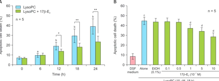
We previously reported that lysoPC induced VSMC apoptosis in a dose-dependent manner at a concentra- tion of 15 mM or higher [10]. Co-treatment of 17β- E2 did not influence lysoPC-induced apoptotic cell death assessed using PI staining (data not shown). In the present study, the effects of 17β-E2 after 24 hours of pre-treatment were investigated. Time-course study showed that 10–6 M of 17β-E2 significantly decreased lysoPC-induced (15 mM) apoptosis after 12 hours (Fig. 1A). In a dose-response experiment at 18 hours of treatment (Fig. 1B), lysoPC significantly increased apoptosis and EtOH (0.1%), a vehicle, did not affect apoptosis. Compared with vehicle-treated controls, 17β-E2 reduced apoptotic cell death in a dose-depen- dent manner and significant reductions were observed at 10–7 M or higher concentration (Fig. 1B).
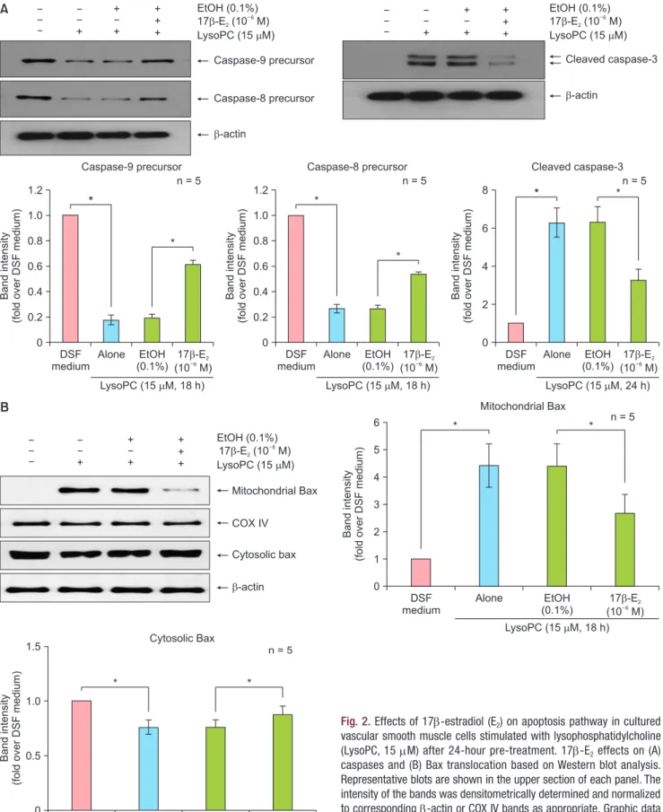
In addition, the effects of 17β-E2 on the apoptosis pathway were investigated. As previously reported [10], Western blot analysis (Fig. 2A) showed that 15 mM of lysoPC decreased caspase-9 and -8 precursors and in- creased the active form of caspase-3; EtOH (0.1%) had no influence on caspase expression. After 24 hours of pre-treatment, 17β-E2 (10–6 M) reversed the changes
in all three caspases compared with vehicle-treated controls (Fig. 2A). We previously showed that although protein levels of Bax and Bcl2 were unchanged, ly- soPC treatment increased Bax translocation from the cytosol to mitochondria [10]. In the present study, Bax and Bcl2 expression under lysoPC treatment was not altered with 17β-E2 based on Western blot analy- sis (data not shown). However, lysoPC-induced Bax translocation was not changed with EtOH (0.1%). Pre- treatment with17β-E2 (10–6 M) for 24 hours significant- ly decreased mitochondrial Bax and simultaneously increased cytosolic Bax compared with vehicle-treated controls (Fig. 2B). The results indicate 17β-E2 inhibits apoptotic cell death in VSMCs treated with lysoPC by down-regulating both extrinsic and intrinsic apoptosis pathways.
Mechanisms of 17β-estradiol action
Mechanisms underlying estrogen actions were fur- ther investigated. In our previous study [10], lysoPC induced VSMC apoptosis via an oxidant mechanism.
Changes in intracellular ROS production in VSMCs are shown in Figure 3A. LysoPC (15 mM) increased ROS 1 hour after treatment and EtOH (0.1%) did not affect ROS production. Compared with vehicle-treated controls, 17β-E2 (10–6 M) significantly decreased ROS production after 24 hours of pre-treatment. LysoPC- induced apoptosis was also reported to be NF-κB- dependent [10]. The NF-κB activity was increased in
response to 15 mM lysoPC treatment for 1 hour and did not change with EtOH (0.1%), as shown in Figure 3B. Pre-treatment with 17β-E2 (10–6 M) for 24 hours significantly suppressed NF-κB activity compared with vehicle-treated controls. In addition, we previously re- ported that estrogen receptor (ER) was expressed in rat VSMCs [16]. PI staining showed that DMSO (0.1%), a vehicle, did not affect VMSC apoptosis, and ICI 182,780 (10–6 M), a specific ER antagonist, significantly blocked estrogen effects (Fig. 3C). The results indicate 17β-E2 reduces apoptosis via receptor-mediated mech- anism and antioxidant activity including NF-κB inhibi- tion.
DISCUSSION
In the present study, direct effects of estrogen on apoptotic cell death of lysoPC-induced VSMCs were investigated. 17β-E2 significantly suppressed apopto- sis via an antioxidant activity and receptor-mediated mechanism.
VSMC apoptosis is induced by various stimuli and stressors including pro-inflammatory cytokines, oxi- dized LDL, high levels of nitric oxide, and mechanical injury [17]. We previously showed that cytotoxic effects of lysoPC are mediated by apoptosis and not by necro- sis [10]. VSMCs treated with oxidized LDL undergo apoptosis via both death receptor (extrinsic) and mi- tochondrial (intrinsic) pathways [17]. Consistently, ly-
0 6 12 18 24
60
50
40
30
20
cellApoptoticdeath(%) 10
Time (h) 0
LysoPC LysoPC + 17 E - 2
a
b
b
b
*
**
n = 5 **
DSF medium
Alone EtOH (0.1%)
0.1 0.5 60
50
40
30
20
cellApoptoticdeath(%) 10
17 E (10 M) 2
- 7
0
n = 5 c
d d
d
1 5 10
LysoPC (15 M, 18 h)
A B
Fig. 1. Effects of 17β-estradiol (E2) on apoptosis in lysophosphatidylcholine (LysoPC, 15 mM)-treated vascular smooth muscle cells after 24-hour pre-treatment. (A) Time-course effects and (B) dose-response effects of 17β-E2 at 18 hours of treatment based on propidium iodide staining. Data are expressed as means ± standard error of the mean. DSF: defined serum-free, EtOH: ethanol. aP < 0.05 versus 0 hour; bP < 0.01 versus 0 hour; cP
< 0.01 versus DSF medium; dP < 0.01 versus EtOH; *P < 0.05; **P < 0.01.
EtOH (0.1%) 17 -E (10 M) 2
6
LysoPC (15 M) Caspase 9 precursor-
Caspase-8 precursor
-actin +
+ +
+ + +
EtOH (0.1%) 17 -E (10 M) 2
6
LysoPC (15 M) +
+ +
+ + +
Cleaved caspase 3-
-actin
DSF medium 1.2
1.0 0.8 0.6 0.4 0.2 Bandintensity (foldoverDSFmedium)
LysoPC (15 M, 18 h) 0
*
*
n = 5
Alone EtOH (0.1%)
17 E (10 M)
- 2
6
Caspase 9 precursor-
DSF medium 1.2
1.0 0.8 0.6 0.4 0.2 Bandintensity (foldoverDSFmedium)
LysoPC (15 M, 18 h) 0
*
Alone EtOH (0.1%)
17 E (10 M)
- 2
6
Caspase 8 precursor-
DSF medium 8
6
4
Bandintensity (foldoverDSFmedium) 2
LysoPC (15 M, 24 h) 0
*
Alone EtOH (0.1%)
17 E (10 M)
- 2
6
n = 5 Cleaved caspase-3
A
B
Mitochondrial Bax
COX IV
Cytosolic bax
actin- EtOH (0.1%)
17 -E (10 M) 2 6 LysoPC (15 M) +
+ +
+ + +
DSF medium 6
5 4 3 2 1
Mitochondrial Bax
0 Bandintensity (foldoverDSFmedium)
n = 5
Alone EtOH
(0.1%)
17 E (10 M)
- 2
6
LysoPC (15 M, 18 h)
DSF medium 1.5
1.0
0.5
Cytosolic Bax
0 Bandintensity (foldoverDSFmedium)
n = 5
Alone EtOH
(0.1%)
17 E (10 M)
- 2
6
LysoPC (15 M, 18 h)
* *
*
*
*
* *
n = 5
Fig. 2. Effects of 17β-estradiol (E2) on apoptosis pathway in cultured vascular smooth muscle cells stimulated with lysophosphatidylcholine (LysoPC, 15 mM) after 24-hour pre-treatment. 17β-E2 effects on (A) caspases and (B) Bax translocation based on Western blot analysis.
Representative blots are shown in the upper section of each panel. The intensity of the bands was densitometrically determined and normalized to corresponding β-actin or COX IV bands as appropriate. Graphic data are depicted in the lower part of the panel. Data are expressed as means ± standard error of the mean. DSF: defined serum-free, EtOH:
ethanol, COX IV: cytochrome c oxidase subunit IV. *P < 0.05.
soPC activated caspase-3 and -8, markers of a common and death receptor pathway, respectively, and increased Bax translocation from the cytosol to mitochondria [10]. An increase in mitochondrial Bax would enhance release of cytochrome c from mitochondria and then activation of caspase-9, a marker of the mitochondrial pathway. These observations support an involvement of dual pathways in lysoPC-induced apoptosis.
In the present study, 17β-E2 significantly suppressed apoptotic cell death of VSMCs treated with lysoPC in a dose-dependent manner based on PI staining. The effects of estrogen occurred at an approximate physi- ologic concentration of 10–7 M. 17β-E2 inhibited cas- pase-3 activation. Furthermore, estrogen treatment attenuated activation of caspase-8 and -9 and decreased Bax translocation to the mitochondria. To the best of our knowledge, this is the first study in which 17β-E2
down-regulated lysoPC-induced VSMC apoptosis by inhibiting both apoptosis pathways.
In the present study, the mechanism of estrogen ac- tion against apoptosis was investigated. 17β-E2 rapidly mitigated ROS production at 1 hour of lysoPC treat- ment. Furthermore, 17β-E2 suppressed NF-κB activity, a major redox-sensitive signaling pathway [18]. The results support the antioxidant effects of estrogen. In addition, estrogen effects against apoptosis were signifi- cantly, but not fully, blocked by ICI 182,780, indicating partial contribution of an ER-mediated mechanism.
Chronic apoptosis of VSMCs accelerates atheroscle- rosis [19]. Furthermore, plaque rupture in early lesions is frequently subclinical because VSMCs repair the rupture and reorganize the associated thrombus. Sub- sequently, this may lead to an obstructive lesion later [1]. In advanced plaques, VSMC apoptosis contributes to plaque thrombogeneity and incites plaque micro- calcification [17]. Furthermore, insufficient clearance of apoptotic VSMCs causes plaque inflammation [20].
These actions account, at least in part, for plaque insta-
DSF medium 5
4
3
2
IntracellularlevelofROS (foldoverDSFmedium) 1
0
Alone EtOH
(0.1%)
17 E (10 M)
- 2
6
LysoPC (15 M, 1 h) n = 5
* *
DSF medium 3
2
1
TranscriptionalactivityNFB (foldoverDSFmedium) - 0
Alone EtOH
(0.1%)
17 E (10 M)
- 2
6
LysoPC (15 M, 1 h) n = 5
* *
DSF medium 60
50
40
30
20
cellApoptoticdeath(%) 10
0
Alone DMSO
(0.1%)
17 E + ICI (10 M)
- 2
6
LysoPC (15 M, 18 h) n = 5
* *
17 E (10 M)
- 2
6
*
*
A B
C
Fig. 3. Mechanisms of 17β-estradiol (E2, 10–7 M) action in lysophosphatidylcholine (LysoPC, 15 mM)-treated vascular smooth muscle cells after 24-hour pre-treatment. (A) Effects of 17β-E2
treatment for 1 hour on intracellular formation of ROS based on flow cytometry analysis using 2´,7´-dichlorofluorescein diacetate. (B) Effects of 17β-E2 for 1 hour on NF-κB-mediated transcriptional activity based on luciferase reporter assay. (C) Effects of estrogen receptor antagonist for 18 hours on 17β-E2 action based on propidium iodide staining. Data are expressed as means ± standard error of the mean. ROS: reactive oxygen species, NF-κB: nuclear factor-κB, DSF: defined serum-free, EtOH: ethanol, DMSO: dimethyl sulfoxide, ICI: ICI 182,780. *P < 0.05.
bility, rupture, and ensuing acute coronary syndrome.
Inhibition of apoptotic cell death of VSMCs due to 17β-E2 could be a key mechanism for direct estrogen actions on arteries.
Post hoc analysis of Women’s Health Initiative data revealed a significant time trend in CHD risk with menopausal hormone therapy (MHT) and the risk was significantly increased by 28% in women 20 years and longer since menopause [12]. Even though the under- lying mechanism is not fully known, arterial status with aging would be attributed considerably to the contrast- ing effects by timing of MHT. In younger menopausal women with early atherosclerosis, MHT is beneficial.
The present study reporting an anti-apoptotic effect of estrogen on VSMCs would provide additional mecha- nism for MHT benefit. In contrast, beginning MHT in late postmenopausal women with established ath- erosclerosis might be harmful by decreased number of functional ER and vasodilation, and increased inflam- matory activation and plaque instability [13].
Oxidative stress also causes apoptosis of VECs and cardiomyocytes [21]. Estrogen prevented VEC apopto- sis induced with hydrogen peroxide [22] and attenuat- ed cardiomyocyte apoptosis induced with hypoxia [23].
Anti-apoptotic actions of estrogen via these mecha- nisms further contribute to the prevention of CHD.
VSMC apoptosis is a notable feature in arterial ag- ing [7] and increased oxidative stress in VSMCs is a causative factor of aortic stiffening [24]. Estrogen could have a favorable impact on arterial aging by suppressing VSMC apoptosis via an antioxidant activity as shown in the present study. Estrogen therapy in postmenopausal women might improve pulse-wave velocity, a common measurement of arterial stiffness [25].
Although using VSMCs derived from rat aortas is an established in vitro model for CHD, cells from human coronary arteries might be more appropriate for further experiments. In addition, caspase-independent path- ways of apoptosis, including apoptosis-inducing factor and endoplasmic reticulum stress, have been reported [21]. Additional studies are needed to further elucidate the mechanism of estrogen action on VSMC apoptosis.
In conclusion, 17β-E2 inhibits lysoPC-stimulated apoptotic cell death in cultured VSMCs mediated by an antioxidant activity and receptor-mediated mechanism.
This action of estrogen would contribute to the preven- tion of CHD with MHT in early menopausal women.
ACKNOWLEDGMENTS
This work was supported in part by the Sungkyunk- wan University Foundation for Corporate Collabo- ration (S-2011-0683-000), the Samsung Biomedical Research Institute grant (C-A0-021-3), the Samsung Medical Center Research Fund (PHX1000071), and the IN-SUNG Foundation for Medical Research (C-A5- 802-1).
CONFLICT OF INTEREST
No potential conflict of interest relevant to this article was reported.
REFERENCES
1. Bennett MR, Sinha S, Owens GK. Vascular smooth muscle cells in atherosclerosis. Circ Res 2016; 118: 692-702.
2. Kattoor AJ, Pothineni NVK, Palagiri D, Mehta JL. Oxidative stress in atherosclerosis. Curr Atheroscler Rep 2017; 19: 42.
3. Pant S, Deshmukh A, Gurumurthy GS, Pothineni NV, Watts TE, Romeo F, et al. Inflammation and atherosclerosis--revisited. J Car- diovasc Pharmacol Ther 2014; 19: 170-8.
4. Elmore S. Apoptosis: a review of programmed cell death. Toxicol Pathol 2007; 35: 495-516.
5. McCarthy NJ, Bennett MR. The regulation of vascular smooth muscle cell apoptosis. Cardiovasc Res 2000; 45: 747-55.
6. Silvestre-Roig C, de Winther MP, Weber C, Daemen MJ, Lutgens E, Soehnlein O. Atherosclerotic plaque destabilization: mechanisms, models, and therapeutic strategies. Circ Res 2014; 114: 214-26.
7. Sawabe M. Vascular aging: from molecular mechanism to clinical significance. Geriatr Gerontol Int 2010; 10 Suppl 1: S213-20.
8. Lee SJ, Park SH. Arterial ageing. Korean Circ J 2013; 43: 73-9.
9. Kita T, Kume N, Minami M, Hayashida K, Murayama T, Sano H, et al. Role of oxidized LDL in atherosclerosis. Ann N Y Acad Sci 2001; 947: 199-205; discussion 205-6.
10. Lee DY, Kang YH, Choi DS, Lee YJ, Rhyu MR, Yoon BK. Cytotox- ic Effects of lysophosphatidylcholine on vascular smooth muscle cells. J Korean Soc Menopause 2012; 18: 139-46.
11. Muka T, Oliver-Williams C, Kunutsor S, Laven JS, Fauser BC, Chowdhury R, et al. Association of age at onset of menopause and time since onset of menopause with cardiovascular outcomes, intermediate vascular traits, and all-cause mortality: a systematic review and meta-analysis. JAMA Cardiol 2016; 1: 767-76.
12. Manson JE, Chlebowski RT, Stefanick ML, Aragaki AK, Rossouw JE, Prentice RL, et al. Menopausal hormone therapy and health outcomes during the intervention and extended poststopping phases of the Women's Health Initiative randomized trials. JAMA
2013; 310: 1353-68.
13. Mendelsohn ME, Karas RH. Molecular and cellular basis of car- diovascular gender differences. Science 2005; 308: 1583-7.
14. Owens GK, Loeb A, Gordon D, Thompson MM. Expression of smooth muscle-specific alpha-isoactin in cultured vascular smooth muscle cells: relationship between growth and cytodiffer- entiation. J Cell Biol 1986; 102: 343-52.
15. Yoon BK, Kang YH, Oh WJ, Park K, Lee DY, Choi D, et al. Impact of lysophosphatidylcholine on the plasminogen activator system in cultured vascular smooth muscle cells. J Korean Med Sci 2012;
27: 803-10.
16. Yoon BK, Oh WJ, Kessel B, Roh CR, Choi D, Lee JH, et al. 17Beta- estradiol inhibits proliferation of cultured vascular smooth muscle cells induced by lysophosphatidylcholine via a nongenomic anti- oxidant mechanism. Menopause 2001; 8: 58-64.
17. Grootaert MOJ, Moulis M, Roth L, Martinet W, Vindis C, Bennett MR, et al. Vascular smooth muscle cell death, autophagy and se- nescence in atherosclerosis. Cardiovasc Res 2018; 114: 622-34.
18. Kabe Y, Ando K, Hirao S, Yoshida M, Handa H. Redox regulation of NF-kappaB activation: distinct redox regulation between the cytoplasm and the nucleus. Antioxid Redox Signal 2005; 7: 395- 403.
19. Clarke MC, Littlewood TD, Figg N, Maguire JJ, Davenport AP, Goddard M, et al. Chronic apoptosis of vascular smooth muscle cells accelerates atherosclerosis and promotes calcification and
medial degeneration. Circ Res 2008; 102: 1529-38.
20. Clarke MC, Talib S, Figg NL, Bennett MR. Vascular smooth mus- cle cell apoptosis induces interleukin-1-directed inflammation: ef- fects of hyperlipidemia-mediated inhibition of phagocytosis. Circ Res 2010; 106: 363-72.
21. Kim NH, Kang PM. Apoptosis in cardiovascular diseases: mecha- nism and clinical implications. Korean Circ J 2010; 40: 299-305.
22. Sudoh N, Toba K, Akishita M, Ako J, Hashimoto M, Iijima K, et al.
Estrogen prevents oxidative stress-induced endothelial cell apop- tosis in rats. Circulation 2001; 103: 724-9.
23. Hsieh DJ, Kuo WW, Lai YP, Shibu MA, Shen CY, Pai P, et al.
17β-estradiol and/or estrogen receptor β attenuate the autophagic and apoptotic effects induced by prolonged hypoxia through HIF- 1α-mediated BNIP3 and IGFBP-3 signaling blockage. Cell Physiol Biochem 2015; 36: 274-84.
24. Canugovi C, Stevenson MD, Vendrov AE, Hayami T, Robidoux J, Xiao H, et al. Increased mitochondrial NADPH oxidase 4 (NOX4) expression in aging is a causative factor in aortic stiffening. Redox Biol 2019; 26: 101288.
25. da Costa LS, de Oliveira MA, Rubim VS, Wajngarten M, Aldrighi JM, Rosano GM, et al. Effects of hormone replacement therapy or raloxifene on ambulatory blood pressure and arterial stiffness in treated hypertensive postmenopausal women. Am J Cardiol 2004;
94: 1453-6.