Author contributions: A.R.H. performed the cell-based experiments.
J.O.N. coordinated the study. Y.J.K. designed and wrote the manu script.
This is an Open Access article distributed under the terms of the Creative Commons Attribution Non-Commercial License, which permits unrestricted non-commercial use, distribution, and reproduction in any medium, provided the original work is properly cited.
Copyright © Korean J Physiol Pharmacol, pISSN 1226-4512, eISSN 2093-3827
INTRODUCTION
Hyperglycemia accelerates the reaction between glucose and proteins and promotes the formation of advanced glycation end products (AGEs). Interacting with its specific receptor, RAGE, AGEs form cross-links with many macromolecules such as col- lagen [1,2]. AGE-related cross-linking of collagen is associated with aortic wall matrix stiffness and AGEs stimulates the release of profibrotic growth factors such as transforming growth factor (TGF)-beta, promote collagen deposition, increase inflammation,
and ultimately lead to tissue fibrosis [3-5]. It has been suggested that connective tissue growth factor (CTGF) is a potent inducer of extracellular matrix (ECM) in diabetes.
CTGF is a widely known as a hallmark of fibrosis in multiple that is implicated in fibroblast proliferation, cellular adhesion, angiogenesis, and ECM synthesis [6]. It has been reported that CTGF promotes vascular smooth muscle cell (VSMC) prolifera- tion, migration, and production of ECM, which may play a role in the development and progression of atherosclerosis [7]. The addition of CTGF to primary mesangial cells induced fibronectin
Original Article
Fluvastatin inhibits advanced glycation end products-induced proliferation, migration, and extracellular matrix accumulation in vascular smooth muscle cells by targeting connective tissue growth factor
Ae-Rang Hwang 1 , Ju-Ock Nam 2 , and Young Jin Kang 1, *
1
Department of Pharmacology, College of Medicine, Yeungnam University, Daegu 42415,
2School of Food Science and Biotechnology, Kyungpook National University, Daegu 41566, Korea
ARTICLE INFO
Received October 19, 2017 Revised November 28, 2017 Accepted December 4, 2017
*Correspondence Young Jin Kang
E-mail: [email protected] Key Words
Advanced glycation end products Cell cycle arrest
Connective tissue growth factor Extracellular matrix
Fluvastatin
Vascular smooth muscle cell
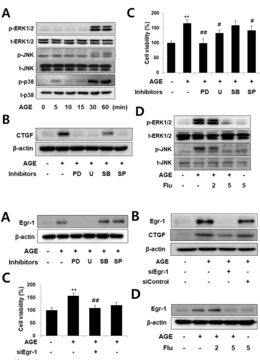
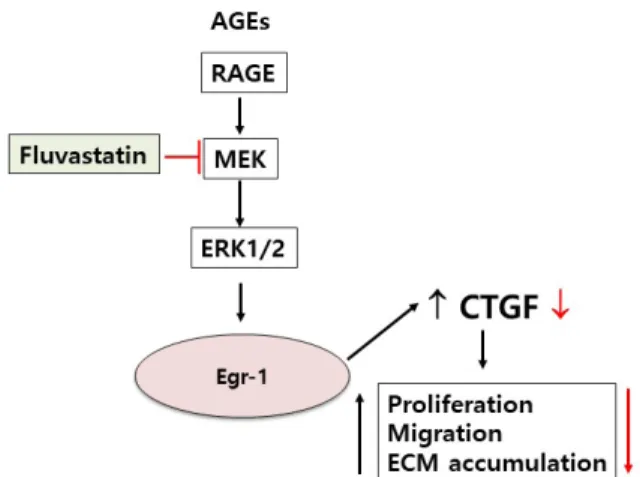
ABSTRACT Connective tissue growth factor (CTGF) is a novel fibrotic mediator, which is considered to mediate fibrosis through extracellular matrix (ECM) synthesis in diabetic cardiovascular complications. Statins have significant immunomodula- tory effects and reduce vascular injury. We therefore examined whether fluvastatin has anti-fibrotic effects in vascular smooth muscle cells (VSMCs) and elucidated its putative transduction signals. We show that advanced glycation end products (AGEs) stimulated CTGF mRNA and protein expression in a time-dependent manner. AGE- induced CTGF expression was mediated via ERK1/2, JNK, and Egr-1 pathways, but not p38; consequently, cell proliferation and migration and ECM accumulation were regulated by CTGF signaling pathway. AGE-stimulated VSMC proliferation, migration, and ECM accumulation were blocked by fluvastatin. However, the inhibitory effect of fluvastatin was restored by administration of CTGF recombinant protein. AGE-in- duced VSMC proliferation was dependent on cell cycle arrest, thereby increasing G1/
G0 phase. Fluvastatin repressed cell cycle regulatory genes cyclin D1 and Cdk4 and augmented cyclin-dependent kinase inhibitors p27 and p21 in AGE-induced VSMCs.
Taken together, fluvastatin suppressed AGE-induced VSMC proliferation, migration,
and ECM accumulation by targeting CTGF signaling mechanism. These findings
might be evidence for CTGF as a potential therapeutic target in diabetic vasculature
complication.
production, cell migration, and cytoskeletal rearrangement which were associated with recruitment of Src and phosphorylation of p42/44 MAPK and protein kinase B [8]. It has been reported that high glucose induces CTGF expression and ECM accumulation in VSMCs via inactivation of ERK1/2 [9].
Statins are a class of drugs used to lower cholesterol levels by inhibiting the enzyme HMG-CoA reductase, which plays an important role in the production of cholesterol in liver [10]. It has been demonstrated that statins have significant immunomodula- tory effects and reduce vascular injury [11]. A previous study by our research team showed that pitavastatin inhibited the effects of angiostetin II (AngII)-induced insulin-like growth factor bind- ing protein (IGFBP)-5, namely, VSMC proliferation, migration, and ECM accumulation [12]. The mechanisms of hyperglycemia- induced vascular remodeling are incompletely understood;
however, metabolic and mechanical factors seem to play impor- tant roles. With this background, we examined whether AGEs induced ERK1/2 and JNK signaling pathways, which could result in the stimulation of CTGF secretion and increase in VSMC pro- liferation and migration, thereby resulting in ECM accumulation.
We observed that these effects were reversed by administration of fluvastatin. Our results demonstrated the inhibitory effect of fluvastatin on AGE-induced VSMC proliferation, migration, and production of ECM, which were associated with secretion of CTGF.
METHODS
Reagents and antibodies
Dulbecco’s modified Eagle’s medium (DMEM), fetal bovine serum (FBS), and antibiotics (penicillin and streptomycin) were purchased from Gibco BRL (Rockville, MD, USA). Anti-β-actin, 3-(4,5-dimethylthiazol-2yl)-2,5-diphenyltetrazolium bromide (MTT) were purchased from Sigma-Aldrich (St. Louis, MO, USA). Anti-CTGF, anti-JNK, anti-pJNK, anti-p38, anti-pp38, an- ti-ERK1/2, anti-pERK1/2, goat anti-rabbit IgG, goat anti-mouse IgG, donkey anti-goat IgG, enhanced chemiluminescence (ECL), Egr-1, and control siRNA were purchased from Santa Cruz Bio- technology (Santa Cruz, CA, USA). AGE-BSA was obtained from Calbiochem (Darmastadt, Germany), PD98059, U0126, SP600125 and SB203580 were purchased from Calbiochem (San Diego, CA, USA). Lipofectamine2000TM was purchased from Invitrogen (Carlsbad, CA, USA). Recombinant CTGF was purchased from R&D system (Minneapolis, ME, USA) and α-tubulin from Sigma (St. Louis, MO).
Primary cell culture
Sprague-Dawley rats were anesthetized with pentobarbital (50 mg/kg). The thoracic aorta removed, cleaned of adhering fat
and connective tissue, and thoracic aortic rings were prepared.
The vascular endothelium was mechanically removed by rub- bing gently with wooden stick to remove endothelium. VSMCs were processed using a 1 mm chop setting in a 100 mm culture dish, and cultured with 50% FBS-DMEM with 1% antibiotics at 37°C in a humidified atmosphere containing 5% CO
2for 7 days.
VSMCs were maintained in DMEM with 10% FBS and 1% anti- biotic (penicillin 10,000 U/ml, streptomycin 10,000 µg/ml). We used VSMCs from passages 4 to 8 at 70-90% confluence in 10 cm dishes, and cell growth was arrested by incubation of the cells in serum-free DMEM for 24 h prior to use.
Transfection of siRNA
The cells were transfected with siRNA using Lipofectamine 2,000 reagent, according to the manufacturer’s instructions.
Aliquots of 1×10
4cells were plated on 60-mm dishes 1 day before transfection, and grown to approximately 70% confluence. The cells were then transfected with siRNA (20 nM Egr-1, 20 nM con- trol) and 3 µl of lipofectamine for 6 h in Opti-MEM
ⓇI reduced serum medium (Invitrogen), followed by incubation for 48 h.
Quantitative real-time polymerase chain reaction (qRT-PCR)
Total RNA was isolated by TRIzol, and a reverse transcription reaction was conducted using TaqMan reverse transcription re- agents, following the manufacturer’s instructions. RT-PCR was conducted with 1 µl of template cDNA and power SYBR Green in ABI PRISM 7500. Quantification was performed using the ef- ficiency-corrected ΔΔCq method. Primers used to amplify DNA sequences were as follows: CTGF, forward 5′-TCAGGCACCCT- CATATAATC-3′ and reverse 5′-GACAATAGTCCACACCA- GA-3′,
Fibronection, forward 5′-TTACTATGGGATGGGGTCCA-3′
and reverse 5′-TGCCAAAACTGTTCACCAAA-3′, collagen I, forward 5′-AAACCACCCTGAAAGCACAG-3′ and reverse 5′-AGTGTTCTGGTGATGCCACA-3′; and collagen III, forward 5′-GGAGCCAAAAGGGTCATCAT-3′ and reverse 5′-GTGATG- GCATGGACTGTGGT-3′, GAPDH, forward 5′-AGGGAAATC- GTCGTGCGTGAG-3′ and reverse 5′-CGCTCATTGCC- GATAGT-3′. The PCR conditions were as follows: preliminary denaturation at 50°C for 2 min; 95°C for 10 min, 95°C for 15 s, and 60°C for 1 min.
Western blotting
The cells were lysed with radioimmunoprecipitation assay
(RIPA) lysis buffer supplemented with 1 mmol/L phenylmeth-
ylsulfonyl fluoride (PMSF) and 0.01 mmol/L protease inhibitor
cocktail (PIC), and incubated on ice for 15 min, followed by cen-
trifugation at 15,000 × g for 10 min at 4°C. Protein concentration
was determined from the supernatant obtained by centrifugation using the Bradford assay. Proteins were separated by SDS-PAGE and transferred onto a polyvinylidene difluoride (PVDF) mem- brane. The membranes were immunoblotted with primary anti- bodies (1:1000), followed by immunoblotting with corresponding secondary antibodies (1:5000). Signals were visualized by using electrochemiluminescence (ECL) detection reagents (Millipore, Temecula, CA), according to the manufacturer’s instructions.
MTT assay
The cells were cultured on 24-well plates. When the cells are approximately 80% confluent, the medium was replaced with serum free DMEM. After starvation, the cells were pretreatment with or without fluvastatin (5 µM) and then stimulated with AGEs (10 µg/ml) for 24 h. MTT reagent was added and incubated for 4 h at 37°C, and then washed with PBS and eluted by adding dimethyl sulfoxide (DMSO). Cell proliferation was measured us- ing microplate reader (Biorad) at 570 nm.
Wound scratch assays
When cells culltured to confluence or near >90% in either 6-well dishes. The medium was replaced with serum free DMEM, and the cells were incubated overnight. Using a sterile 200 µl pipet tip, one separate wound was scratched through the cells moving perpendicular to the line drawn in the step above. After scratching, the cells were pretreated with fluvastatin in the pres- ence or absence of recombinant CTGF (100 nM) for 1 h and then
exposed to AGEs (10 µg/ml) for 36 h. The images were acquired using a phase contrast microscope (40×).
Flow cytometric analysis
The cells (1×10
5) were trypsinized and fixed in 95% ethanol overnight. Fixed cells were stained with propidium iodide (PI; 50 µg/ml) for 30 min at 37°C. PI enters the cells and stains the nucle- us. PI-stained cells were filtered using a 5 mL polystyrene round bottom tube with a cell-strainer cap prior to flow cytometry. All flow cytometry measurements were done using a FACSCalibur (Becton Dickinson, San Jose, CA, USA). Cell cycle analysis was performed using Cell-Quest Pro-Software.
Statistics
Data in the bar graph was expressed as mean±S.D. Statistical significance of difference was measured by Student’s t test. A p values of <0.05 was considered significant. p values less than 0.05 are indicated by *, and p values less than 0.01 are indicated by **.
RESULTS
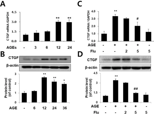
Fluvastatin inhibits AGE-induced CTGF expression in VSMCs
To determine whether AGEs induce CTGF expression in VSMCs, the cells were treated with 10 µg/ml AGEs at various
Fig. 1. Fluvastatin inhibits AGE-in- duced CTGF expression in VSMCs.
Cells were treated with AGE 10 µg/ml for 0, 6, 12, 18, 24 h. CTGF mRNA level was determined by qRT-PCR analysis (A) CTGF protein level was determined by Western blot (C). Cells were treated with 2 or 5 µM fluvastatin for 1 h before incubation with AGEs for 24 h. CTGF mRNA level was determined by qRT-PCR analysis (B) and CTGF protein level was determined by Western blot (D). Data are representative of three independent experiments with similar results *p<0.05
**p<0.01 vs. untreated cells,
#p<0.05
##