ISSN 1226-8704 http://dx.doi.org/10.6118/jksm.2012.18.3.139 J Korean Soc Menopause 2012;18:139-146
Lysophosphatidylcholine의 혈관평활근세포에 대한 세포 독성
이동윤1·강영희2·최두석1,2·이영주3·류미라4·윤병구1,2
1성균관대학교 의과대학 삼성서울병원 산부인과, 2삼성생명과학연구소, 3세종대학교 생명공학과, 4한국식품연구원 대사기능연구본부
Cytotoxic Effects of Lysophosphatidylcholine on Vascular Smooth Muscle Cells
Dong-Yun Lee, M.D.1, Young-Hee Kang, M.S.2, Doo Seok Choi, M.D.1,2, Young Joo Lee, Ph.D.3, Mee-Ra Rhyu, Ph.D.4, Byung-Koo Yoon, M.D., Ph.D.1,2
1Department of Obstetrics and Gynecology, Samsung Medical Center, Sungkyunkwan University School of Medicine; 2Samsung Biomedical Research Institute; 3Department of Bioscience and Biotechnology, Sejong University, Seoul; 4Divisions of Metabolism and Functionality Research, Korea Food Research Institute, Seongnam, Korea
Objectives: To investigate the cytotoxic effects of lysophosphatidylcholine (lysoPC), an active component of oxidized low-density lipoproteins (LDL), on vascular smooth muscle cells (VSMCs).
Methods: VSMCs were derived from rat aorta. Cell death was assessed by 3-(4,5-dimethylthiazol-2-yl)-2,5-diphenyl-tetrazolium bromide (MTT) assay, lactic dehydrogenase (LDH) assay, and DNA fragmentation assay. Apoptosis was quantified by propidium iodide staining and fluorescent activated cell sorting (FACS) analysis, and intracellular free radical production was determined using 2’,7’-dichlorofluorescin diacetate (DCF-DA). In addition, the changes in caspases, bcl-2 and bax proteins were evaluated by western blot analysis.
Results: LysoPC over 25 μM induced more than 50% of the cell death at 10 hours on MTT assay with no change in the level of LDH. The DNA ladder pattern showed that cell death induced by lysoPC was caused by apoptosis, which was associated with increased free radical production. Vitamin E, a potent antioxidant and caffeic acid phenylethyl ester (CAPE), an inhibitor of nuclear factor-kappaB (NF-kB), blocked apoptosis. The casepase-3 precursor decreased and the active form of caspase-8 increased.
Total bcl-2 and bax proteins did not change with lysoPC treatment, but translocation of bax from cytosole to the mitochondria membrane was observed.
Conclusion: LysoPC induces apoptosis in VSMCs via an oxidant mechanism, dependent on NF-kB. (J Korean Soc Menopause 2012;18:139-146)
Key Words: Apoptosis, Atherosclerosis, Lysophosphatidylcholines, Vascular smooth muscle cell
Received: June 29, 2012 Revised: September 4, 2012 Accepted: September 19, 2012
Address for Correspondence: Byung-Koo Yoon, Department of Obstetrics and Gynecology, Samsung Medical Center, Sungkyunkwan University School of Medicine, 81 Irwon-ro Gangnam-gu, Seoul 135-710, Korea
Tel: +82-2-3410-3519, Fax: +82-2-3410-0630, E-mail: [email protected]
Original Article
J KSM
심혈관질환은 폐경 여성의 주요 사망 원인으로, 죽상동맥 경화증 (atherosclerosis)은 대표적 심혈관질환 중 하나이다.
혈관평활근세포 (vascular smooth muscle cell)는 죽상동맥경
화증 발생에 있어 중요한 역할을 담당하는데, 이 세포는 파 열된 죽상경화반 (atherosclerotic plaque)에서 가장 흔하게 발견되는 세포이며, 죽상동맥경화증의 진행 과정 중에 혈관
J Korean Soc Menopause 2012;18:139-146
J KSM
J Korean Soc Menopause 2012;18:139-146 평활근세포의 세포사가 일어난다.1산화 스트레스가 죽상동맥경화증 발생에 기여하는데, 이 중 저밀도지단백 (low-density lipoprotein, LDL)이 산 화 저밀도지단백 (oxidized LDL)으로 변화되는 것이 중요 하다. 산화 저밀도지단백은 심혈관질환의 병태 생리에 있 어 핵심적인 역할을 하는 것으로 알려져 있는데, 특히 혈 관평활근세포에 대한 세포 독성 (cytotoxicity)을 유도한다.2 한편, lysophosphatidylcholine (lysoPC)은 죽상화된 지단백 (atherogenic lipoprotein)의 주요한 인지질 (phospholipid) 성 분으로,3 내인성 포스포리파아제 (phospholipase) A2에 의한 저밀도지단백의 산화 과정에서 생성되며,4 죽상경화 병변 에서 높은 농도로 발견된다.5 혈관평활근세포의 세포자멸사 (apoptosis)는 고혈압과6 혈관 협착7에 관여하고, 초기 동맥 경화병변 (fatty streak)의 죽상경화반으로의 진행에 영향을 주며,8 죽상동맥경화반의 파열 및 이후의 혈전 (thrombosis) 형성에도 기여한다. 기존 연구에서 lysoPC의 혈관평활근세 포에 대한 독성 효과가 보고된 바 있으나,2 정확한 기전에 대한 연구는 아직 부족한 상황이다.
본 연구의 목적은 lysoPC에 의한 혈관평활근세포의 세포 사 기전을 밝히는 것이다.
연구대상 및 방법
1. 혈관 평활근세포의 분리와 배양
3개월 된 Sprague-Dawley rat (Charles River Japan, Hino, Japan)에서 흉부대동맥을 떼어 내어 혈관 평활근세포를 분 리하였다.9 세포는 GIBCO BRL (Grand Island, NY, USA)로 부터 구입한 세포배양액 (Dulbecco’s Modified Eagle Medium [DMEM]/F12 supplemented with 10% fetal bovine serum [FBS], 100 mg/mL penicillin, 0.1 mg/mL streptomycin)에 서 37oC humidified 5% CO2/95% air 환경에서 배양하였으 며, 5-10번 계대 배양된 세포를 실험에 사용하였다. 세포들 이 almost confluence 상태에 도달하면 defined serum-free medium (DMEM/F12 medium containing insulin 5×10⁻⁷ M, transferrin 5 mg/mL, ascorbic acid 0.2 mM)에서 48시간 배 양하여 quiescent 상태의 세포를 사용하였다.
본 연구는 성균관대학교 의과대학의 동물실험윤리위원회 의 승인을 받아 진행되었다 (승인 번호 H-A9-003).
2. 3-(4,5-dimethylthiazol-2-yl)-2,5-diphenyl- tetrazolium bromide (MTT) assay
세포를 96 well plate에서 배양 후 MTT (Sigma Chemical
Co., St. Louis, MO, USA) 용액을 0.25 mg/mL의 농도로 처리한 다음 37oC에서 4시간 동안 반응 시킨 후 enzyme- linked immunosorbent assay (ELISA) reader (Bio-Rad, Hercules, CA, USA)를 이용하여 540 nm에서 흡광도를 측정 하였다.
3. Lactate dehydrogenase (LDH) assay
세포독성 측정을 위한 실험으로 세포가 괴사 (necrosis)를 일으키면 LDH의 분비가 증가되는 것을 이용하였다. 세포를 96 well plate에서 배양한 후 well에서 배지 (media)를 얻었다.
2M Hepes buffer (pH 7.4)를 미리 넣은 둔 다른 96 well plate 에 모아둔 시료와 substrate solution (10 mg/mL lactate, 6.7 mg/mL iodonitrotetrazolium violet, 1 mg/mL new antigenic determinant [NAD]+, 0.01% bovine serum albumin [BSA], 0.4% sucrose, 0.2 unit/mL diaphorase)을 넣고 60분간 배 양하고 17 mg/mL oxamate로 반응을 끝낸 후 ELISA reader 492 nm로 측정했다.
4. DNA fragmentation assay
세포 용해물 (cell lysate)에 digestion buffer (10 mM TrisCl, pH 8.0, 100 mM NaCl, 25 mM ethylenediaminetetraacetic acid [EDTA], 0.5% sodium dodecyl sulphate [SDS], 0.2 mg/
mL Proteinase K)를 넣고 50oC에서 14-18시간 정도 반응시 켰다. 그 후 phenol 및 phenol/chloroform extraction 방법을 통하여 핵산만을 추출하고 추출된 물질은 다시 에탄올 침전 후, 원심분리에 의해 pellet 상태로 모았다. 모아진 pellet을 다시 Tris-EDTA (TE) buffer로 녹이고, DNase-free RNase (0.2 mg/mL) (Sigma Chemical Co., St. Louis, MO, USA)를 37oC에서 약 2시간 반응시켜 DNA만을 얻었다. 반응이 끝 난 후 10 μg 정도의 DNA sample을 1.5% agarose gel 상에 서 분리시켰고, ethidium bromide (Sigma Chemical Co., St.
Louis, MO, USA) 용액을 이용하여 염색한 다음, 자외선 하 에서 fragmentation 정도를 확인했다.
5. Fluorescent activated cell sorting (FACS) analysis:
propidium iodide (PI) staining
12 well plate에서 배양된 세포를 trypsin-EDTA를 이용 하여 cell pellet을 얻었다. Pellet을 1 mM EDTA가 함유된 phosphate buffered saline (PBS)로 현탁 시킨 후 70% 에탄올 로 고정시켰다. 일정 시간 4oC에서 보관 후 3500 rpm에서 5 분간 원심 분리 하고 100 μg/mL RNase A, 100 μg/mL PI가 포함 된 용액에 30분간 상온에서 반응 시킨 후 FACS Calibur flow cytometer (BD Biosciences, San Jose, CA, USA)를 이용
J Korean Soc Menopause 2012;18:139-146
J Korean Soc Menopause 2012;18:139-146 이동윤 외. Lysophosphatidylcholine의 세포독성
7. Western blotting
세포 용해물을 protease inhibitor cocktail (Sigma Chemical Co., St. Louis, MO, USA)이 포함된 lysis buffer (50 mM Tris pH 7.4, 150 mM NaCl, 1% Triton X-100, 1% NP-40, 0.1%
SDS)에 용해 (lysis)시켰다. 단백질을 SDS-polyacrylamide gel electrophoresis (PAGE)로 분리한 후 nitrocellulose membrane에 transfer시켰다. 1차 항체를 반응 시킨 후 horseradish peroxidase conjugated 2차 항체를 반응시켰다.
ECL kit (Amersham Pharmacia Biotech, Piscataway, NJ, USA)를 이용하여 단백질의 발현 정도를 X-ray상에서 확인 하였다.
Western blot은 caspase와 bcl-2 및 bax 발현 분석에 사용 하였으며, vitamin E 및 caffeic acid phenylethyl ester (CAPE) 투여 효과 분석에는 사용하지 않았다.
8. Preparation of subcellular fractionations 세포 내 단백질의 이동을 확인하기 위하여 subcellular fractionation을 시행하였다. 일정 시간 100 mm 접시에 하여 세포의 수와 DNA 양의 상관관계를 분석하였다.
6. 세포내 reactive oxygen species (ROS) 생성 측정 세포내 free radical 생성은 Molecular Probes 사 (Eugene, OR, USA)에서 구입한 2’,7’-dichlorofluorescin diacetate (DCF-DA)를 이용하여 측정하였다. DCF-DA는 세포막을 통과할 수 있는 oxidation-sensitive probe로서 세포내 ROS 와 반응하여 세포막을 통과 할 수 없는 형광 물질인 DCF로 전환된다. 혈관평활근세포를 chamber slide에서 배양하고 quiescent 상태의 세포에 lysoPC와 함께 50 μM DCF-DA를 1 시간 처리하였다. PBS로 세척한 후 confocal-laser scanning microscope (DIAPHOT 300, Nikon, Japan)로 관찰하였다.
ROS의 생성을 정량화 하기 위해서 배양한 혈관평활근세포 에 lysoPC와 함께 10 μM DCF-DA를 1시간 처리한 후, PBS 로 세척하고 Trypsin/EDTA를 이용하여 얻은 pellet을 1 mM EDTA를 함유한 PBS에 현탁시켜 FACS analysis와 CellQuest software (Becton-Dickinson, San Jose, CA, USA)를 이용하 여 형광의 정도를 측정하였다.
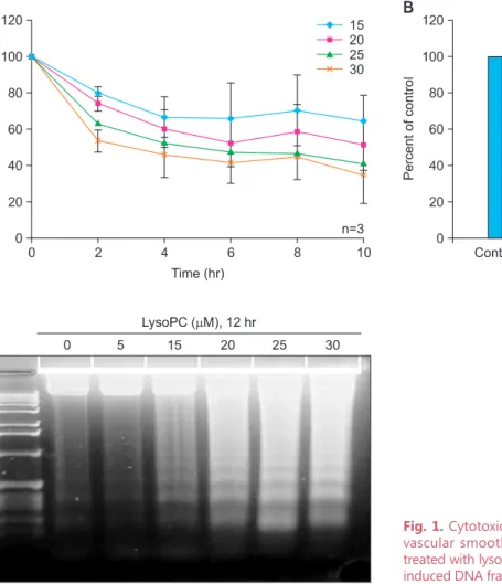
Fig. 1. Cytotoxic effects of lysophosphatidylcholine (lysoPC) on vascular smooth muscle cells (VSMCs). (A) Viability of VSMC treated with lysoPC, (B) cell necrosis induced by lysoPC, (C) lysoPC induced DNA fragmentation.
J Korean Soc Menopause 2012;18:139-146
J KSM
J Korean Soc Menopause 2012;18:139-146 서 배양 된 세포를 trypsin-EDTA를 이용하여 cell pellet 을 얻었다. Lysis buffer (20 mM 4-[2-hydroxyethyl]-1- piperazineethanesulfonic acid [HEPES], pH 7.5 10mM KCl, 1.5 mM MgCl2, 1 mM ethylene glycol-bis[2-aminoethyl]- N,N,N’N’-tetraacetic acid [EGTA], 1 mM EDTA, 1 mM dithiothreitol [DTT], protease inhibitors)를 넣고 homogenizer 에서 5번 이상 homogenize하였다. 4oC에서 750 g로 5분간 원심 분리 한 후 상층액을 다시 4oC에서 10,000 g로 15분간 원심 분리하여 침전물인 mitochondrial membrane fraction이 얻어 지고 상층액은 cytosol fraction이 된다. 각각을 western blotting하였다.결 과
결과 분석을 위해 동일 실험을 3회 반복하여 얻은 값의 평 균을 이용하였다.
Figure 1A는 배양된 혈관평활근세포에 lysoPC를 처리하 였을 때 처리 시간 및 농도에 따른 세포의 생존율을 MTT assay로 분석한 결과로, lysoPC를 처리 후 2시간이 경과하면 서부터 세포의 생존율이 급격하게 감소하여 25 및 30 μM 농 도의 경우에는 10시간 경과 후 50% 미만의 세포들만이 생존 하였다.
Figure 1B는 lypoPC를 처리하였을 때 농도에 따른 LDH 분 비의 변화로, 실험 결과 negative control인 defined serum- free medium (DSF) 사용과 비교하여 차이가 없었으며 이러
한 결과는 세포가 죽은 원인이 괴사와는 다른 기전임을 의 미한다. 반면, lysoPC의 농도가 20 μM 이상인 경우 세포자 멸사에 특징적인 DNA ladder 양상의 발현을 확인할 수 있었 으며 (Fig. 1C), 이를 종합하면 lysoPC에 의한 혈관평활근세 포의 세포사는 세포자멸사를 통하여 유도되었음을 확인할 수 있었다.
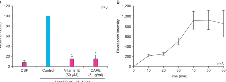
Figure 2A는 lysoPC와 함께 reactive oxygen species (ROS) 를 억제하는 작용을 하는 vitamin E 또는 nuclear factor- kappaB (NF-kB) 억제제인 CAPE를 함께 처리하였을 때 혈 관평활근세포의 세포자멸사를 FACS를 이용한 PI 염색으 로 분석한 결과이다. Figure 1의 결과에 따라 25 μM 농도 의 lysoPC를 이용하여 실험한 결과 vitamin E와 CAPE 모 두 lysoPC에 의해 유도된 혈관평활근세포의 세포자멸사를 유의하게 감소시키는 것으로 나타났다. 한편, figure 2B는 lysoPC 처리 후 혈관평활근세포의 ROS 생성이 시간이 지남 에 따라 증가하는 양상을 보여준다.
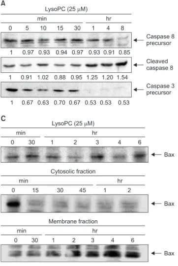
LysoPC 처리 후 세포자멸사의 표지자인 caspase의 변화를 관찰하였다. Caspase-3 전구체의 발현은 5분 이후부터 감 소하기 시작하여 1시간 이후에는 거의 발견 되지 않았는데, 이는 활성화된 caspase-3가 증가되었음을 의미한다 (Fig.
3A). 또한 caspase-8 전구체의 발현이 4시간 이후에 감소하 였으며, 이와 동시에 caspase-8의 활성형 (active form)이 증 가하였다. 한편, lysoPC 처리는 총 bcl-2 단백의 발현 및 미 토콘드리아 세포막에 존재하는 bcl-2 단백의 영향을 미치지 않았으며 (Fig. 3B), 총 bax의 발현 역시 변화가 없었다 (Fig.
3C). 하지만, subcellular fraction을 이용한 실험 결과에서는
Fig. 2. Mechanisms for vascular smooth muscle cell (VSMC) apoptosis induced by lysophosphatidylcholine (lysoPC). (A) Involvement of reactive oxygen species and nuclear factor-kappaB (NF-kB), as assessed by propidium iodide (PI) staining, (B) kinetics of reactive oxygen species (ROS) production determined by 2’,7’-dichlorofluorescin (DCF) staining. DSF: defined serum-free medium, CAPE: caffeic acid phenylethyl ester. *P < 0.001 vs. control.
J Korean Soc Menopause 2012;18:139-146
J Korean Soc Menopause 2012;18:139-146 이동윤 외. Lysophosphatidylcholine의 세포독성
세포질 분획 (cytosolic fraction)의 bax의 발현은 감소한 반 면, 미토콘드리아 세포막 분획에서의 bax 발현이 증가한 것 으로 나타났는데, 이는 lysoPC 처리 후 세포질에서 미토콘드 리아 세포막으로 bax가 이동한다는 증거이다 (Fig. 3C).
고 찰
본 연구 결과 lysoPC를 배양된 혈관평활근세포에 처리하 였을 때 세포자멸사 발생이 관찰되었다. 세포자멸사는 강력 한 항산화제인 vitamin E에 의하여 억제되었는데 이는 산화 기전이 중요한 역할을 함을 시사하며, CAPE에 의한 세포자 멸사 억제는 NF-kB가 downstream mechanism으로 작용하 는 것을 의미한다.
아직까지 lysoPC에 의한 세포 독성 기전은 명확하게 밝혀 지지 않았지만, Kume 등10은 lysoPC가 극성이 있는 인지질 로 마치 세제와 같이 작용할 수 있음을 보고하였다. 즉, 특
Fig. 3. Mechanisms for vascular smooth muscle cell (VSMC) apoptosis induced by lysophosphatidylcholine (lysoPC), evaluated by western blot analyses. (A) expression of caspases, (B) expression of bcl-2, (C) expression of bax.
정 농도 이상에서는 세포막의 구조에 영향을 주고, 세포막 내 고분자 (macromolecule)의 기능을 저해할 수 있다는 것 이다.11 또한 Ohara 등12은 lysoPC가 혈관내 단백 키나아제 (protein kinase) C를 활성화시켜 산화 라디칼 생성 증가를 초래하여 세포막이 손상을 받을 수 있다는 기전을 제시하 였으며, 이는 산화 기전의 관련성을 제시한 본 연구의 결과 와 유사한 소견이다. 이와 함께 칼슘 통로의 활성화를 통한 세포내 칼슘 농도의 증가가 연관되었을 가능성도 제시되었 으며,13 최근 extracellular signal-regulated kinase (ERK)1/2, Jun N-terminal kinase (JNK), 그리고 p38와 같은 mitogen- activated protein kinase의 인산화 역시 lysoPC에 의한 혈관 평활근세포의 자멸사에 영향을 주는 것이 관찰되었다.14 결 과적으로 lysoPC가 혈관평활근세포를 손상시켜 세포자멸사 를 초래하는 과정에는 다양한 기전들이 관여하고 있는 것으 로 생각된다.
본 연구 결과 lysoPC에 의한 세포사는 괴사보다는 세포자 멸사에 의해 발생하였는데, 이는 기존 연구에서 죽상경화
J Korean Soc Menopause 2012;18:139-146
J KSM
J Korean Soc Menopause 2012;18:139-146반내의 손상된 혈관평활근세포의 대부분이 괴사에 의해 죽 는다고 보고한 것과 상이한 결과이다.1 하지만, 기존 연구가 파열 병력이 있는 사람으로부터 채취한 죽상경화반 조직 내 손상된 세포를 이용하여 진행된 반면, 본 연구는 쥐로부터 얻은 세포를 배양한 후 lysoPC를 처리하여 실험을 진행하였 다는 차이점이 있다. 하지만, 마찬가지로 쥐의 혈관평활근 세포에 대한 lysoPC의 독성을 측정한 다른 연구에서도 세포 괴사에 의한 세포사가 관찰된 바 있다.2 본 연구에서 사용 된 쥐 (12주)와 기존 연구 (20주)에서 사용된 쥐의 월령이 다 르다는 점에서 결과의 차이가 기인하였을 가능성이 있으며, 향후 월령에 따른 혈관의 상태를 고려한 비교 연구가 이를 해석하는데 있어서 도움이 될 것으로 사료된다.
기존 연구 결과 배양된 혈관평활근세포에서 산화 저밀도 지단백에 의해 유도되는 세포자멸사는 bcl-2 단백의 조절을 통해서 일어난다고 보고된 바 있으나,15 본 연구에서는 bcl- 2 단백의 변화가 관찰되지 않아 이와는 다른 결과를 나타내 었다. 하지만, bcl-2 단백 변화뿐만 아니라, 본 연구 결과와 같이 bax의 translocation이 세포자멸사를 유도한다는 연구 결과들도 있으므로,16,17 정확한 조절 기전에 대해서는 향후 추가 연구가 필요할 것으로 사료된다.
아직까지 lysoPC에 의해 유도되는 세포자멸사의 발생 기 전은 정확하게 알려지지 않았으나 본 연구에서 intrinsic pathway와 함께 extrinsic pathway의 활성화를 시사하는 caspase-8 증가가 관찰된 점은 다양한 조절 경로가 존재함 을 시사한다 (Fig. 4).
요약하면, 심혈관질환 발생 과정에서 중요한 역할을 할
것으로 생각되는 lysoPC는 혈관평활근세포의 세포자멸사를 유도하며, 산화 기전 및 NF-kB 경로가 관련되어 있다. 에스 트로겐이 강력한 항산화 효과가 있으며 NF-kB 경로를 조절 하는 작용을 하는 것을 고려할 때, 본 연구는 향후 에스트로 겐에 의한 심혈관질환 보호 효과의 기전 규명을 위한 의미 있는 기초 연구 결과로 사료된다.
감사의 글
본 연구는 삼성생명과학연구소 (#SBRI C-A0-021), 성균 관대학교 산학협력단 (S-2008-0021-000), 그리고 농림수 산식품부 농림기술개발사업 (109127-03-3-SB010)의 지원 으로 시행되었음.
참고문헌
1. Crisby M, Kallin B, Thyberg J, Zhivotovsky B, Orrenius S, Kostulas V, et al. Cell death in human atherosclerotic plaques involves both oncosis and apoptosis. Atherosclerosis 1997; 130: 17-27.
2. Hsieh CC, Yen MH, Liu HW, Lau YT. Lysopho- sphatidylcholine induces apoptotic and non-apoptotic death in vascular smooth muscle cells: in comparison with oxidized LDL. Atherosclerosis 2000; 151: 481-91.
3. Steinberg D, Parthasarathy S, Carew TE, Khoo JC, Witztum JL. Beyond cholesterol. Modifications of low- density lipoprotein that increase its atherogenicity. N Engl J Med 1989; 320: 915-24.
4. Steinbrecher UP, Parthasarathy S, Leake DS, Witztum JL, Steinberg D. Modification of low density lipoprotein by endothelial cells involves lipid peroxidation and degradation of low density lipoprotein phospholipids. Proc Natl Acad Sci U S A 1984; 81: 3883-7.
5. Keaney JF Jr, Xu A, Cunningham D, Jackson T, Frei B, Vita JA. Dietary probucol preserves endothelial function in cholesterol-fed rabbits by limiting vascular oxidative stress and superoxide generation. J Clin Invest 1995; 95: 2520-9.
6. Hamet P, Richard L, Dam TV, Teiger E, Orlov SN, Gaboury L, et al. Apoptosis in target organs of hypertension.
Hypertension 1995; 26: 642-8.
7. Isner JM, Kearney M, Bortman S, Passeri J. Apoptosis in human atherosclerosis and restenosis. Circulation 1995; 91:
2703-11.
8. Kockx MM, De Meyer GR, Muhring J, Jacob W, Bult H, Fig. 4. Pathway of apoptosis.
J Korean Soc Menopause 2012;18:139-146
J Korean Soc Menopause 2012;18:139-146 이동윤 외. Lysophosphatidylcholine의 세포독성
Herman AG. Apoptosis and related proteins in different stages of human atherosclerotic plaques. Circulation 1998;
97: 2307-15.
9. Owens GK, Loeb A, Gordon D, Thompson MM. Expression of smooth muscle-specific alpha-isoactin in cultured vascular smooth muscle cells: relationship between growth and cytodifferentiation. J Cell Biol 1986; 102: 343-52.
10. Kume N, Cybulsky MI, Gimbrone MA Jr. Lysopho- sphatidylcholine, a component of atherogenic lipoproteins, induces mononuclear leukocyte adhesion molecules in cultured human and rabbit arterial endothelial cells. J Clin Invest 1992; 90: 1138-44.
11. Bergmann SR, Ferguson TB Jr, Sobel BE. Effects of amphiphiles on erythrocytes, coronary arteries, and perfused hearts. Am J Physiol 1981; 240: H229-37.
12. Ohara Y, Peterson TE, Zheng B, Kuo JF, Harrison DG.
Lysophosphatidylcholine increases vascular superoxide anion production via protein kinase C activation. Arterioscler Thromb 1994; 14: 1007-13.
13. Massaeli H, Pierce GN. Involvement of lipoproteins, free
radicals, and calcium in cardiovascular disease processes.
Cardiovasc Res 1995; 29: 597-603.
14. Hsu JH, Wu JR, Liou SF, Chen HM, Dai ZK, Chen IJ, et al. Labedipinedilol-a prevents lysophosphatidylcholine- induced vascular smooth muscle cell death through reducing reactive oxygen species production and anti-apoptosis.
Atherosclerosis 2011; 217: 379-86.
15. Nishio E, Watanabe Y. Oxysterols induced apoptosis in cultured smooth muscle cells through CPP32 protease activation and bcl-2 protein downregulation. Biochem Biophys Res Commun 1996; 226: 928-34.
16. De Giorgi F, Lartigue L, Bauer MK, Schubert A, Grimm S, Hanson GT, et al. The permeability transition pore signals apoptosis by directing Bax translocation and multimerization. FASEB J 2002; 16: 607-9.
17. Gao CF, Ren S, Zhang L, Nakajima T, Ichinose S, Hara T, et al. Caspase-dependent cytosolic release of cytochrome c and membrane translocation of Bax in p53-induced apoptosis. Exp Cell Res 2001; 265: 145-51.
J Korean Soc Menopause 2012;18:139-146
J KSM
국문초록
연구목적: 혈관평활근세포에 대한 lysophosphatidylcholine (lysoPC)의 세포 독성 효과를 조사하였다.
연구재료 및 방법: 쥐의 대동맥으로부터 혈관평활근세포를 얻어 배양한 후 lysoPC에 의해 세포사가 유도되는지의 여 부를 3-(4,5-dimethylthiazol-2-yl)-2,5-diphenyl-tetrazolium bromide (MTT)와 lactic dehydrogenase (LDH) 측정 및 DNA 사다리 발현 양상 비교를 이용하여 관찰하였다. 세포자멸사의 변화를 propidium iodide (PI) 염색 및 FACS를 이 용하여 조사하였고, 2’,7’-dichlorofluorescin diacetate (DCF-DA)를 이용하여 세포내 reactive oxygen species (ROS) 생 성을 측정하였다. 또한 caspase, bcl-2 및 bax의 발현을 western blot 분석으로 측정하였다.
결과:MTT 측정 결과 25 μM 이상 농도의 lysoPC 처리 후 10시간이 경과하였을 때 50% 이상의 혈관평활근세포가 죽는 것이 관찰되었으며, LDH 수치는 변화가 없었다. 반면, DNA 사다리 양상 관찰 결과 세포자멸사에 특징적인 양상이 확인되었다. 항산화제인 비타민 E와 NF-kB 억제제인 caffeic acid phenylethyl ester (CAPE) 투여는 모두 lysoPC에 의 한 세포자멸사를 억제하였다. Western blot 분석 결과 lysoPC 처리시 caspase-3 및 -8의 증가가 관찰되었다. 총 bcl-2 및 bax 단백 발현에는 변화가 없었으나, bax의 세포질에서 미토콘드리아 세포막으로의 위치 변화가 관찰되었다.
결론: LysoPC는 혈관평활근세포의 세포자멸사를 유도하며, 산화 기전 및 nuclear factor-kappaB (NF-kB) 경로가 관련
되어 있다.
중심단어: 세포자멸사, 죽상동맥경화증, Lysophosphatidylcholine, 혈관평활근세포