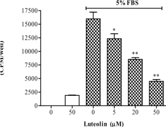
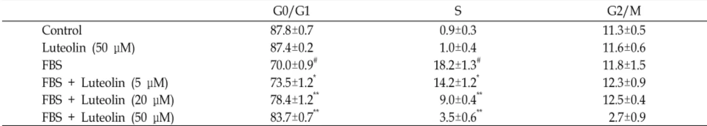
Effects of Luteolin on Fetal Bovine Serum-induced Events in Cultured Rat Vascular Smooth Muscle Cells
5
0
0
전체 글
(2)
(3)
(4)
(5)
수치

관련 문서
Effects of Nitroglycerin(Ni) on Spontaneous Activity(S.A) and Oxytocin (OT) Induced Contractions in the Uterine Smooth Muscle. Data are expressed as mean
The purpose of this study was to examine the effects of 8 weeks of Zumba dance exercise for obese middle-aged women on blood lipid index and vascular aging.. The subjects of
In this study, we investigated the effects of low-power CO 2 laser on proliferation on human gingival fibroblast cells so that determine laser
Effects of pulse frequency of low-level laser thrapy (LLLT)on bone nodule formation in rat calvarial cells.. Low-level laser therapy stimulats
In the current study, I investigated the association between vascular risk factors as chronic ischemia of the prostate and BPH by conducting experiments to elucidate
PI3K inhibition decreased antioxidants/GD-induced apoptosis in A549 cells, and PI3K inhibitor LY294002 had inhibitory effect on antioxidants/GD-induced caspase-3
In the present study we investigated the proliferation effect of human oral cancer KB cell treated with pulsatilla koreana extract.. We analyzed the effects of this
In this study, we investigated the surface characteristics of hydroxyapatite film on the micro-pore structured Ti-35Ta-xNb alloys by
