http://dx.doi.org/10.11620/IJOB.2016.41.4.217 pISSN 1226-7155, eISSN 2287-6618
Porphyromonas gingivalis, a foremost periodontal pathogen, has been known to cause periodontal diseases.
Epidemiologic evidences have indicated the involvement of P.
gingivalis in the development of cardiovascular diseases. In this study, we show that the P. gingivalis lipopolysaccharide increases the mRNA expression and protein secretion of interleukin-6 in vascular smooth muscle cells. We demonstrate that P. gingivalis LPS activates the extracellular signal- regulated kinase 1/2 (ERK1/2), p38 mitogen-activated protein kinase (MAPK), and Akt, which mediate the IL-6 expression in vascular smooth muscle cells. Also, P. gingivalis LPS stimulates the vascular smooth muscle cell migration, which is a critical step for the progression of atherosclerosis.
Moreover, neutralization of the IL-6 function inhibits the migration of vascular smooth muscle cells induced by P.
gingivalis LPS. Taken together, these results indicate that P.
gingivalis LPS promotes the expression of IL-6, which in turn increases the migration of vascular smooth muscle cells.
Key words: Porphyromonas gingivalis LPS, vascular smooth muscle cells, Interleukin-6
서 론
혐기성, 그람음성세균인 P. gingivalis는 만성 염증성 질환인 구강 내 치주질환을 일으키는 주요 세균으로 잘 알려져 있다 [1]. P. gingivalis가 가진 lipopolysaccharide (LPS), 펩티도글리칸, fimbriae, gingipain과 같은 독성인 자들이 급성 또는 만성 치주염의 시작 및 진행을 촉진 하는 병인인자들로 작용하게 된다 [2].
최근에는 치주염이 동맥경화증, 관상동맥질환, 뇌졸중, 당뇨병, 그리고 조산 및 미숙아 출산 등의 전신성 질환 의 발병에 관련되어 있다는 사실이 많은 역학적 연구로 제시되고 있다[3]. 특히 동맥경화증 환자의 구강 외 혈 관 및 혈장에서 치주질환 병인균이 발견되고 있으며, 심 혈관계 질환의 진행과정에 치주질환 병인균 감염이 중 요한 위험인자로 알려지고 있다 [4]. 이들 연관성을 설 명할 수 있는 기전에 대해서는 명확히 규명이 되어 있 진 않지만, P. gingivalis 및 P. gingivalis LPS가 전신성 혈류에 침투하여 혈관세포의 항상성을 붕괴하고 염증반 응의 증가를 초래할 것으로 제안되고 있다 [5,6].
혈관평활근세포는 특히 혈관평활근세포의 증식 및 이 동은 혈관성형술후 재협착증, 관상심장질환, 동맥경화증 을 포함한 혈관계 질환의 진행과정에서 필수적인 단계
Porphyromonas gingivalis Lipopolysaccharide Regulates Migration of Vascular Smooth Muscle Cells
Yeon Kim
1, So-Jeong Kim
1, Mi-Kyoung Kim
1, Hyun-Joo Park
1,2, Hyung Joon Kim
1, Soo-Kyung Bae
2and Moon-Kyoung Bae
1,*1
Department of Oral Physiology, BK21 PLUS Project, School of Dentistry, Pusan National University, Yangsan 626-870, South Korea
2
Department of Dental Pharmacology, BK21 PLUS Project, School of Dentistry, Pusan National University, Yangsan 626-870, South Korea
(received November 24, 2016; revised December 08, 2016; accepted December 09, 2016)
*Correspondence to: Moon-Kyoung Bae, Department of Oral Physiology, School of Dentistry, Pusan National University, Yangsan 626-770, South Korea;
Tel: 82-51-510-8239; Fax: 82-51-510-8238;
E-mail: [email protected] ORCID : 0000-0003-3948-4922
This is an Open-Access article distributed under the terms of the Creative Commons Attribution Non-Commercial License (http://creati- vecommons.org/licenses/by-nc/3.0) which permits unrestricted non- commercial use, distribution, and reproduction in any medium, pro- vided the original work is properly cited.
217
로 여겨지고 있다 [7]. 정상적인 혈관평활근세포는 수축 성 형질의 특징을 가지고 있지만, 혈관손상이 있는 경우 는 염증성 형질로 전환이 된다. 이러한 변화는 혈관평활 근세포에서 혈관평활근 특이적 표식자들의 발현을 감소 시키고, 염증유발물질을 분비하게 된다 [8]. 이렇게 분비 된 염증유발물질들이 혈관평활근세포의 증식, 이동, 화 학주성등을 조절하게 된다고 알려져 있다 [9].
본 연구에서는
P. gingivalis LPS가 혈관평활근세포의
이동에 미치는 영향을 조사하고 그 기전을 조사하였다.P. gingivalis LPS는 혈관평활근세포에서 대표적인 염증
성 사이토카인인 interleukin-6 (IL-6)의 발현 및 분비를 증가시키며, 이것이 혈관평활근세포의 이동에 관여하고 있음을 확인하였다.실험재료 및 방법
실험재료
P. gingivalis LPS는 Invivogen (USA)에서 구입하여 사
용하였으며, Phospho-ERK1/2, ERK1/2, phospho-Akt, Akt, phospho-p38MAPK, p38MAPK 항체들은 Cell signaling technology (USA)에서 구입하여 사용하였다. Anti-α-tubulin 항체는 Biogenex (USA)에서 구입하였다.혈관평활근세포배양
Sprague-Dawley 쥐 (45~55 g) 3주령 수컷을 에테르로 마취 시켜 희생시킨 다음 대동맥을 적출하여 지방조직과 남은 혈액을 제거한 후 6-well plate에 각각 조직을 적절하게 잘라 혈관의 안쪽 면이 아래쪽으로 향하도록 놓고 cover slip (22mm*22mm)을 덮어 조직을 지지시켰다. 여기에 10% FBS (GibcoBRL, USA)과 1% Penicillin-streptomycin (GibcoBRL, USA), 5 ㎍/ml Plasmocin (invivogen, USA) 이 첨가된 DMEM (GibcoBRL, USA) 배지를 첨가하여 5% CO2가 공급되는 3 7℃ 세포 배양기에서 7일간 배양하였다. 이후 현미경으로 혈관평활근세포가 자라는 것을 확인한 후 3-5일마다 계대 배양하며 5-8 세대 사이의 세포를 실험에 사용하였다.
RNA 분리와 중합효소연쇄반응 (Reverse transcription- polymerase chain reaction; RT-PCR)
혈관평활근세포에서 Total RNA를 추출하기 위해 RiboEx reagent kit (GeneAll Biotechnology, korea)를 사용 하였다. Reverse transcription kit (Promega, USA)를 사용 하여 2 ㎍의 RNA로부터 cDNA를 합성하였다. 합성된 cDNA 는 β-actin (sense: 5′-GACTACCTCATGAAGATC- 3′, antisense: 5′-GATCCACATCTGCTGGAA-3′), rat IL-6
(sense: 5′-CAAGAGACTTCCAGCCAGTTGC3′, antisense:
5′-TTGCCGAGTAGAC
CTCATAGTGACC-3′)의 primer를 이용하여 역전사중 합효소연쇄반응을 시행하였다. Real-time PCR 은 power SYBR Green (Applied Biosystems, USA) 시약을 사용하 였으며, 7500 Real-Time PCR System (Applied Biosystems, USA) 기기를 사용하여 실시하였다. PCR 반응 조건은 95℃에서 10분간 1주기, 변성 (denaturation) 반응은 95℃
에서 15초, 결합 (annealing) 반응을 60℃에서 60초, 중합 (extension) 반응을 72℃에서 7초간 40주기를 반복하여 반응시켰다. 각각의 primer는 다음과 같다. GAPDH (sense: 5′-GGCACAGTCAAGGCTGAGAATG-3′, antisense:
5′-ATGGTGGTGAAGACGCCAGTA-3′), rat IL-6 (sense: 5′
-AGAGACTTCCAGCCAGTTGC-3′, antisense: 5′-AGCCTCC GACTTGTGAAGTG-3′).
효소결합면역흡착분석법 (Enzyme-linked immunosorbent assays: ELISA)
Sprague-Dawley 쥐 6주령 수컷의 대동맥을 적출하여 지방조직과 남은 혈액을 제거한 후 작은 조각으로 잘라 10% FBS와 1% Penicillin-streptomycin, 5 ㎍/ml Plasmocin 이 첨가된 DMEM 배지에 넣은 후 P. gingivalis LPS (5
㎍/mL)을 2, 8시간 동안 처리하여 배양액을 수집하였다.
이 배양액을 4°C에서 12,000 rpm, 1분 원심 분리하여 상 층액을 얻어 효소결합면역흡착검사키트 (BioLegend, USA) 및 포함된 제품 설명서에 의거하여 배양액에 분비된 IL-6 단백질의 농도를 측정하였다.
Western immunoblot analysis
수확한 혈관평활근세포를 3회 차가운 PBS로 세척한 후 세포용해 용액 (RIPA buffer, Proteinase inhibitor, 1 mM PMSF, 10 mM NaF 및 10 mM Na3VO4)으로 용해시 켰다. 각 단백질의 양은 BCA (Sigma Aldrich, USA) 정 량법으로 측정하였다. 25 ㎍의 전체 단백질을 SDS- PAGE (poly acrylamide gel electrophoresis)로 변성 분리하 였고, 이를 nitrocellulose membrane (Amersham Pharmacia Biotech, USA) 막에 전이시켰다. Membrane의 blocking을 5% skim milk가 함유된 TBS-T (TBS, 0.1% Tween 20) 용액으로 상온에서 1시간 동안 실시한 후, 단백질의 발 현을 측정하기 위해 적절한 1차 항체를 4°C에서 16시간 동안 반응시켰다. 이어서 TBS-T용액으로 10분 간격으로 3회 세척한 다음 Horseradish peroxidase가 결합된 이차항 체를 실온에서 2시간 반응시키고, 다시 TBS-T용액으로 10 분 간격으로 3회 세척한 다음 화학발광제 (ECL: Amersham PharmaciaBiotech, USA)를 반응시킨 후 LAS4000 (GE
Healthcare Life Sciences, Sweden) 기기로 단백질 발현을 확인하였다.
혈관평활근세포의 이동성 분석 (Transwell migration assay)
세포이동의 측정은 transwell chamber법을 사용하였다.
Transwell migration chamber (Corning Life Science, USA)의 low chamber에 1% FBS를 함유한 DMEM에 혈관평활근세 포의 이동을 유도하는 P. gingivalis LPS (5 ㎍/mL)와 IL-6 중화항체 (Abcam, UK) (20 ㎍/mL)을 넣었다. Upper chamber의 insert에는 IL-6 중화항체 (20 ㎍/mL)와 각 well 당 5 × 10⁴개의 혈관평활근세포를 첨가하여 37℃, 5%
CO2 배양기에서 24시간 동안 배양하였다. 배양 후 filter의 아랫면을 메탄올 (SK chemical, Korea)로 고정시키고, hematoxilin 및 eosin (DAKO, Germany)으로 염색시킨 후 insert 내면을 면봉으로 닦아 이동하지 않은 세포를 제거 한 뒤 염색된 세포를 광학현미경 (Nikon, Japan)으로 관찰 하며 계수하였다.
통계처리
모든 실험치는 세 번 실험하여 얻어진 평균 및 평균의 표준 편차로 표시하였고 그룹간의 통계적 차이는 Student's t-test를 적용하여 분석하였다. 대조군과 비교하여 P < 0.1인 경우에 통계적으로 유의성이 있는 것으로 판정하였다.
결 과
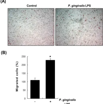
혈관평활근세포 이동에 미치는 P. gingivalis LPS의 효과 혈관평활근세포의 이동은 다양한 혈관계 질환의 진행 과정에서 필수적인 단계로 여겨지고 있다[7]. 따라서 본 실험에서는
P. gingivalis LPS가 혈관평활근세포의 이동
에 어떠한 영항을 미치는 지 transwell chamber를 이용하 여 조사하였다. Low chamber에 P. gingivalis LPS를 5 ㎍ /mL 처리하여 24시간 배양 후 transwell chamber에 이동 된 세포를 hematoxilin과 eosin 염색을 통해 확인하였다 (그림 1A). 대조군에 비해 P. gingivalis LPS를 처리하였 을 때 혈관평활근세포의 이동이 확연히 증가함을 확인 하였고, 이동한 세포의 수를 계수한 결과 2배 이상 증가 함을 확인하였다 (그림 1B).혈관평활근세포에서 P. gingivalis LPS가 IL-6 발현 에 미치는 영향
혈관평활근세포의 이동에 IL-6가 관여한다는 여러 사실 이 보고되고 있다 [10,11]. 따라서 P. gingivalis LPS가 IL-6의 발현을 변화시키는 지를 조사하였다. P. gingivalis LPS가 혈
Fig. 1. Effect of P. gingivalis LPS on the migration of vascular smooth muscle cells (A) Vascular smooth muscle cells were seeded into the upper chamber and incubated on Transwell chambers for 24 hrs in the absence or presence of P. gingivalis LPS (5 ㎍/mL). Migrated cells were stained with hematoxylin/
eosin and photographed. (B) The numbers of migrated cells were counted. *P < 0.01 vs. control.
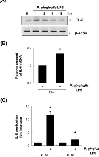
관평활근세포에서 IL-6 mRNA 발현에 어떠한 영향을 미치 는지를 관찰하기 위해 P. gingivalis LPS를 시간별로 처리하 여 IL-6의 mRNA 발현을 RT-PCR법으로 조사하였다. 그 결 과, 그림 2A에서 보는 바와 같이 P. gingivalis LPS를 시간대 별로 처리하였을 때, IL-6의 mRNA의 발현이 1시간과 2시간 에서 대조군에 비해 증가되었음을 확인하였다. 뿐만 아니라 유전자들의 발현 차이를 정량적으로 비교하고자 Real-time PCR을 수행하였다. GAPDH의 발현양에 대해 상대적인 발 현차이를 비교하였을 때 IL-6의 경우 P. gingivalis LPS 2시간 처리시 눈에 띄는 발현차이를 보였다 (그림 2B). 따라서 RT-PCR 과 Real-time PCR을 통해 P. gingivalis LPS가 혈관평 활근세포에서 IL-6의 mRNA 발현을 증가시키고 있음을 확 인하였다. 그렇다면 P. gingivalis LPS가 분비성 IL-6 단백질 의 양을 증가시키는지 알아보기 위해 쥐로부터 대동맥을 얻어 효소결합면역흡착법 (ELISA)을 실시하였다. 쥐 대동 맥에 P. gingivalis LPS을 처리하여 얻은 배양액을 수집하여 배양액에 분비된 IL-6 단백질의 농도를 측정하였다. 측정 결과 대조군에 비해
P. gingivalis LPS 2시간 처리한 실험군
에서 10배 이상 증가된 IL-6 분비성 단백질량이 관찰되었고, 8시간 처리하였을 때 역시 대조군에 비해 IL-6 단백질의 농 도가 2배 가까이 증가함을 확인하였다 (그림 2C). 따라서 효소결합면역흡착법을 통해 P. gingivalis LPS가 분비성 IL-6Fig. 2. Effect of P. gingivalis LPS on the IL-6 mRNA expression and protein production. (A) Vascular smooth muscle cells were treated with P. gingivalis LPS (5 ㎍/mL) with indicated time points. Total RNAs were isolated and then analyzed by RT-PCR using specific primers to rat IL-6. β-actin served as an internal control. (B) After incubation with P. gingivalis LPS (5 ㎍/mL) for 2 hrs, total RNAs were isolated. By real-time PCR assay, the expression levels of rat IL-6 mRNA were quantified. The expression level of the control (untreated) was set to 1.0, and the values are normalized to the GAPDH mRNA levels. *P < 0.01 vs.
control. (C) Rat aortic segments were treated with P. gingivalis LPS (5 ㎍/mL) for 2 hrs or 8 hrs. IL-6 in the supernatants was determined by ELISA analysis (OD at 450 nm). *P < 0.01 vs.
control; #P < 0.01 vs. control.
단백질의 양을 증가시킴을 알 수 있다.
P. gingivalis LPS가 혈관평활근세포의 신호전달경로 의 활성에 미치는 영향
치주인대세포에서
P. gingivalis LPS가 ERK1/2 및 Akt
신호전달 경로를 활성화 시켜서 IL-7 및 IL-23의 발현을 유도한다고 알려져 있다 [12]. 본 연구에서도 P. gingivalis LPS가 혈관평활근세포에서 이러한 신호전달경로를 활성 화시키는 지에 대하여 조사하였다. P. gingivalis LPS가 혈Fig. 3. Effect of P. gingivalis LPS on the activation of ERK1/2, p38MAPK, and AKT (A) Vascular smooth muscle cells were treated with P. gingivalis LPS (5 ㎍/mL) for the indicated times.
Western blots were probed with anti-phospho-ERK1/2, anti- ERK1/2, anti-phospho-p38, anti-p38, anti-phospho-Akt and anti-Akt antibodies. α-tubulin served as the loading control. (B) Vascular smooth muscle cells were pretreated for 30 min with or without LY294002 20 uM, SB203580 20 uM, or U0126 20 uM prior to stimulation with P. gingivalis LPS (5 ㎍/mL). Total RNAs were isolated and then analyzed by RT-PCR using specific primers to rat IL-6. β-actin served as an internal control.
(C) Using real-time PCR, the expression level of NMB-R mRNA was quantified. The expression level of the control (untreated) was set to 1.0, and the values are normalized to the GAPDH mRNA levels. *P < 0.01 vs. control; #P < 0.01 vs. P.
gingivalis LPS
관평활근세포에서 ERK1/2, p38 MAPK, Akt의 활성을 자 극하는지를 각각의 특정 항체를 이용한 Western blot 법으 로 확인하였다. P. gingivalis LPS는 ERK1/2와 p38MAPK, Akt의 총단백질량에는 큰 영향을 끼치치 않고, 인산화를 유도하였음을 확인하였다 (그림 3A). 이는 ERK, p38MAPK, Akt의 인산화는 혈관평활근세포를 자극을 준지 10분 이 내에 증가하는 양상을 보였다. 따라서 혈관평활근세포에 서
P. gingivalis LPS가 유도하는 IL-6의 발현에 ERK,
p38MAPK, Akt 신호전달경로가 관여하는 지를 이들의 신 호전달 인산화 효소 저해제를 처리하여 조사하였다. 그림 3B에서 보는 바와 같이 phosphatidylinositol 3-kinase 저해 제인 LY294002, MEK 특이적 저해제인 U0126, p38MAPK 의 선택적 저해제인 SB203580를 혈관평활근세포에 전처 리하였을 때P. gingivalis LPS가 증가시켰던 IL-6 mRNA
발현이 감소되었다. 그리고 신호전달 인산화 효소 저해제 들에 의한 IL-6 mRNA 유전자들의 발현 변화를 정량적으 로 확인하기 위해 real-time PCR을 수행하여 발현 변화를 다시 한 번 확인하였다 (그림 3C).P. gingivalis LPS가 촉진하는 혈관평활근 세포의 이동 능에 IL-6의 관련성
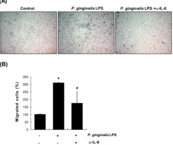
P. gingivalis LPS가 유도하는 혈관평활근세포의 이동에 P. gingivalis LPS가 증가시킨 분비성 IL-6가 매개하는 지를
Fig. 4. Involvement of IL-6 in P. gingivalis LPS-induced vascular smooth muscle cell migration (A) Vascular smooth muscle cells seeded into the upper chamber were pretreated with or without the IL-6 neutralizing antibody (20 ㎍/ml) before exposure to P. gingivalis LPS (5 ㎍/mL) and incubated on transwell chambers for 24 hrs. Migrated cells were stained with hematoxylin/eosin and photographed. (B) The numbers of migrated cells were counted. *P < 0.01 vs. control; #P < 0.1 vs.
P. gingivalis LPS
확인하기 위해 transwell의 low chamber 및 upper chamber에 IL-6 중화항체 (neutralizing antibody)를 20 ㎍/mL 처리하여
P. gingivalis LPS가 촉진시키는 이동능에 어떠한 영향을 끼
치는지 조사하였다. 대조군과 비교하여 P. gingivalis LPS에 의해 3배 이상 증가된 혈관평활근세포의 이동능이 IL-6 중 화항체를 전처리 하였을 때 절반 이상 감소됨을 확인하였다 (그림 4A와 4B). 이러한 결과로 미루어 보아 IL-6가 P.gingivalis LPS가 유도하는 혈관평활근세포의 이동에 관여
하고 있음을 알 수 있다.고 찰
여러 가지 역학조사에서
P. gingivalis가 심혈관계 질환,
당뇨병, 미숙아 출산, 류마티스성 관절염등과 같은 전신성 질환의 진행에 관여하고 있는 것으로 보고 되어 있다 [13].특히
P. gingivalis를 포함한 치주질환 병인균들이 동맥경
화증, 관상심장질환, 하지동맥질환, 심내막염의 심장판막 등을 포함한 여러 가지 심혈관질환을 가진 환자들에게서 발견되고 있다 [14]. 또한 ApoE 결손생쥐와 같은 콜레스테 롤혈증 동물모델 및 고지방식 섭취 동물모델에서P.
gingivalis 감염에 의해 동맥경화증이 촉진된다는 사실이
밝혀졌다 [15]. 이러한 결과들은 P. gingivalis 감염이 동맥 경화증의 형성 및 진행에 중요한 위험요인으로 작용할 것 이라는 가정을 강력하게 뒷받침해주고 있으나 이를 설명 할 수 있는 정확한 기전에 대해서는 잘 알려져 있지 않다 . 동맥경화증의 초기단계에서는 사이토카인이 단핵구를 모집을 조절하게 된다 [16]. P. gingivalis와 연관된 치주염 환자의 염증성 병변 유래 염증성 세포에서 prostaglandins, interleukin-1β, interleukin-8, C-reactive protein, tumor necrosis factor 같은 여러 가지 사이토카인을 분비되며, 이 들은 전신성 순환계에서도 높은 수준으로 관찰된다 [17].혈류에 있는 높은 농도의 수용성 염증성 매개물질들이 더 많은 염증성 세포를 활성화 시키고 불러들이게 된다. 활성 화된 염증성 세포가 활성산소를 생산케 되고 이것이 산화 된 저밀도 리포단백질 (low-density lipoprotein) 및 거품세 포를 혈관 내에 생성하게 한다 [18]. 예전 보고에 따르면 혈관내피세포가 상처를 입은 이후
P. gingivalis가 혈관평
활근세포에 노출되어 S100 calcium-binding protein A9 (S100A9) 발현이 높아진 후 혈관평활근세포의 수축성 형 질이 증식능이 활발해진 염증성 형질로 바뀌게 된다고 알 려져 있다[19]. 따라서 본 연구에서 밝혀진 P. gingivalis LPS가 유도한 IL-6와 S100A9와의 상관관계를 규명하고 이것이 혈관평활근세포의 이동능에 어떠한 영향을 미치는 지에 대한 추가 연구가 필요할 것이다.IL-6는 주요 염증성 사이토카인중 하나로써 단핵구, T- 세포, 섬유아세포, 혈관내피세포와 같은 여러 가지 종류의 세포에서 합성되고 분비된다 [20]. 15-lipoxygenase 2 대사 물질인 15(S)-HETE가 유도한 혈관평활근세포의 이동에 IL-6가 필요하다는 사실이 보고된 바 있다 [11]. 또한 최근 에는 toll-like receptor 4 (TLR4) 또는 TLR2 활성화에 의해 유도된 IL-6가 혈관평활근세포의 이동을 촉진시키며, 이 과정 중에 혈관평활근세포 내 p38MAPK 및 ERK1/2이 활 성화되어 있음이 보고되었다 [21,22]. IL-6는 막결합 수용 체 (IL-6R) 또는 수용성 수용체 (sIL-6R)를 통해 세포내 신 호전달을 활성화시키며, 이들 과정에서 막단백질인 gp130 의 결합을 필요로 하게 된다 [23]. 막결합 수용체를 통한 고전적 IL-6 신호기전은 주로 재생 및 보호기능을 유발하 게 되고, 반면에 수용성 수용체를 통한 IL-6 신호기전은 염 증성 반응을 일으킨다고 알려져 있다 [24]. 수용체 결합에 의한 IL-6의 세포내 신호기전은 주로 STAT (signal transducer and activator of transcription)-의존적 또는 -비의존적으로 일어난다고 한다 [25]. 본 연구에서는 P. gingivalis LPS가 혈관평활근세포에서 IL-6 발현 및 분비를 증가시킨다고 보고하고 있다. 따라서 P. gingivalis LPS에 의해 유도된 IL-6는 혈관평활근세포에 존재하는 어떠한 IL-6 수용체를 통해 혈관평활근세포의 이동을 조절하고 있는지는 향후 추가 연구가 필요할 것으로 생각된다. 조절장애에 의한 IL-6의 계속적인 합성은 자가면역질환, 만성염증질환, 암 과 같은 여러 질환들의 진행과정에 관여하게 된다[26]. 최 근 많은 보고에서 IL-6 및 IL-6 수용체를 표적으로 하는 항 체 및 약물을 이용하여 IL-6 활성을 임상적으로 차단함으 로써 만성염증성 질환 및 고형암의 치료에 적용되고 있으 며 그와 관련된 연구가 활발히 진행 중이다 [27]. 따라서 본 연구를 기반으로 한 심화연구에서는 IL-6 및 IL-6 수용 체 표적약물을 이용하여 치주염 및 치주염 관련 혈관계 질 환에 적용이 가능한 지에 대해 조사해 볼 가치가 있을 것 으로 생각된다.
감사의 글
이 논문은 부산대학교 기본연구지원사업(2년)에 의하 여 연구되었음 (to Bae M-K).
Conflict of interest
The authors declare that they have no conflicting interest.
References
1. Socransky SS, Haffajee AD. The bacterial etiology of destructive periodontal disease: current concepts. J Periodontol. 1992;63:322-331. doi: 10.1902/jop.1992.63.
4s.322
2. Bainbridge BW, Darveau RP. Porphyromonas gingivalis lipopolysaccharide: an unusual pattern recognition receptor ligand for the innate host defense system. Acta Odontol Scand. 2001;59:131-138.
3. Amar S, Han X. The impact of periodontal infection on systemic diseases. Med Sci Monit. 2003;9:RA291-299.
4. Wada K, Kamisaki Y. Molecular dissection of Porphyromonas gingivalis-related arteriosclerosis: a novel mechanism of vascular disease. Periodontol 2000. 2010;54:222-234. doi:
10.1111/j.1600-0757.2009.00336.x.
5. Hokamura K, Inaba H, Nakano K, Nomura R, Yoshioka H, Taniguchi K, Ooshima T, Wada K, Amano A, Umemura K.
Molecular analysis of aortic intimal hyperplasia caused by Porphyromonas gingivalis infection in mice with endothelial damage. J Periodontal Res. 2010;45:337-344.
doi: 10.1111/j.1600-0765.2009.01242.x.
6. Kim SR, Park HJ, Bae SK, Park JH, Kim HS, Koo TH, Bae MK. Porphyromonas gingivalis lipopolysaccharide increases monocyte adhesion to microvascular endothelium by induction of adhesion molecules. Int J Oral Biol.
2008;33:149-154.
7. Owens GK. Molecular control of vascular smooth muscle cell differentiation and phenotypic plasticity. Novartis Found Symp. 2007;283:174-191.
8. Bennett MR, Sinha S, Owens GK. Vascular Smooth Muscle Cells in Atherosclerosis. Circ Res. 2016;118:
692-702. doi: 10.1161/CIRCRESAHA.115.306361.
9. Chistiakov DA, Orekhov AN, Bobryshev YV. Vascular smooth muscle cell in atherosclerosis. Acta Physiol (Oxf).
2015;214:33-50. doi: 10.1111/apha.12466
10. Wang Z, Newman WH. Smooth muscle cell migration stimulated by interleukin 6 is associated with cytoskeletal reorganization. J Surg Res. 2003;111:261-266.
11. Chava KR, Karpurapu M, Wang D, Bhanoori M, Kundumani-Sridharan V, Zhang Q, Ichiki T, Glasgow WC, Rao GN. CREB-mediated IL-6 expression is required for 15(S)-hydroxyeicosatetraenoic acid-induced vascular smooth muscle cell migration. Arterioscler Thromb Vasc Biol.
2009;29:809-815. doi: 10.1161/ATVBAHA.109.185777 12. Park YD, Kim YS, Jung YM, Lee SI, Lee YM, Bang JB,
Kim EC. Porphyromonas gingivalis lipopolysaccharide regulates interleukin (IL)-17 and IL-23 expression via SIRT1 modulation in human periodontal ligament cells.
Cytokine. 2012;60:284-293. doi: 11.1016/j.cyto.2012.05.021.
13. Li X, Kolltveit KM, Tronstad L, Olsen I. Systemic diseases caused by oral infection. Clin Microbiol Rev. 2000;13:
547-558.
14. Hayashi C, Gudino CV, Gibson FC,3rd, Genco CA.
Review: Pathogen-induced inflammation at sites distant
from oral infection: bacterial persistence and induction of cell-specific innate immune inflammatory pathways. Mol Oral Microbiol. 2010;25:305-316. doi: 10.1111/j.2041-1014.
2010.00582.x
15. Lalla E, Lamster IB, Hofmann MA, Bucciarelli L, Jerud AP, Tucker S, Lu Y, Papapanou PN, Schmidt AM. Oral infection with a periodontal pathogen accelerates early atherosclerosis in apolipoprotein E-null mice. Arterioscler Thromb Vasc Biol. 2003;23:1405-1411. doi: 10.1161/01.
ATV.0000082462.26258.FE
16. Libby P. Inflammation in atherosclerosis. Arterioscler Thromb Vasc Biol. 2012;32:2045-2051. doi: 10.1161/
ATVBAHA.108.179705
17. Aarabi G, Eberhard J, Reissmann DR, Heydecke G, Seedorf U. Interaction between periodontal disease and atherosclerotic vascular disease-Fact or fiction? Atherosclerosis. 2015;241:
555-560. doi: 10.1016/j.atherosclerosis.2015.04.819 18. Qi M, Miyakawa H, Kuramitsu HK. Porphyromonas
gingivalis induces murine macrophage foam cell formation.
Microb Pathog. 2003;35:259-267.
19. Inaba H, Hokamura K, Nakano K, Nomura R, Katayama K, Nakajima A, Yoshioka H, Taniguchi K, Kamisaki Y, Ooshima T, Umemura K, Murad F, Wada K, Amano A.
Upregulation of S100 calcium-binding protein A9 is required for induction of smooth muscle cell proliferation by a periodontal pathogen. FEBS Lett. 2009;583:128-134.
doi: 10.1016/j.febslet.2008.11.036
20. Nishimoto N, Kishimoto T. Interleukin 6: from bench to bedside. Nat Clin Pract Rheumatol. 2006;2:619-626. doi:
10.1038/ncprheum0338.
21. Lee GL, Wu JY, Tsai CS, Lin CY, Tsai YT, Lin CS, Wang YF, Yet SF, Hsu YJ, Kuo CC. TLR4-Activated MAPK- IL-6 Axis Regulates Vascular Smooth Muscle Cell Function. Int J Mol Sci. 2016;17:E1394. doi: 10.3390/ijms 17091394
22. Lee GL, Chang YW, Wu JY, Wu ML, Wu KK, Yet SF, Kuo CC. TLR 2 induces vascular smooth muscle cell migration through cAMP response element-binding protein-mediated interleukin-6 production. Arterioscler Thromb Vasc Biol. 2012;32:2751-2760. doi: 10.1161/
ATVBAHA.112.300302
23. Taga T, Hibi M, Hirata Y, Yamasaki K, Yasukawa K, Matsuda T, Hirano T, Kishimoto T. Interleukin-6 triggers the association of its receptor with a possible signal transducer, gp130. Cell. 1989;58:573-581.
24. Rose-John S. IL-6 trans-signaling via the soluble IL-6 receptor: importance for the pro-inflammatory activities of IL-6. Int J Biol Sci. 2012;8:1237-1247. doi: 10.7150/ijbs.4989 25. Schaper F, Rose-John S. Interleukin-6: Biology, signaling
and strategies of blockade. Cytokine Growth Factor Rev.
2015;26:475-487. doi: 10.1016/j.cytogfr.2015.07.004 26. Tanaka T, Kishimoto T. The biology and medical
implications of interleukin-6. Cancer Immunol Res. 2014;
2:288-294. doi: 10.1158/2326-6066.CIR-14-0022
27. Jones SA, Scheller J, Rose-John S. Therapeutic strategies for the clinical blockade of IL-6/gp130 signaling. J Clin Invest. 2011;121:3375-3383. doi: 10.1172/JCI57158