구강상피세포의 배양환경의 차이에 의한 마이크로어레이 기반 유전자 발현의 융복합 분석
손화경
영남이공대학교 치위생과 교수
The Convergence Analysis of Microarray-Based Gene Expression by Difference of Culture Environment in Human Oral Epithelial Cells
Hwa-Kyung Son
Professor, Department of Dental Hygiene, Yeungnam University College
요 약 이 연구는 HPV 16 E6/E7 도입 불멸화 구강상피세포의 배양 미세환경과 세포 분화간의 관계를 분석하였다. 배양환 경을 변화시켜서 IHOK-EF 세포와 IHOK-EFKGM 세포를 얻었고, 이들 세포의 특성변화를 세포증식분석, 면역형광분석 및 마이크로어레이와 실시간 정량 PCR분석으로 알아보았다. IHOK-EF 세포는 상피세포의 특성을 상실하고 간엽세포의 특성을 획득하였고, 마이크로어레이 분석결과, 분화억제 유전자인 ID2, IL6, TWIST1이 과발현 되었다. 이러한 변화는 초기 의 배양환경으로 회복되었을 때, 특별히, ID2와 IL6에서 유전자발현의 복귀를 나타내면서 세포의 특성이 부분적으로 회복되 었다. 이 연구는 세포의 특성을 결정하는 연구에서 배양 미세환경의 변화에 따른 세포의 생존을 위한 적응양상을 이해하는 데 공헌할 것이며, 향후, 암세포의 미세환경변화에 따른 생존연구에 적용하여 질병에 대한 치료적 접근을 가능하게 할 것이 다.
주제어 : 구강상피세포, 마이크로어레이, 분화, 미세환경, 융복합
Abstract This study was analyzed about the relationship between culture microenvironment and cell differentiation of HPV 16 E6/E7-transfected immortalized oral keratinocyte(IHOK). By the alteration of culture environment, IHOK-EF and IHOK-EFKGM were obtained, and the modulation of cell properties was observed by cell proliferation assay, immunofluorescence, microarray, and quantitative real-time PCR analysis. IHOK-EF losed the properties of epithelial cells and obtained the properties of mesenchymal cells, and in the result of microarray analysis, genes related to the inhibition of differentiation such as IL6, TWIST1, and ID2 were highly expressed in IHOK-EF. When the culture environment was recovered to initial environment, these changes were recovered partially, presenting the return of genes involved in the inhibition of differentiation such as IL6, and ID2, particularly. This study will contribute to understand adjustment aspect for cell surviving according to the change of culture microenvironment in the study for determining the cell characteristic, and facilitate therapeutic approach for human disease by applying surviving study according to the change of cancer microenvironment.
Key Words : Oral Keratinocyte, Microarray, Differentiation, Microenvironment, Convergence
This study was supported by the Yeungnam University College Research Grants in 2016.
*Corresponding Author : Hwa-Kyung Son([email protected]) Received March 12, 2019
Accepted April 20, 2019
Revised April 4, 2019 Published April 28, 2019
1. 서론
세포의 미세환경은 다양한 종류의 세포에서 복잡한 신호전달 경로와 세포 간 상호작용을 통하여 상피세포 분화를 조절하는 중요한 요소이다. 미세환경의 신호는 지엽적인 환경에 적응하기 위한 세포의 능력에 영향을 준다[1]. 그것들은 세포형태에 영향을 주고 기관의 발생 과 같은 생리학적 기능과 밀접하게 연관되어 왔다[2,3].
정상조직의 항상성은 상피세포와 그들의 미세환경간의 역동적인 상호작용에 의해 유지된다. 그러나, 항상성의 파괴는 비정상적인 세포의 성장이나 부착, 이동을 유도 할 수 있고, 악성종양으로의 변화를 촉진시킬 것이다 [4,5]. 암세포에서 종양 미세환경은 세포와 세포간의 상호 작용, 세포와 세포 밖 환경과의 접촉, 세포에 의해 분비되 는 가용성인자와 다양한 영양분과 같은 화학적 환경, 기 계적 양상에 의해 만들어진다[6]. 실제로 종양의 환경은 종양의 전개, 전이, 종양의 개시세포의 형성에 영향을 준 다[7]. 그러므로, 세포 미세환경의 연구는 상피세포의 생 리와 암에 대한 치료적 접근의 이해를 위해 중요하다[8].
각화세포는 역동적인 환경에 의해 영향을 받으며, 각 화세포의 증식과 분화, 형태형성을 조절한다[9]. 사람각 화세포는 각화세포와 미세환경간의 관계를 조사하기 위 해 다양한 성장배지에 의해 배양되어왔다. 초기의 각화 세포 배양에서는 feeder layer가 각화세포의 성장을 개선 시키기 위해 사용되거나 섬유모세포 조건배지가 함께 사 용되었다[10]. 또한 혈청이 없는 배지나 혈청이 없이 칼 슘이 저농도로 포함된 배지가 각화세포 배양을 위해 사 용되었다[11]. 이러한 다양한 배양조건은 각화세포의 증 식과 분화, 형태형성에 영향을 주었다.
HPV(Human Papillomvirus) E6/E7 불멸화 사람각화 세포는 각화세포분화와 암연구에 사용되었고, 상피세포 연구를 위한 매우 좋은 모델세포주로 고려되었다. HPV E6/E7의 발현은 사람각화세포를 불멸화시켰고, serum이 나 칼슘에 의한 세포분화를 억제시켰다[12]. 국내연구에 서, serum이 포함되지 않은 배양조건에서 확립된 HPV E6/E7 도입 불멸화 구강상피세포를 serum이 포함된 배 양환경에 노출시켰을 때, 세포의 침윤능력이나 이동성의 증가와 같은 암세포의 특성이 연구된 바 있다[13].
배양조건은 환경적 네트웍에 의해 다양한 형태변경을 촉진시키고, 재형성된 골격은 세포의 이동과 성장에 중 요한 영향을 줄 수 있다. 여러 연구에서 다양한 자극에
의한 세포 형태의 변화를 보여주었다. 예를 들면, 신경세 포는 신경성장인자와 호르몬을 포함한 몇몇 인자에 반응 하여 긴 신경돌기로 변화되었고[14], 림프구는 화학주성 인자에 의해 이동성을 보여주었다[15]. 많은 경우에서, 다 양한 형태변화는 미세환경적 상호작용에 의해 개시되었 고, 골격재형성이 수반되었다.
세포분화는 덜 전문화된 세포가 더 전문화된 세포로 변화되는 과정이다. 분화는 세포의 크기, 모양, 대사활성 과 신호에 대한 반응을 변화시킬 수 있다. 대부분의 경우 에, 세포분화는 결코 DNA 단편을 변화시키지 않는다. 그 래서 각기 다른 세포들은 유전자가 같더라도 다양한 생 리적 특성을 가질 수 있다. 암세포에서 분화는 얼마나 성 숙한 암세포가 종양 속에 포함되어있는가를 말한다. 분 화된 종양세포는 정상세포를 닮았고, 분화되지 않거나 분화가 안 좋은 종양세포보다 더 느리게 퍼지며, 분화가 안 좋은 종양세포는 정상세포의 기능과 구조가 결핍되어 있고, 성장이 통제가 안 된다. 이러한 분화의 억제는 EMT(Epithelial-to-mesenchymal transition)와 관련된 다[16]. EMT는 상피세포의 특성을 잃고 간엽세포의 특 성을 획득하는 과정이며, 배아 발생과정과 암세포의 침 윤과 전이과정에서 중요한 역할을 한다.
이 연구에서, IHOKs(Immortalized Human Oral Keratinocytes, 불멸화사람각화세포)는 배양환경에 따라 성장, 형태, 유전자발현의 변화와 같은 다양한 세포분화 의 조절을 보여주었다. 그러므로, 이 연구는, 상피세포가 미세환경의 변화에 의해 생존하기 위한 세포적응의 방법 을 이해하는데 도움을 줄 것이다.
2. 연구재료 및 방법
2.1 세포 배양
IHOKs는 경희대학교 김은철교수로부터 제공받았고, 이후에 IHOKs와 관련된 논문이 저술 된 바 있다[17]. 몇 종류의 배지에 의한 세포의 형태변화를 분석하기 위하여 두 가지 종류의 각화세포 배양배지가 사용되었다. IHOKs 를 몇 가지 첨가물을 추가한 각화세포 성장배지(KGM, Lonza, MD, USA)로 배양한 것은, IHOK-KGM이라 명 명하였다. IHOK-KGM을 EF배지(DMEM, F12, 10%
FBS, 1% penicillin/streptomycin, 0.01 ㎍/㎖ cholera toxin, 0.04 ㎍/㎖ hydrocortisone, 0.5 ㎍/㎖ insulin, 0.5 ㎍/㎖
apo-transferrin, 0.2 ㎍/㎖ 3'-5-triodo-l-thyronine)에 배양한 것은 IHOK-EF라 명명하였고, IHOK-EF를 다시 KGM 배지로 배양한 것을 IHOK-EFKGM으로 명명하였 다. 정상 구강의 치은 섬유모세포는 병소가 없는 사랑니 환자로부터 채취하였으며, 정보동의를 받았다 (IRB-2-2009-0002). 치은섬유모세포는 hTERT를 감염 시켜서 불멸화시킨 것이며[18], F배지(DMEM, F12, 10%
FBS, 1% penicillin/streptomycin)에 의해 배양되었다.
2.2 세포증식 분석
세 가지 종류의 IHOKs의 성장은 MTT [3-(4,5-methylthiazol-2-yl)-2,5-diphenyltetrazolium bromide]분석에 의해 측정되었다. 흡광도는 72시간동안 측정되었고, 이 분석은 세 번 실시하였다.
2.3 역전사(Reverse transcriptase, RT)-중합효 소연쇄반응(Polymerase Chain Reaction, PCR)
RNeasy kit (Qiagen, Hilden, Germany)를 사용하여 RNA(Ribonucleic acid)를 추출하였고, cDNA는 전체 RNA 1mg과 Accu Power Hot Start PCR Pre Mix (Bioneer, Daejeon, South Korea)을 사용하여 합성하였 고, 제조자의 사용법에 따라 시행하였다. PCR 결과물은 1% agarose gel에서 ethidium bromide를 사용하여 확인 하였다.
2.4 면역형광분석
IHOKs는 4-well chamber 슬라이드에 주입하고, 4%
formaldehyde에서 15분 동안 고정하였으며, 0.5% triton X-100 solution에서 투과성을 획득하였으며, 5분마다 PBS로 세척하였다. 그 후 E-cadherin 이나 snail (Cell Signaling Technology, 1:100)에 대한 항체로 세포를 표 지하였고 2차 항체인 Alexa Fluor 488-conjugated anti-rabbit IgG 로 배양하였다. Cytokeratin과 vimentin 염색을 위해, 세포를 FITC monoclonal anti-Pan Cytokeratin conjugate (Sigma, 1:200)와 monoclonal anti-vimentin cy3 conjugate (Sigma, 1:200)로 각각 배양 하였다. DNA를 검출하기 위해 세포를 DAPI로 염색하였 으며, 슬라이드를 마운팅 시약(Vectashield, Vector Laboratories, Burlingame, CA)으로 봉입하였다. 면역형
광 이미지는 LSM 510 laser scanning confocal microscope와 software(Carl Zeiss Inc., Oberkochen, Germany)로 캡쳐해서 400배로 분석하였다.
2.5 마이크로어레이 데이터 분석
IHOK-KGM과 IHOK-EF, IHOK-EFKGM는 각각 70% 채워진 상태까지 배양하였고. Total RNA는 RNeasy kit (Qiagen, Hilden, Germany)를 이용하여 제조 자의 사용법에 따라 분리하였다. 각 total RNA sample (200ng)을 Low Input Quick Amp labeling kit (Agilent technologies, CA)를 이용하여 표지하고 증폭하였다.
Cy3가 표지된 aRNAs를 교잡시약(Agilent technologies, CA) 50㎕로 처리한 후 표지된 aRNAs를 Gasket 8-plex slide 위에 놓았다. Agilent scanner를 이용하여 마이크로 어레이가 분석되었다. 유전자발현 수준은 Feature Extraction v10.7.3.1 (Agilent technologies, CA)에 의해 산출되었다. 각 유전자의 상대적인 신호강도가 Robust Multi-Array Average algorithm에 의해 생산되었다. 데 이터는 Gene Spring GX 7.3.1을 사용하여 median polish normalization method에 기초하여 처리되었고, 표준화된 로그변환 강도가 분석되었다. 배수 변화는 상향 조절된 유전자 중에서는 대조구에 비해 거의 200% 높게 조절된 유전자들을 걸러내었고, 하향 조절된 유전자 중에서는 대조구에 비해 50% 낮게 조절된 유전자들을 걸러내어 나타내었다. Hierarchical clustering data는 유사하게 행 동하는 그룹을 모아놓은 것이다.
2.6 실시간 정량 PCR
RNeasy kit (Qiagen, Hilden, Germany)를 사용하여 IHOK-KGM과 IHOK-EF, IHOK-EFKGM로부터 Total RNA를 추출하였고, cDNA는 Accu Power Hot Start PCR Pre Mix (Bioneer, Daejeon, South Korea)을 사용 하여 total RNA의 1mg을 가지고 제조자의 사용법에 따 라 합성하였다. 마이크로어레이 데이터를 검증하기 위해 서, SYBER green I Master (Roche applied science, Switzerland)를 이용하여 실시간 정량 PCR을 시행하였 고 GAPDH 유전자로 결과를 표준화하였다. 각 PCR반응 은 세 번 시행하였고 결과는 Light Cycler 480 software 를 이용하여 분석하였다.
3. 연구결과
3.1 배양조건의 변경에 의한 IHOKs의 변화 Fig. 1의 A-a과 같이 HPV E6/E7 IHOKs는 KGM 배 지에서 배양하며 (IHOK-KGM) 조약돌모양의 형태를 나 타낸다. 변화된 배양환경에서 각화세포의 특성 변화를 조사하기 위하여 IHOK-KGM을 EF배지에서 배양하였 다(IHOK-EF). 배양한지 약 60일 째 되었을 때, 세포들은 세포 간 간격이 넓어지면서 흩어지기 시작했고, 길게 연 장된 세포형태를 나타내었으며, Fig. 1의 A-b과 같이 90 일째 되었을 때는 IHOK-KGM과 비교했을 때, 긴 방추 모양의 섬유모세포와 유사한 형태로 변화되었다. 한편, IHOK-KGM에서 IHOK-EF로의 변화가 가역적인 변화 인가를 알아보기 위해서 IHOK-EF를 다시 KGM 배지에 서 배양(IHOK-KEFKGM) 하였으며, 배양한지 약 60일 째 되었을 때, 조약돌모양의 세포와 IHOK-EF 보다도 더 길게 연장된 세포들이 공존하였으며, 90일째는 조약돌모
양의 세포들이 방추모양의 세포보다 더 많이 존재하였으 며, Fig. 1의 A-c와 같이 120일째 되었을 때는 방추모양 의 세포들이 모두 사라지고 IHOK-KGM과 유사한 형태 의 세포들로 채워졌다. 이러한 세포형태의 변화가 세포 증식과 관련이 있는지 알아보기 위하여, 변경된 세포를 이용하여 세포 간 증식률을 분석하였다. 배양 후 1일째부 터 세포증식에 차이가 나기 사작하였는데, IHOK-KGM 의 증식률이 세 가지 세포 중에서 가장 높았으며, 이것은 IHOK-EF보다 1.3배 높은 증식률이었고, Fig. 1의 B와 같이 IHOK-EFKGM에서는 IHOK-KGM에 가까운 증식 률의 회복을 보여주었다. 이 결과는 배양조건의 변화가 세포의 형태 및 증식률을 변화시킬 수 있으며, 다시 초기 의 조건이 주어졌을 때는 본래의 특성을 회복할 수 있다 는 것을 나타낸다.
3.2 IHOK-EF로부터 IHOK-KGM로의 특성 회복 분석
Relative Cell Growth (unit: Absorbance)
Day no.
Cells Day0 Day1 Day2 Day3
KGM 1 3.98 7.53 17.72
EF 1 2.32 5.15 13.75
EFKGM 1 2.76 6.72 16.97
Fig. 1. The alteration of IHOK by change of culture conditions
(A) Control IHOK-KGM cells were cultured in two media. a, Control IHOK-KGM cells, b, IHOK-KGM cells were cultured in EF media for 90days, c, IHOK-EF cultured for 90days were re-cultured in KGM for 120 days. (B) The cell growth rate of control IHOK-KGM and two altered cell lines was analyzed.
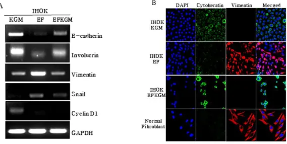
IHOK-EF로부터 IHOK-KGM으로의 가역적인 특성 변화를 RNA수준과 단백질 수준에서 확인하고자 RT-PCR 과 면역형광염색을 시행하였다. RT-PCR에서, 상피세포 분화의 표지자인 E-cadherin과 involucrin의 발현을 알아 보니, IHOK-KGM과 IHOK-EFKGM에서 발현이 높고, IHOK-EF에서 발현이 낮았다. 간엽세포분화의 표지자인 vimentin과 snail의 발현은 IHOK-EF에서 높고, IHOK-KGM 과 IHOK-EFKGM에서 낮았다. IHOK-KGM과 IHOK-EFKGM에서 보여준 발현정도를 비교했을 때, IHOK-EFKGM이 IHOK-KGM에서 보다 다소 낮거나 높은 발현률을 나타내기는 하였으나, IHOK-EF에 비교 하면 분명히 IHOK-EFKGM은IHOK-KGM으로 회복되 어가고 있는 세포임을 알 수 있었다. 반면, 세포주기 조절 의 표지자인 cyclin D1의 발현은 IHOK-KGM에서만 볼 수 있었고, IHOK-EFKGM에서는 IHOK-EF에서처럼 확 인할 수 없었다. Fig. 2 A 면역형광염색을 통해 상피세포 분화의 표지자인 cytokeratin과 간엽세포분화의 표지자 인 vimentin 의 발현을 알아보았다. IHOK-EF에서는 IHOK-KGM과 비교하여 cytokeratin의 발현이 낮게 조 정되었고 정상 섬유모세포(Normal Fibroblast)에서처럼 vimentin의 발현이 높게 조정되었다. IHOK-EFKGM에 서는 IHOK-KGM과 유사하게 발현이 회복되었다. 그러 므로 배양환경의 변화는 구강상피세포의 분화를 낮게 조 절하고 간엽세포로서의 특성을 높게 조절하기도 하였으
며, 이러한 특성 변화는 다시 초기의 배경환경에 처 했을 때 회복됨으로써 구강상피세포 분화의 가역적인 역동성 을 나타내었다.
3.3 마이크로어레이에 의한 계층군집과 기능적 분류 배양조건에 의한 세 가지 세포주의 유전자발현 패턴 을 확인하기 위하여 마이크로어레이 분석을 시행하였다.
세 가지 세포주에서 24244개의 사람유전자를 포함하는 전체 유전체어레이를 사용하여 mRNA가 분석되었다.
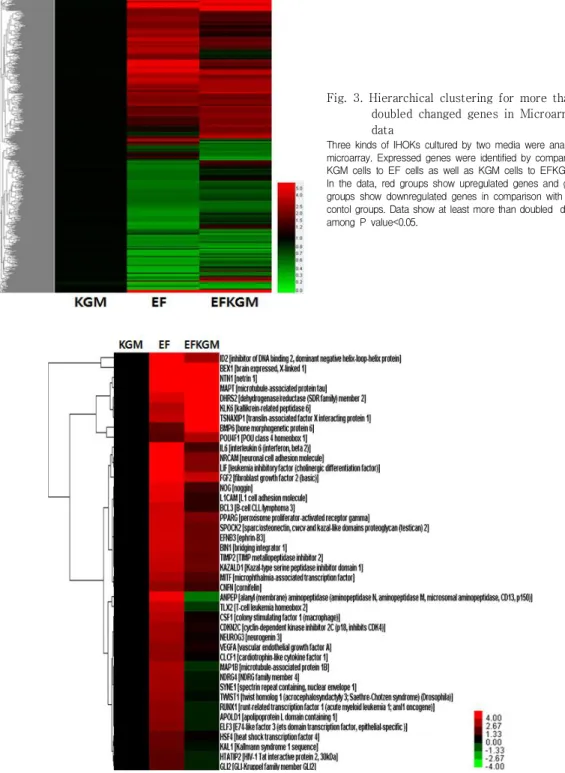
Fig. 3은 IHOK-KGM에 비교하여 IHOK-EF나 IHOK-EFKGM에서 과발현되거나 저발현된 유전자 중 2 배 이상 변화된 유전자에 대한 계층군집을 나타낸다. 기 능적 분류 분석은 전체 유전자발현에서 중요한 변경을 보여주었다. IHOK-EF에서는 IHOK-KGM에 비교하여, 3080개의 유전자들이 변경되었는데, 그 중 IHOK-KGM 에 비교하여 과발현된 유전자가 1518개, 저발현된 유전 자가 1562개였다. IHOK-EFKGM의 발현변화를 보았을 때, IHOK-EF에서 변화된 유전자 중 1414개의 유전자들 이 IHOK-KGM 수준으로 되돌아왔다. Fig. 4는 세포분화 와 관련하여 IHOK-KGM과 IHOK-EF간에 2배 이상 변 화된 유전자 중에서 다시 2배 이상 발현이 회복된 유전자 들을 모은 계층군집이며, 가장 강도가 높은 유전자들을 나타낸다.
Fig. 2. The Analysis of Restoration from IHOK-EF to IHOK-KGM
(A) RT-PCR was analized by EMT(E-cadherin, vimentin, snail), differentiation(involucrin,) and cell cycle regulation(cyclin D1) markers.
(B) Cells were stained by vimentin and cytokeratin antibodies during 2 hr, respectively, for analyss of Immunofluoresence
Fig. 3. Hierarchical clustering for more than doubled changed genes in Microarray data
Three kinds of IHOKs cultured by two media were analyzed by microarray. Expressed genes were identified by comparisons of KGM cells to EF cells as well as KGM cells to EFKGM cells.
In the data, red groups show upregulated genes and green groups show downregulated genes in comparison with KGM contol groups. Data show at least more than doubled difference among P value<0.05.
Fig. 4. Hierarchical clustering of more than doubled changed genes for cell differentiation
Up-regulated or down-regulated genes, which expressed the highest intensity among returned genes more than doubled from genes changed more than doubled between IHOK-KGM control cells and IHOK-EF cells for cell differentiation, were shown.
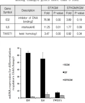
3.4 마이크로어레이 데이터의 검증
EMT과정 동안 상피세포는 섬유모세포와 유사한 형 태를 획득하고 세포 간 부착이 감소되고, 이동성을 나타 낸다. 각화세포의 역동적인 특성변화는 세포 분화의 억 제를 포함한다. 그래서 세포분화와 관련하여 변화된 유 전자 중 세포분화를 억제하는 유전자인 ID2, IL6, TWIST1을 선택하였다. Table 1
이들 분화억제표지자를 이용하여 실시간 정량 PCR로 마이크로어레이 데이터를 검증한 결과, ID2와 IL6의 mRNA 발현이 IHOK-KGM과 비교하여 IHOK-EF에서 각각 63배와 55배 높게 나타났으며, TWIST1은 8배 정도로 그쳤으며, 이것은 IHOK-EFKGM에서는 IHOK-KGM과 유사하거나 조금 높게 나타남으로써, 배양조건의 변경에 따라 상피세포의 특성이 변경되었다가 회복될 수 있음을 보여주었다. Fig. 5
Gene
Symbol Description EF/KGM EFKGM/KGM
Fold P-value Fold P-value ID2 inhibitor of DNA
binding2 76.98 0.00 3.89 0.19
IL6 interleukin6 11.25 0.01 1.77 0.09
TWIST1 twist homolog1 3.47 0.00 0.92 0.34 Table 1. List of genes which inhibit epithelial
differentiation in comparison for
IHOK-KGM cells to IHOK-EF-cells and IHOK-KGM cells to IHOK-EFKGM cells among changed genes more than 3 fold.
Fig. 5. Validation of micoarray data for
differentiation inhibitors by real time PCR mRNA expression of ID2, IL6, and TWIST1 was verified by real time PCR in the three cell lines.
4. 고찰 및 결론
이 연구는 두 가지 배양조건을 이용하여 불멸화각화 세포의 특성 변화와 회복을 통하여 배양 환경에 의한 상 피세포 생물학의 역동적인 변화를 보여주었다. 이러한 세포의 특성변화는 상피세포에서 뿐 아니라, 다양한 배 양조건에 의해 배양된 암세포와 섬유모세포에 대해서도 보고되었다. 결장암세포주에서 다양한 배양조건에 의해 연구된 약물의 민감도변화가 보고되었고[19], 사람 피부 의 섬유모세포를 다양한 조건에서 배양하여 단백질 발현 의 변화양상을 연구하기도 하였다[20]. 구강각화세포의 불멸화를 연구하기 위해 사용해 온 KGM 배지는 혈청이 없는 저농도(0.15mM)의 칼슘을 포함하는 배지이며 사람 이나 동물의 일차세포 증식을 위해 고안되었고, 각화세 포의 특성이 잘 보존되는 배지이다[21]. KGM 배지와 비 교하여, EF배지에는 cholera toxin과 triiodothyronin이 포함되어 있으며, 이 연구에서는 cholera toxin을 세포의 중요한 특성 변화 유발요인으로서 고려하고 있다. 왜냐 하면, IHOK-EF에서 EMT와 세포증식억제를 보여주었 는데, cholera toxin은 여러 논문의 다양한 세포에서 EMT를 포함한 세포의 형태변화[22,23]와 세포증식억제 [24,25]에 관해 보고되었기 때문이다. 본연구의 RT-PCR 분석(Fig. 2A)에서, EMT를 겪은 IHOK-EF의 cyclin D1 의 발현이 억제된 것은, cholera toxin이 cyclin D1과 CDK2와 같은 세포주기 조절 단백질의 발현을 억제하는 것에 의해 세포주기를 정지시키는 것과 연관이 있다고 판단된다[26].
이 연구에서는 마이크로어레이를 이용하여 불멸화각 화세포의 특성을 변화시킨 매개체로서, ID2, IL6, Twist1 과 같은 분화억제표지자의 과발현에 주목하였다. ID2는 다양한 종류의 세포에서 세포의 분화를 억제하고, EMT 를 촉진하는 것으로 알려져 있다[27]. IL6는 유방암세포 를 비롯한 많은 암세포에서 E-cadherine의 억제를 통하 여 EMT를 유도하는 것으로 알려져 있다[28]. 또한 Twist1은 vimentin과 같은 EMT 표지자의 발현을 증가 시키는 중요한 EMT유도인자로서 알려져 있다[29]. 실시 간 정량 PCR 결과, 실제로 ID2와 IL6 및 Twist1이 IHOK-EF에서 과발현되는 것을 확인함으로써, IHOK-KGM은 배양조건의 변화에 의해 과발현된 분화 억제 표지자들에 의해 각화세포로서의 분화가 억제되고 EMT를 겪었으며, 이러한 특성은 다시 초기의 배양환경 에 처했을 때, 회복된다는 것을 보여주었다.
결론적으로, HPV E6/E7 불멸화 사람각화세포는 배양 환경의 변화에 의해 세포형태의 변화와 세포증식의 억제 를 보여주었으며, 그 변화요인이 세포분화의 억제표지자 를 가역적으로 과발현시킴으로써 세포의 형태를 변화시 킨 것으로 고려된다. 향후에는 이 변화요인과 억제표지 자간의 직접적인 관계를 분석하기 위한 연구가 필요하다 고 사료된다.
이 연구는, 세포의 생물학적 특성을 결정하는 연구에 서 배양 미세환경의 변화에 따른 세포의 생존을 위한 적 응양상을 이해하는데 도움을 줄 것이며, 향후 암세포의 미세환경변화에 따른 생존연구에 적용하여 질병에 대한 치료적 접근을 가능하게 할 것이다.
REFERENCES
[1] Y. L. Dorland & S. Huveneers. (2017). Cell-Cell junctional mechanotransduction in endothelial remodeling.
Cell Mol Life Sci 74(2), 279-292.
DOI: 10.1007/s00018-016-2325-8
[2] M. Arocena et al. (2019). Using a variant of coverslip hypoxia to visualize tumor cell alterations at increasing distances from an oxygen source. J Cell Physiol, 1-8.
DOI: 10.1002/jcp.28507
[3] T. L. Lee. (2018). The Convergence effect of medical industry through stem cell implant treatment. Journal of Convergence for Information Technology, 8(2), 61-65.
DOI: 10.22156/CS4SMB.2018.8.2.061
[4] Y. F. Wang et al. (2017). G9a regulates breast cancer growth by modulating iron homeostasis through the repression of ferroxidase hephaestin. Nat Commun 8(1), 274.
DOI: 10.1038/s41467-017-00350-9
[5] H. K. Oh, E. Y. Do, H.R. Park. (2015). Convergence Studies of NO homeostasis in Cellualar Signalling.
Journal of Digital Convergence, 13(12), 461-467.
DOI : 10.14400/JDC.2015.13.12.461
[6] F. S. Varn, Y. Wang, D. W. Mullins, S. Fiering, C.
Cheng. (2017). Systematic Pan-Cancer Analysis Reveals Immune Cell Interactions in the Tumor Microenvironment. Cancer Res, 77(6), 1271-1282.
DOI: 10.1158/0008-5472.CAN-16-2490
[7] M. J. Oudin & V. M. Weaver. (2016). Physical and Chemical Gradients in the Tumor Microenvironment Regulate Tumor Cell Invasion, Migration, and Metastasis. Cold Spring Harb Symp Quant Biol, 81,
189-205.
DOI: 10.1101/sqb.2016.81.030817
[8] S. Chandrasekaran, U. B. Giang, M. R. King & L. A.
DeLouise. (2011). Microenvironment induced spheroid to sheeting transition of immortalized human keratinocytes (HaCaT) cultured in microbubbles formed in polydimethylsiloxane. Biomaterials, 32, 7159-7168.
DOI: 10.1016/j.biomaterials.2011.06.013
[9] X. Luo, M. K. Ruhland, E. Pazolli, A. C. Lind & S. A.
Stewart. (2011). Osteopontin stimulates preneoplastic cellular proliferation through activation of the MAPK pathway. Mol Cancer Res, 9, 1018-1029.
DOI: 10.1158/1541-7786.MCR-10-0472
[10] J. G. Rheinwald & Green H. (1975). Serial cultivation of strains of human epidermal keratinocytes: the formation of keratinizing colonies from single cells. Cell, 6, 331-343.
[11] R. Takagi, M. Yamato, D. Murakami, H. Sugiyama & T.
Okano. (2011). Low calcium culture condition induces mesenchymal cell-like phenotype in normal human epidermal keratinocytes. Biochem Biophys Res Commun, 412, 226-231.
DOI: 10.1016/j.bbrc.2011.07.069
[12] J. J. Li, J. S. Rhim, R. Schlegel, K. H. Vousden & N. H.
Colburn. (1998). Expression of dominant negative Jun inhibits elevated AP-1 and NF-kappaB transactivation and suppresses anchorage independent growth of HPV immortalized human keratinocytes. Oncogene, 16, 2711-2721.
DOI: 10.1038/sj.onc.1201798
[13] H. K. Son & J. Kim. (2012). Alteratiojn of epithelial properties by culture condition in HPV16 E6/E7-immortalized human oral keratinocytes. Kor J Oral Maxillofac Pathol, 36(6), 309-316.
[14] T. Tojima & E. Ito . (2004). Signal transduction cascades underlying de novo protein synthesis required for neuronal morphogenesis in differentiating neurons. Prog Neurobiol, 72, 183-193.
DOI: 10.1016/j.pneurobio.2004.03.002
[15] L. Stephens, L. Milne & P. Hawkins. (2008). Moving towards a better understanding of chemotaxis. Curr Biol, 18, 485-494.
DOI: 10.1016/j.cub.2008.04.048
[16] S. Ohashi1, M. Natsuizaka, S. Naganuma & S. Kagawa.
(2011). A NOTCH3-Mediated Squamous Cell Differentiation Program Limits Expansion of EMT-Competent Cells That Express the ZEB Transcription Factors. Cancer Research, 71(21), 6836-47.
DOI: 10.1158/0008-5472.CAN-11-0846.
[17] H. J. Lee et al. (2005). Effects of nicotine on proliferation, cell cycle, and differentiation in immortalized and malignant oral keratinocytes. J Oral Pathol Med, 34, 436-443.
DOI: 10.1111/j.1600-0714.2005.00342.x
[18] R. P. Illeperuma et al. (2011). Immortalized gingival fibroblasts as a cytotoxicity test model for dental materials. J Mater Sci Mater Med., 23(3), 753-62.
DOI: 10.1007/s10856-011-4473-6
[19] J. Sakamoto et al. (1989). Alteration of phenotype, morphology and drug sensitivity in colon cancer cell lines under various culture conditions. Gan to Kagaku Ryoho Cancer Chemotherapy, 16, 1864-1873.
[20] F. Boraldi, G. Annovi, C. Paolinelli-Devincenzi, R. Tiozzo
& D. Quaglino. (2008). The effect of serum withdrawal on the protein profile of quiescent human dermal fibroblasts in primary cell culture. Proteomics, 8, 66-82.
DOI: 10.1002/pmic.200700833
[21] K. Lorenz, T. Rupf, J. Salvette & A. Bader. (2009).
Enrichment of human beta 1 bri/alpha 6 bri/CD71 dim keratinocytes after culture in defined media. Cells Tissues Organs, 189, 382-390.
DOI: 10.1159/000151291
[22] X. Han, A. J. Papadopoulos, T. Jones, O. Devaja & Raju KS. (1999). Cholera toxin-induced alteration of the phenotype and behaviour of an ovarian carcinoma cell line, SR8. Immunol Cell Biol, 77, 377-384.
DOI: 10.1046/j.1440-1711.1999.00840.x
[23] N. Morinaga, Y. Kaihou, N. Vitale, J. Moss & M. Noda.
(2001). Involvement of ADP-ribosylation factor 1 in cholera toxin-induced morphological changes of Chinese hamster ovary cells. Journal of Biological Chemistry, 276(25), 22838–22843.
DOI: 10.1074/jbc.M101184200
[24] T. J. Shaw, E. J. Keszthelyi, A. M. Tonary, M. Cada, B.
C. Vanderhyden. (2002). Cyclic AMP in ovarian cancer cells both inhibits proliferation and increases c-KIT expression. Exp Cell Res, 273(1), 95-106.
DOI: 10.1006/excr.2001.5426
[25] J. Viallet, Y. Sharoni, H. Frucht, R. T. Jensen, J. D.
Minna, E. A. Sausville. (1990). Cholera toxin inhibits signal transduction by several mitogens and the in vitro growth of human small-cell lung cancer. J Clin Invest, 86(6), 1904-12.
DOI: 10.1172/JCI114923
[26] Y. Li, W. Yin, X. Wang, W. Zhu, Y. Huang, G. Yan.
(2007). Cholera toxin induces malignant glioma cell
differentiation via the PKA/CREB pathway. Proc Natl Acad Sci U S A., 104(33), 13438-43.
DOI: 10.1073/pnas.0701990104
[27] Y. U. Kamata, T. Sumida, Y. Kobayashi, A. Ishikawa, W. Kumamaru & Mori Y. (2016). Introduction of ID2 Enhances Invasiveness in ID2-null Oral Squamous Cell Carcinoma Cells via the SNAIL Axis. Cancer Genomics
& Proteomics., 13(6), 493-497.
[28] N, J. Sullivan et al. (2009). Interleukin-6 induces an epithelial-mesenchymal transition phenotype in human breast cancer cells. Oncogene, 33, 2940-7.
DOI: 10.1038/onc.
[29] J. Meng et al. (2018). Twist1 Regulates Vimentin through Cul2 Circular RNA to Promote EMT in Hepatocellular Carcinoma. Cancer Research,. 15, 4150-4162.
DOI: 10.1158/0008-5472.CAN-17-3009.
손 화 경(Son, Hwa Kyung) [정회원]
․1999년 2월 : 서울여자대학교 생물 학과(이학사)
․2005년 2월 : 연세대학교 생명공학 과(공학석사)
․2012년 2월 : 연세대학교 치과대학 (치의학박사)
․2015년 1월 ~ 현재 : 영남이공대학교 치위생과 교수
․관심분야 : 종양생물학, 구강병리학, 구강미생물학
․E-Mail : [email protected]