Cholesterol Depletion in Cell Membranes of Human Airway Epithelial Cells Suppresses MUC5AC Gene Expression
Kee Jae Song,
1Na Hyun Kim,
1Gi Bong Lee,
1Ji Hoon Kim,
1Jin Ho Kwon,
1and Kyung-Su Kim
1,21Department of Otorhinolaryngology, 2Human Barrier Research Institute, Yonsei University College of Medicine, Seoul, Korea.
Received: March 23, 2012 Revised: July 18, 2012 Accepted: July 24, 2012
Corresponding author: Dr. Kyung-Su Kim, Department of Otorhinolaryngology, Gangnam Severance Hospital, Yonsei University College of Medicine, 211 Eonju-ro, Gangnam-gu, Seoul 135-720, Korea.
Tel: 82-2-2019-3463, Fax: 82-2-3463-4750 E-mail: [email protected]
∙ The authors have no financial conflicts of interest.
© Copyright:
Yonsei University College of Medicine 2013 This is an Open Access article distributed under the terms of the Creative Commons Attribution Non- Commercial License (http://creativecommons.org/
licenses/by-nc/3.0) which permits unrestricted non- commercial use, distribution, and reproduction in any medium, provided the original work is properly cited.
Purpose: If cholesterol in the cell membrane is depleted by treating cells with
methyl-β-cyclodextrin (MβCD), the activities of transmembrane receptors are al- tered in a cell-specific and/or receptor-specific manner. The proinflammatory cyto- kines, IL-1β is potent inducers of MUC5AC mRNA and protein synthesis in human airway epithelial cells. Cells activated by IL-1β showed increased phosphorylation of extracellular signal regulated kinase (ERK) and p38 mitogen-activated protein kinase (MAPK). Thus, we investigated the effects of cholesterol depletion on the expression of MUC5AC in human airway epithelial cells and whether these altera- tions to MUC5AC expression were related to MAPK activity.
Materials and Methods: After NCI-H292 cells were pretreated with 1% MβCD before addingIL-1β for 24 hours, MUC5AC mRNA expression was determined by reverse tran- scription-polymerase chain reaction (RT-PCR) and real time-PCR. Cholesterol de- pletion by MβCD was measured by modified microenzymatic fluorescence assay and filipin staining. The phosphorylation of IL-1 receptor, ERK and p38 MAPK, was analyzed by western blot. Results: Cholesterol in the cell membrane was sig- nificantly depleted by treatment with MβCD on cells. IL-1β-induced MUC5AC mRNA expression was decreased by MβCD and this decrease occurred IL-1-re- ceptor-specifically. Moreover, we have shown that MβCD suppressed the activa- tion of ERK1/2 and p38 MAPK in cells activated with IL-1β. This result suggests that MβCD-mediated suppression of IL-1β-induced MUC5AC mRNA operated via the ERK- and p38 MAPK-dependent pathway. Conclusion: Cholesterol deple- tion in NCI-H292 cell membrane may be considered an anti-hypersecretory meth- od since it effectively inhibits mucus secretion of respiratory epithelial cells.
Key Words:
Cholesterol, cell membrane, cultured cells, mucins, MAP kinases
INTRODUCTION
Cholesterol is an essential component of the plasma membranes of eukaryotic
cells and plays important roles in membrane fluidity, permeability, receptor func-
tion, and ion channel activity.
1-4The concentration of cholesterol is focally high in
the submicroscopic areas enriched with sphingolipids and gangliosides. These mi-
crodomains, known as lipid rafts, are associated with the regulation of transmem-
brane receptors, especially tyrosine kinase receptors and G protein-coupled recep-
tems, Minneapolis, MN, USA) treatment. Some cultures were pretreated with MβCD (Sigma Chemical Co., St. Lou- is, MO, USA) for 1 hour before being exposed to IL-1β. IL- 1β was dissolved with PBS containing 0.1% bovine serum albumin.
Materials
MβCD was purchased from Sigma Chemical Co.; IL-1β was purchased from R&D Systems; anti-phospho-Type I IL-1 receptor (phospho-Y496) antibody (IL-1RI) was pur- chased from Abcam Co. (Cambridge, MA, USA); anti- phospho-p44/42 MAPK (Thr
202/Tyr
204) antibodies and anti- phospho-p38 MAPK (Thr
180/Tyr
182) antibodies, were purch- ased from Cell Signaling Co. (Beverly, MA, USA).
Determination of cell viability (MTS assay)
Cell viability was determined by MTS [3-(4,5-dimethylthi- azol-2-yl)-5-(3-carboxymethoxyphenyl)-2-(4-sulfophenyl)- 2H-tetrazolium, inner salt] and electron coupling reagent (phenazine ethosulfate) assays. The cells were seeded on 96 well plates at 2000 cells/well. After serum starvation for 24 hours, cells were then treated MβCD with at various con- centrations (0, 0.5, 1, 2%) for 1 hour. After the exposure pe- riod, the media were removed. Thereafter, the medium was changed and incubated with reagent (CellTiter96 AQueous One Solution Proliferation Assay: Promega, Madison, WI, USA) for 1 hour at 37°C, 5% CO
2. Optical density was measured by spectrophotometer at 492 nm.
Measurement of intra-membranous cholesterol
Cells were rinsed twice with 1 mL cold PBS, and lipids were extracted with chloroform-methanol 2 : 1 (v/v). Ho- mogenized cell lysates were centrifuged for 10 min at 14000 rpm. The organic phase was transferred to a clean tube, dried under vacuum, and re-dissolved in 20 μL 2-pro- panol and 10% Triton X-100. One μL per assay was used, and it was adjusted to 50 μL with cholesterol reaction buf- fer in the wells of a 96-well plate. Cholesterol levels were measured using a modified microenzymatic fluorescence assay (Cayman Chemical Company, Ann Arbor, MI, USA) according to the manufacturer’s protocol. Samples were in- cubated at 37°C for one hour. A spectrofluorometer (excita- tion 544 nm and emission 590 nm) was used for measure- ment. The protein concentrations of the supernatant were measured by bicinchronic acid protein assay using bovine serum albumin. The results were described as the ratio of cholesterol/cell protein (μg/mg).
tors. If cholesterol in lipid rafts is depleted by treating cells with methyl-β-cyclodextrin (MβCD), the activities of re- ceptors are altered in a cell-specific and/or receptor-specific manner.
5,6Mucin hypersecretion results in inflammatory airway dis- eases such as rhinitis, sinusitis, and bronchitis. Various genes and cytokines are related to mucin secretion. Among hu- man mucin genes, MUC5AC is recognized as the major air- way mucin gene in the airway epithelium.
7-9Recently, it has been determined that IL-1β, a well-known proinflammatory cytokine, induces MUC5AC gene over-expression in NCI- H292 cells via extracellular signal regulated kinase (ERK)/
p38 mitogen-activated protein kinase (MAPK) cascade.
10,11As MAPK signal transduction is associated with regulation of MUC5AC, it has been postulated that cholesterol deple- tion can influence MUC5AC gene expression in NCI-H292 cells by changing the activity of MAPK. Other studies dem- onstrated that MAPK activation was regulated by MβCD treatment in keratinocyte, NIH3T3 cells, and cultured hu- man dermal fibroblasts.
12-14However, the relationship be- tween MAPK and MUC5AC in human airway epithelial cells is still unknown. Therefore, we sought to investigate the effects of cholesterol depletion on the expression of
MUC5AC in NCI-H292 cells. If the expression of MU- C5AC is related to cholesterol depletion in the cell membraneof airway epithelial cells, we also sought to investigate whether the alteration of MUC5AC expression is related to MAPK activity.
MATERIALS AND METHODS
Cell culture
Human pulmonary mucoepidermoid carcinoma cell lines
(NCI-H292 cells) from American Type Culture Collection
(Rockville, MD, USA) were cultured respectively in RPMI
1640 (Gibco BRL, Grand Island, NY, USA) and DMEM
(Gibco BRL, Grand Island, NY, USA) supplemented with
10% fetal bovine serum (Cellgro, Hemdon, VA, USA) in
the presence of 2 mM L-glutamine, penicillin (100 μg/mL)
and streptomycin (100 μg/mL) at 37°C in a humidified
chamber with 95% air and 5% CO
2. When cultures reached
60-80% confluence, the cells were incubated in each medi-
um containing 0.5% fetal bovine serum for 24 hours, after
which they were rinsed with phosphate buffered saline
(PBS) and exposed to the indicated concentrations of re-
agents sub-sequent human recombinant IL-1β (R&D Sys-
sured via bicinchronic acid protein assay and bovine serum albumin was also added. Next, the protein was placed into lanes by 30 μg and electrophoresis was performed. We re- spectively used 4-12% gel (Invitrogen, Grand Island, NY, USA) in IL-1RI analysis and 8% SDS-polyacrylamide gel in MAPK analysis for electrophoresis. Afterwards, they were transferred to the nitrocellulose membrane. We then performed Western blot analysis using a previously de- scribed method.
11We used p-IL-1RI (1 : 500), p-ERK (1 : 1000), and p-p38 MAPK (1 : 1000) antibodies.
Statistics
The experiment was performed at least three times and the mean value and the standard deviation for all experiments were calculated. The repeated measures ANOVA test was used to analyze differences, and multiple comparisons were added. Cases in which the p-value was less than 0.05 were defined as significant.
RESULTS
Cell viability by cholesterol depletion of cell membranes in NCI-H292 cells
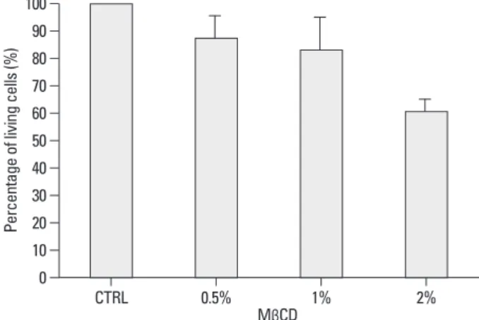
We treated cells with MβCD at various concentrations (0, 0.5%, 1%, 2%) for 1 hour, and examined cell viability. 0.5%
MβCD showed 88±8% cell viability compared to the control group (no treatment with MβCD) and 1% MβCD showed 83
±10% and 2% MβCD showed 60±5% (Fig. 1). Cells ex- posed to 1% MβCD exhibited normal viability, but those exposed to 2% MβCD showed loss of cell viability. There- Filipin staining
Cells, cultured in the polysin-coated cover slip, were divid- ed into two groups; a control group and an experimental group, and treated with MβCD. Subsequently, cells were rinsed with cold PBS and fixed on ice using 4% parafor- maldehyde. The cells were rinsed again with cold PBS for 10 min, and stained at room temperature with 100 μg/mL of filipin (Sigma Chemical Co., St. Louis, MO, USA) for 2 hours. After being rinsed once with PBS, the cells were ob- served through a fluorescence microscope with a UV filter set (340-380 nm excitation, 40 diachronic, 430 nm long pass filter).
Reverse transcription-polymerase chain reaction (RT-PCR) analysis of MUC5AC mRNA
The total RNA was isolated from cells under each condi- tion using TRI-reagent (Molecular Research Center, Cin- cinnati, OH, USA). Total RNA (1 μg/20 μL) was reverse transcribed into cDNA using random hexanucleotide prim- ers and Moloney murine leukemia virus reverse transcrip- tase (Gibco-BRL), and MUC5AC cDNA was amplified by PCR using a Perkin-Elmer Cetus DNA Thermal Cycler (Perkin-Elmer, Norwalk, CT, USA) using the previously described method.
11β
2-microglobulin (β
2M) was used as a control gene for RT-PCR.
Real-time polymerase chain reaction of MUC5AC mRNA
Primers and probes were designed using Applied Biosys- tems Primer Express software and purchased from Applied Biosystems (Carlsbad, CA, USA). Commercial reagents (TaqMan Universal PCR Master Mix; Applied Biosystems) and conditions were applied in accordance with the manu- facturer’s protocol. One μg of cDNA (reverse transcription mixture) and oligonucleotides with final concentrations of 800 nM for primers and 200 nM for TaqMan hybridization probes were analyzed in a 25 µL volume using a previously described method.
11Relative quantities of MUC5AC mRNA were obtained using a comparative cycle threshold method and normalized using β
2M as an endogenous control. Ex- pression was reported as a ratio of MUC5AC intensity to β
2M intensity.
Western blot analysis
Cell lysates were made into cultured cells using a radioim- munoprecipitation assay buffer (1% NP-40, 0.5% sodium deoxycholate, 0.1% SDS). Protein quantities were mea-
Fig. 1. Cell viability by cholesterol depletion in cell membranes of NCI-H292 cells. Cell viability was examined after treatment with various concentra- tions of MβCD, and the data were compared to that of the control group (CTRL). Cell survival is maintained above 80% with 1% MβCD treatment.
Data are presented as mean±SD of six replicates from three independent experiments. MβCD, methyl-β-cyclodextrin; SD, standard deviation.
MβCD 0
10 20 30 40 50 60 70 80 90 100
Percentage of living cells (%)
CTRL 0.5% 1% 2%
along the cell membrane. However, in the MβCD-treatment group, the intensity of fluorescence decreased significantly (Fig. 2B).
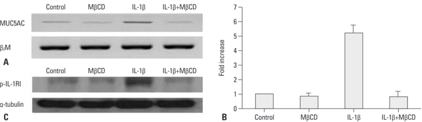
Suppression of MUC5AC expression after treatment with MβCD
We evaluated whether MβCD suppressed IL-1β-induced
MUC5AC gene expression using RT-PCR and real time-PCR, as well as whether this suppression occurred IL-1-re- ceptor-specifically. Cells were pretreated with 1% MβCD for 1 hour, after which the media was freshly changed and cells were incubated with IL-1β (10 ng/mL) for 24 hours.
We then picked MUC5AC mRNA and examined its expres- sion through RT-PCR and real-time PCR. The expression of p-IL-1RI was examined by Western analysis. We found that IL-1β induced the expression of MUC5AC, but this increase was significantly decreased by treatment with MβCD in RT- fore, we used 1% MβCD in the following experiments.
Verification of cholesterol depletion by MβCD treatment
To verify whether intra-membranous cholesterol was deplet- ed after 1 hour of treatment with 1% MβCD, intra-membra- nous cholesterol level analysis and filipin staining were per- formed. After treatment with 1% MβCD for 1 hour, the intra-membranous cholesterol/protein ratio was calculated and the data for the experimental group was compared to that of the control group, which had not been treated with MβCD. As a result, the relative ratio of intra-membrane cholesterol/protein was 0.17±0.07. This result indicates that MβCD significantly decreased the cholesterol level in the cell membranes (p<0.05) (Fig. 2A).
With regards to the filipin staining, in the control group, filipin was observed to adhere specifically to cholesterol
Fig. 2. Determination of intra-membranous cholesterol level by a modified microenzymatic fluorescence assay (A) and filipin staining (B). (A) The intra-mem- branous cholesterol level was determined using a modified microenzymatic fluorescence assay. The amount was expressed in cholesterol/protein ratio, and the data of the experimental group were compared with that of the control group (no treatment of MβCD). The cholesterol/protein ratio is significantly de- creased by 1% MβCD treatment in NCI-H292 cells (p<0.05). Data are presented as mean±SD of three independent experiments. (B) Filipin staining in NCI-H292 cells after treatment with MβCD. Filipin staining was done in cells after one-hour pre-treatment of 1% MβCD with a fluorescence microscope. Compared to the control group, the staining intensity along the cell membrane was significantly weaker. MβCD, methyl-β-cyclodextrin; SD, standard deviation.
Fig. 3. MUC5AC gene expression after treatment with MβCD. NCI-H292 cells were pretreated with 1% MβCD for 1 hour, after which the media was freshly changed and cells were incubated with IL-1β (10 ng/mL) for 24 hours. MUC5AC gene expression was examined by RT-PCR (A) and real time-PCR (B) analy- sis. β2M served as the internal control. The expression of p-IL-1RI was examined by Western analysis (C). α-tubulin served as the internal control. IL-1β in- duces expression of MUC5AC, but this increased expression is significantly decreased by MβCD treatment (IL-1β+MβCD) (p<0.05). Data are presented as mean±SD of three independent experiments. The expression of p-IL-1RI increased according to IL-1β treatment, but this increase was decreased by MβCD treatment (IL-1β+MβCD). Data are presented from three independent experiments. MβCD, methyl-β-cyclodextrin; RT-PCR, reverse transcription-polymerase chain reaction; IL, interleukin.
0.0 0.5 1.0
Relative ratio of cholesterol/protein
Control MβCD
A
BMUC5AC
p-IL-1RI β2M
α-tubulin
Control
Control
IL-1β
IL-1β MβCD
MβCD
IL-1β+MβCD
IL-1β+MβCD
0 1 2 3 4 5 6 7
Fold increase
Control MβCD IL-1β IL-1β+MβCD
A
C B
Control MβCD
MβCD depleted the cholesterol of the plasma membrane.
However, cholesterol is distributed both intramembranous- ly and intracellularly, and intra-membranous cholesterol level analysis examines the relative ratio of cholesterol/pro- tein.
1,20,21Therefore, we determined whether the cholesterol of the cell membrane was depleted through filipin staining.
By staining filipin that selectively combines with cholesterol, we verified that filipin on the membrane was further de- creased in MβCD-treated cell lines as compared to the con- trol group. From this result, we confirmed that MβCD selec- tively depletes the cell membrane’s cholesterol.
Because MUC5AC expression is induced by IL-1β in NCI-H292 cells, we applied MβCD before IL-1β treatment.
11According to our results, MUC5AC expression was signifi- cantly decreased. Next, we wondered whether these results were induced by the disruption of a ligand-specific receptor such as IL-1 receptor or not. Therefore, we examined wheth- er the phosphorylation of IL-1 receptor is induced by IL-1β and whether such activation of the ligand-specific receptor is decreased by MβCD. As a result, the expression of p-IL-1RI was increased by IL-1β treatment, but this increase was sig- nificantly decreased by MβCD treatment. Thus, we dis- cerned the suppression of MUC5AC expression by MβCD to be an IL-1 receptor-specific reaction.
In order to reveal the possible signal pathway by which
MUC5AC is decreased by MβCD, we examined the MAPKcascade. In our previous study of NCI-H292 cells, IL-1β maximally induced activation of p-ERK and p-p38 in the 20 min after treatment.
11Thus, our experiments were per- formed 20 min after IL-1β treatment. We found ERK and PCR and real-time PCR analysis (control : MβCD : IL-1β :
IL-1β+MβCD=1 : 0.9±0.2 : 5.3±0.8 : 0.8±0.3) (p<0.05) (Fig. 3A and B). The expression of p-IL-1RI increased ac- cording to the IL-1β treatment, but MβCD treatment signifi- cantly decreased the expression of p-IL-1RI (Fig. 3C).
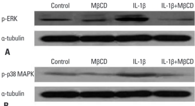
MAPK’s changes by MβCD treatment
To figure out whether the decrease in MUC5AC expression induced by MβCD treatment was dependent upon the de- pression of MAPK activity, we pre-treated cells for 1 hour with 1% MβCD. Then the media was freshly changed and cells were incubated with IL-1β for 20 min. A no MβCD or IL-1β treatment group was used as a negative control group and only IL-1β was administered in the positive control group. After each experiment, protein was obtained and a Western blot analysis of p-ERK and p-p38 MAPK was per- formed. Elevated expression of p-ERK in only IL-1β-treated cells decreased markedly in both IL-1β and MβCD-treated cells (Fig. 4A). Expression of p-p38 was also reduced to the negative control level in both IL-1β and MβCD-treated cells (Fig. 4B).
DISCUSSION
MβCD is a compound that specifically combines with cho- lesterol, deletes it, and then disrupts the lipid raft. Due to this effect, MβCD affects kinases of plasma membrane receptors and may cause alterations to cell proliferation.
15,16These facts mean that MβCD induces inhibition of cell prolifera- tion according to cell type and concentration of MβCD. It is thus necessary to determine the appropriate concentration of MβCD. Our results regarding cell viability were above 80% when using 1% MβCD; therefore, this concentration was considered safe for maintaining cell survival. Although we found the appropriate concentration of MβCD, we fixed the treatment time at one hour. In other studies, the duration of treatment was 1 or 2 hours and, after reviewing previous results, we fixed our duration as 1 hour.
17-19In the preceding experiments, we treated cells for 2 hours at a 1% concentra- tion, but cell proliferation was remarkably inhibited (data not shown), and we decided to use one hour as the experi- mental time.
Our next step was to verify whether cholesterol was de- pleted by MβCD in NCI-H292 cells via intra-membranous cholesterol level analysis and filipin staining. Through the intra-membranous cholesterol analysis, we found that
Fig. 4. Suppression of IL-1β-induced activation of ERK and p38 MAPK by treatment with MβCD. NCI-H292 cells were pretreated for 1 hour with 1%
MβCD, after which the media was freshly changed, cells were treated for 20 min with IL-1β, and Western blot analysis was performed. Expression of both p-ERK (A) and p-p38 MAPK (B) is increased by IL-1β treatment (IL- 1β), but this increased expression of both p-ERK and p-p38 MAPK is de- creased to the control level by MβCD treatment (IL-1β+MβCD). Data are presented from three independent experiments. ERK, extracellular signal regulated kinase; MAPK, mitogen-activated protein kinase; MβCD, methyl- β-cyclodextrin; IL, interleukin.
p-ERK
p-p38 MAPK α-tubulin
α-tubulin
Control
Control
IL-1β
IL-1β MβCD
MβCD
IL-1β+MβCD
IL-1β+MβCD
A
B
effectively inhibit mucus secretion in human airway epithe- lial cells.
ACKNOWLEDGEMENTS
This work was supported by a grant (6-2007-0099) from the Academic Research Fund of Yonsei University College of Medicine, Seoul, Korea.
REFERENCES
1. Brown DA, London E. Functions of lipid rafts in biological mem- branes. Annu Rev Cell Dev Biol 1998;14:111-36.
2. Burger K, Gimpl G, Fahrenholz F. Regulation of receptor function by cholesterol. Cell Mol Life Sci 2000;57:1577-92.
3. Simons K, Toomre D. Lipid rafts and signal transduction. Nat Rev Mol Cell Biol 2000;1:31-9.
4. Edidin M. The state of lipid rafts: from model membranes to cells.
Annu Rev Biophys Biomol Struct 2003;32:257-83.
5. Simons K, Ikonen E. Functional rafts in cell membranes. Nature 1997;387:569-72.
6. Pike LJ. Lipid rafts: bringing order to chaos. J Lipid Res 2003;44:
655-67.
7. Hovenberg HW, Davies JR, Herrmann A, Lindén CJ, Carlstedt I.
MUC5AC, but not MUC2, is a prominent mucin in respiratory se- cretions. Glycoconj J 1996;13:839-47.
8. Thornton DJ, Howard M, Khan N, Sheehan JK. Identification of two glycoforms of the MUC5B mucin in human respiratory mu- cus. Evidence for a cysteine-rich sequence repeated within the molecule. J Biol Chem 1997;272:9561-6.
9. Wickström C, Davies JR, Eriksen GV, Veerman EC, Carlstedt I.
MUC5B is a major gel-forming, oligomeric mucin from human salivary gland, respiratory tract and endocervix: identification of glycoforms and C-terminal cleavage. Biochem J 1998;334(Pt 3):685-93.
10. Song KS, Lee WJ, Chung KC, Koo JS, Yang EJ, Choi JY, et al.
Interleukin-1 beta and tumor necrosis factor-alpha induce MU- C5AC overexpression through a mechanism involving ERK/p38 mitogen-activated protein kinases-MSK1-CREB activation in hu- man airway epithelial cells. J Biol Chem 2003;278:23243-50.
11. Kim JH, Chang JH, Yoon JH, Kwon SH, Bae JH, Kim KS.
[6]-Gingerol suppresses interleukin-1 beta-induced MUC5AC gene expression in human airway epithelial cells. Am J Rhinol Al- lergy 2009;23:385-91.
12. Lambert S, Ameels H, Gniadecki R, Hérin M, Poumay Y. Inter- nalization of EGF receptor following lipid rafts disruption in kera- tinocytes is delayed and dependent on p38 MAPK activation. J Cell Physiol 2008;217:834-45.
13. Calleros L, Lasa M, Toro MJ, Chiloeches A. Low cell cholesterol levels increase NFkappaB activity through a p38 MAPK-depen- dent mechanism. Cell Signal 2006;18:2292-301.
14. Kim S, Han J, Lee DH, Cho KH, Kim KH, Chung JH. Cholester- ol, a Major Component of Caveolae, Down-regulates Matrix Me- talloproteinase-1 Expression through ERK/JNK Pathway in Cul- tured Human Dermal Fibroblasts. Ann Dermatol 2010;22:379-88.
p38 MAPK to be associated with a decrease in MUC5AC expression by MβCD treatment. Previous studies have in- vestigated the relationship between MAPK and cholesterol depletion. Jans, et al.
21found that cholesterol depletion of the cell membrane in keratinocyte induced prolonged phos- phorylation of p38 MAPK. Kim, et al.
14reported that cho- lesterol depletion brought about activation of ERK and JNK in human dermal fibroblasts. Calleros, et al.
13showed that low cell cholesterol levels increased NFκB activity by in- creasing p38 activity. The difference of MAPK activation be- tween our study and others seems to be related to alterations in respective receptors. MβCD treatment triggers MAPK cascade, and changes lipid raft in keratinocyte, NIH3T3 cells, and cultured human dermal fibroblasts.
12-14Thereafter, the activation of MAPK is followed in a cell-specific man- ner. On the contrary, NCI-H292 cells express a very low level of MAPK in unstimulated state. The cells stimulated with IL-1β show MAPK activation.
11Disintegration of lift raft leads to alterations in IL-1β receptor, and the activation of MAPK is attenuated even though IL-1β is challenged.
This phenomenon is also found in human promonocytic THP-1 cells. If THP-1 cells are challenged with lipopolysac- charide, MAPK such as ERK 1/2 is activated. With MβCD pre-treatment, this MAPK activation is attenuated in spite of lipopolysaccharide treatment.
22These results coincide with our results. Modulation of lipid raft may represent a mechanism for regulation of MAPK activaton. In addition, two studies reported that EGFR ligands induce MUC5AC production through EGFR signaling kinase cascade in the airway system, and are also related to MAPK activation.
23,24Thus, further studies on IL-1β receptor and EGFR signal- ing cascades are needed.
Mucin is hypersecreted in many respiratory diseases such as rhinitis, sinusitis, nasal allergy, and chronic bronchitis.
10Hypersecretion of mucin results in clinical problems such as rhinorrhea, nasal stuffiness, and sputum. Accordingly, regulation of MUC5AC can be a new strategy for treating respiratory diseases because it decreases mucus hypersecre- tion. Furthermore, IL-1 receptor was recently considered a novel potential therapeutic target for bronchial asthma.
25In this study, we showed that MUC5AC expression in NCI-H292 cells is significantly decreased by treatment with MβCD, which depletes cholesterol in the cell membrane.
We also found that the decrease of MUC5AC expression is
dependent upon the activation of ERK and p38 MAPK. In
conclusion, cholesterol depletion in the cell membrane may
be considered as anti-hypersecretory method because they
20. Maxfield FR, Tabas I. Role of cholesterol and lipid organization in disease. Nature 2005;438:612-21.
21. Jans R, Atanasova G, Jadot M, Poumay Y. Cholesterol depletion upregulates involucrin expression in epidermal keratinocytes through activation of p38. J Invest Dermatol 2004;123:564-73.
22. Cuschieri J. Implications of lipid raft disintegration: enhanced anti- inflammatory macrophage phenotype. Surgery 2004;136:169-75.
23. Chen X, Resh MD. Cholesterol depletion from the plasma mem- brane triggers ligand-independent activation of the epidermal growth factor receptor. J Biol Chem 2002;277:49631-7.
24. Zuo W, Chen YG. Specific activation of mitogen-activated protein kinase by transforming growth factor-beta receptors in lipid rafts is required for epithelial cell plasticity. Mol Biol Cell 2009;20:
1020-9.
25. Lee JH, Wang LC, Yu HH, Lin YT, Yang YH, Chiang BL. Type I IL-1 receptor (IL-1RI) as potential new therapeutic target for bronchial asthma. Mediators Inflamm 2010;2010:567351.
15. Muppidi JR, Siegel RM. Ligand-independent redistribution of Fas (CD95) into lipid rafts mediates clonotypic T cell death. Nat Im- munol 2004;5:182-9.
16. Garofalo T, Misasi R, Mattei V, Giammarioli AM, Malorni W, Pontieri GM, et al. Association of the death-inducing signaling complex with microdomains after triggering through CD95/Fas.
Evidence for caspase-8-ganglioside interaction in T cells. J Biol Chem 2003;278:8309-15.
17. Gniadecki R. Depletion of membrane cholesterol causes ligand- independent activation of Fas and apoptosis. Biochem Biophys Res Commun 2004;320:165-9.
18. Lambert S, Vind-Kezunovic D, Karvinen S, Gniadecki R. Ligand- independent activation of the EGFR by lipid raft disruption. J In- vest Dermatol 2006;126:954-62.
19. Bang B, Gniadecki R, Gajkowska B. Disruption of lipid rafts causes apoptotic cell death in HaCaT keratinocytes. Exp Dermatol 2005;14:266-72.