INTRODUCTION
Mucin is a macromolecular glycoprotein produced in secre- tory epithelial cells of the respiratory, gastrointestinal, and the reproductive tract (1). Mucin is an important component of airway mucus secretion. Several mucin genes regulate mucin secretion in human body. Mucus hypersecretion is a major problem of the inflammatory airway diseases such as chronic bronchitis, asthma, cystic fibrosis, and chronic rhinosinusitis (1). Various cytokines and inflammatory mediators stimulate mucus hypersecretion, directly or indirectly (2, 3). Among these cytokines and inflammatory mediators, tumor necrosis factor- (TNF- ) (4, 5), interleukin-1 (IL-1 ) (6), and lipo- polysaccharide (LPS) (7) activate mucin secretion by up-reg- ulating expression of the mucin genes (2, 3, 7-9). Thirteen mucin genes have been identified. Among these mucin genes, the respiratory mucin genes are MUC2, MUC4, MUC5AC, MUC5B, MUC6, MUC7, and MUC8 (10-14). The MUC5AC gene is one of the most important genes in the respiratory
tract. The MUC2 gene is expressed at a low level in a normal condition, but the expression is increased in chronic bronchitis and cystic fibrosis (15).
Among the cytokines that have various inflammatory func- tions in acute and chronic upper respiratory tract infection, IL- 1 is related with the pathogenesis of respiratory tract infection in upper airway diseases such as asthma and bronchitis (5, 16, 17). Our recent study (6) demonstrated that IL-1 stimulates the expression of MUC2 gene and mucin secretion by tran- scriptional regulation. However, we could not delineate the specific signal pathway involved in the mediation of mucin secretion in that study.
Since it was reported that Pseudomonas aeruginosa-induced mucin overproduction requires nuclear factor B (NF- B) activation in epithelial cells (3), some researchers have stud- ied to elucidate the signal transduction pathway involved in the expression of the mucin gene (3, 4, 7, 9, 12). Although a previous study (18) showed that IL-1 activates mitogen-acti- vated protein kinase (MAPK) subgroups c-Jun NH2 terminal
Yong-Dae Kim, Jae-Yun Jeon, Hyun Jae Woo, Jung Cheul Lee*, Jin Hong Chung�, Si Youn Song, Seok-Keun Yoon, Suk-Hwan Baek�
Department of Otorhinolaryngology-Head and Neck Surgery, Department of Thoracic and Cardiovascular Surgery*, Department of Internal Medicine�, Department of Biochemistry and Molecular Biology�, College of Medicine, Yeungnam University, Daegu, Korea
Address for correspondence Yong-Dae Kim, M.D.
Department of Otorhinolaryngology-Head and Neck Surgery, College of Medicine, Yeungnam University, 317-1 Daemyung-dong, Nam-gu, Daegu 705-717, Korea
Tel : +82.53-620-3784, Fax : +82.53-628-7884 E-mail: [email protected]
765
Interleukin-1 Induces MUC2 Gene Expression and Mucin Secretion via Activation of PKC-MEK/ERK, and PI3K in Human Airway
Epithelial Cells
Interleukin 1 (IL-1 ), a proinflammatory cytokine, is related with inflammatory dis- eases and it up-regulates MUC2 gene expression and mucin secretion. This study was designed to investigate the signal transduction pathway of the IL-1 -mediat- ed MUC2 gene expression and mucin secretion in human airway epithelial cells.
In cultured human airway NCI-H292 epithelial cells, the steady state of the mRNA level of MUC2 gene expression and mucin secretion induced by IL-1 were deter- mined by reverse transcriptase-polymerase chain reaction (RT-PCR), enzyme immunoassay, and immunoblot analysis. To observe the signal pathway of the IL-1 -mediated MUC2 gene expression and mucin secretion, we used several specific inhibitors. PD98059 (MEK/ERK inhibitor) suppressed IL-1 -mediated MUC2 gene expression and mucin secretion, while SB203580 (p38 inhibitor) did not.
Ro31-8220 (PKC inhibitor) inhibited IL-1 -mediated MUC2 gene expression and mucin secretion. It inhibited ERK phosphorylation, but did not inhibit p38 phosphory- lation. LY294002 (PI3K inhibitor) also suppressed MUC2 expression, but did not inhibit any MAPKs phosphorylation. These results suggest that the IL-1 -medi- ated MUC2 gene expression and mucin secretion in NCI-H292 cells are regulat- ed through activation of the PKC-MEK/ERK pathway, and that PI3K is also involved in the IL-1 -mediated MUC2 gene expression and mucin secretion.
Key Words : Mucins; Interleukin-1; Cytokines; Epithelial Cells; Signal Transduction
Received : 16 May 2002 Accepted : 22 July 2002
kinase (JNK) and extracellular signal-regulated kinase (ERK) in human articular chondrocyte, the signal pathway involved in the IL-1 -mediated MUC2 gene expression and mucin secretion has not yet been understood clearly.
The present study was undertaken to investigate the signal transduction pathway involved in the IL-1 -mediated MUC2 gene expression and mucin secretion. We observed whether protein kinase C (PKC), MAPKs (ERK, p38 and JNK), and phosphatidylinositol 3-kinase (PI3K) are involved in the IL- 1 -mediated MUC2 gene expression and mucin secretion in cultured human airway NCI-H292 epithelial cells.
MATERIALS AND METHODS Cell Culture
The NCI-H292 airway epithelial cells (human pulmonary mucoepidermoid carcinoma cell line, American Type Culture Collection, Rockville, MD) were seeded at a density of 1×10⁶ cells into 6 well plates. Cultures were maintained in the RPMI 1640 medium (Gibco BRL, Grand Island, NY) supplement- ed with 10% fetal calf serum (Gibco BRL), penicillin (100 U/mL), and streptomycin (100 g/mL) at 37℃in a humid- ified atmosphere of 95% air and 5% CO2.When the cultures were confluent, the cells were incubated with RPMI 1640 medium containing 0.5% fetal calf serum for 24 hr. They were rinsed with phosphate buffered saline (PBS) and exposed to human recombinant IL-l (R&D Systems Inc., Minneapolis, MN) treatment. To study which signal transduction pathway is related to the MUC2 expression, some cultures were pre- treated with specific inhibitors such as PD98059 (MEK/ERK inhibitor, Biomol Research Laboratories, Inc., Plymouth Meeting, PA), SB203580 (p38 inhibitor, Biomol Research Laboratories), Ro31-8220 (protein kinase C inhibitor, Biomol Research Laboratories), and LY294002 (PI3K inhibitor, Biomol Research Laboratories) at 1 hr before exposed to IL-1 and then incubated for 8 hr after treatment of IL-1 . Control cultures remained untreated. Total cellular RNA was extracted using a Tri-Reagent (Molecular Research Center, Cincinnati, OH). Cell lysates were prepared in PBS.
IL-1 was dissolved with PBS containing 0.1% bovine serum albumin, and each specific inhibitor was dissolved in dimethyl sulfoxide (DMSO) prior to addition to cell cultures.
The final concentrations of DMSO or other vehicle solvents in the medium were less than 0.1%.
RT-PCR Analysis of the MUC2 Gene
The method used to detect and quantify the MUC2 mRNA level employed a modified technique of RT-PCR and has been described previously in detail (6). Briefly, total RNA was reverse transcribed into cDNA using random hexanucleotide primers and MULV reverse transcriptase (Perkin-Elmer, Mor-
risville, NC). The oligonucleotide primers for the PCR part of the procedure were designed on the basis of the published sequences of human MUC2 (GenBank Accession No. L21998, 5′primer: TGC CTG GCC CTG TCT TTG: 3′primer:
CAG CTC CAG CAT GAG TGC). The PCR consisted of 35 cycles of denaturation (95℃, 1 min), annealing (60℃, 1 min), and extension (72℃, 1 min) in the presence of 2.5 mM MgC12, and a final extension at 72℃for 20 min. The MUC2 DNA fragment generated was 440-bp in size as expected. The oligonucleotide primers for the 2 microglobu- lin ( 2M, used as a control gene for the RT-PCR) were pur- chased from Clontech (Palo Alto, CA) and generated 335-bp PCR fragment. Specific amplification of MUC2 was con- firmed by sequencing (dsDNA Cycle Sequencing System, Gibco BRL). PCR products were separated by electrophoresis through a 2% agarose gel in 1% Tris-boric acid-EDTA (TBE) buffer containing 50 ng/mL of ethidium bromide and pho- tographed using a Polaroid type 55 film. The intensity of the bands was analyzed with a densitometer.
Immunoassay of the MUC2 Mucin
Cell lysates were prepared in PBS at multiple dilutions, and 50 L of each sample was incubated at 40℃in a 96-well plate until dry. Plates were then washed three times with PBS, blocked with 2% bovine serum albumin for 1 hr at room temperature, washed again three times with PBS, and then incubated with a MUC2 antibody (Santa Cruz Biotechnology, Santa Cruz, CA) that has been diluted (1:100) with PBS con- taining 0.05% Tween-20. After 1 hr, the wells were washed three times with PBS, and then horseradish peroxidase (HRP)- conjugated anti-rabbit IgG (Santa Cruz Biotechnology) was dispensed into each well, and after 4 hr, the plates were washed three times with PBS. Color was developed with a 3,3′,5,5′- tetramethylbenzidine peroxidase solution and stopped with 2N-H2SO4. Absorbance was read at 450 nm. The amount of estimated MUC2 mucin was represented as % above control, which indicates the excess proportion over control.
Immunoblot Analysis for MAPKs and phospho-MAPKs
For analysis of the MAPKs and their activated form, phos- pho-MAPKs, the treated cells were washed with cold-PBS, scraped off, pelleted at 700×g at 4℃, and the cell pellet was resuspended in lysis buffer (50 mM Tris-HCl, pH 8.0, 5 mM EDTA, 150 nM NaCl, 0.5% Nonidet P-40, 1 mM phenylmethylsulfonyl fluoride (PMSF), and protease inhibitor cocktail). The preparation was then clarified by centrifugation, and the supernatant was saved as a whole-cell lysate. Proteins (50 g) were separated using 10% reducing SDS-polyacryl- amide gel and electroblotted in 20% methanol, 25 mM Tris, and 192 mM glycine onto a nitrocellulose membrane. The membrane was then blocked with 5% non-fat dry milk in TTBS (25 mM Tris-HCl, 150 mM NaCl, and 0.2% Tween-20)
and then incubated with the indicated antibodies for 4 hr.
ERK, phospho-ERK antibodies and p38, phospho-p38 anti- bodies were purchased from New England BioLabs (Beverly, MA). Subsequently, the membrane was washed and incubated for 1 hr with secondary antibodies conjugated to HRP, rewashed, and developed using an enhanced chemiluminescence system (ECL, Amersham Pharmacia Biotech, Inc., Buckinghamshire, England).
RESULTS
Induction of the MUC2 Gene Expression and Mucin Secretion by IL-1
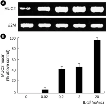
When the cultures were confluent, the cultured cells were incubated with IL-1 in the range of 0.02-20 ng/mL at 37℃ for 8 hr. The total mRNA was extracted and subjected to RT- PCR analysis. As the dose of IL-1 was increased from 0.02 to 20 ng/mL, there was a parallel increase in the MUC2 mRNA level. RT-PCR products of 2M mRNA were used as internal control (Fig. 1A). To define the regulation of mucin secretion by IL-1 at the protein level, we used immunoassay with a
MUC2 monoclonal antibody. MUC2 mucin secretion was also increased in a dose-dependent manner, and to a maximal level at 20 ng/mL of IL-1 (Fig. 1B). These results of mucin secre- tion were consistent with gene expression data. These data were similar to those of our previous study (6).
Effect of MAPKs on the IL-1 -mediated MUC2 Gene Expression and Mucin Secretion
To examine whether IL-1 activates MAPKs (ERK1/2, p38, JNK), the phosphorylation level of MAPKs was estimated by examining their phosphorylation in immunoblot with phos- pho-specific antibodies. All the three MAPKs phosphorylation by IL-1 were similar in their extent (Fig. 2). The maximum levels of phosphorylation induced by IL-1 , as indicated by immunoblotting, were observed at 20-30 min for three MAPKs. These results show that IL-1 activates ERK, p38, and JNK.
We evaluated whether the specific inhibitors of MAPKs appropriately blocked the phosphorylation of different MAPKs and which MAPK is involved in the MUC2 gene expression and mucin secretion induced by IL-1 in NCI-H292 cells.
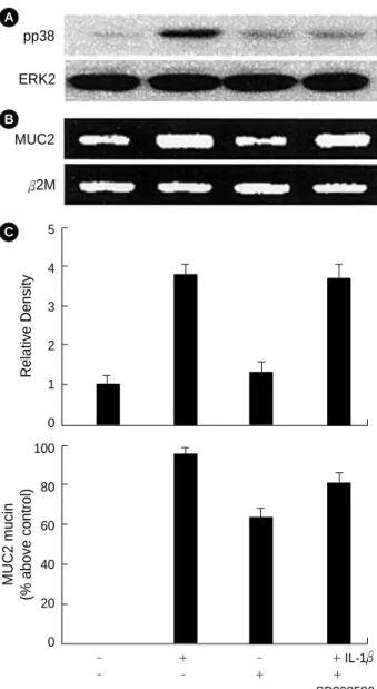
After the cultured cells were pretreated with PD98059 (50 M) or SB203580 (10 M) for 1 hr, they were stimulated with IL-1 (20 ng/mL) for 8 hr. PD98059 completely inhibited IL-1 -stimulated ERK1/2 phosphorylation (Fig. 3A). Also, pretreatment of PD98059 inhibited the MUC2 gene expression (Fig. 3B) and mucin secretion (Fig. 3C). SB203580 inhibited p38 phosphorylation (Fig. 4A), but did not suppress the IL-1 - mediated MUC2 gene expression (Fig. 4B) or mucin secretion (Fig. 4C).
Effect of PKC on the IL-1 -mediated MUC2 Gene Expression and Mucin Secretion
To observe whether PKC activation is related with the signal pathway of the IL-1 -induced MUC2 gene expression and mucin secretion, Ro31-8220 (PKC inhibitor) was pretreated.
MUC2 mucin (% above control) 100
80 60 40 20
0
0 0.02 0.2 2 20
Fig. 1. Induction of the IL-1 -mediated MUC2 gene expression and mucin secretion. (A) The NCI-H292 cells were treated with various concentrations of IL-1 (0.02, 0.2, 2, 20 ng/mL). The mRNA levels of MUC2 and 2M were determined by RT-PCR. 2M is a positive internal control. The amounts of the RT-PCR products of MUC2 were quantified using densitometry and expressed relative to the densities of 2M by the ratio of MUC2/ 2M DNA bands.
The data is representative of three independent experiments. (B) The MUC2 mucin was determined by immunoassay. The data represent average values of three independent experiments plus standard deviation.
IL-1 (ng/mL) MUC2
2M A
B
0 5 10 20 30 60
Fig. 2. Phosphorylation of three MAPKs (pERK, pp38, pJNK) according to time of IL-1 treatment. NCI-H292 cells were treat- ed with 20 ng/mL of IL-1 for 5, 10, 20, 30, 60 min. Analysis of phosphorylation of each protein was done by immunoblot. ERK2 is a positive internal control.
pERK pp38 pJNK ERK2 Time (min)
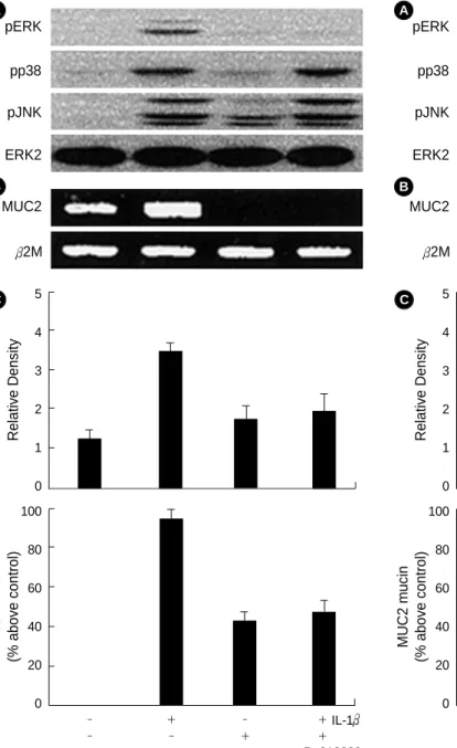
Pretreatment with Ro31-8220 (20 M) for 1 hr inhibited the IL-1 -mediated MUC2 gene expression and mucin secretion.
To further determine whether PKC acts at upstream or down- stream molecule of MAPKs, we tested the effect of PKC inhibitor on the IL-1 -activated MAPKs phosphorylation.
When cells were pretreated with Ro31-8220 (20 M) for 1 hr, only ERK1/2 phosphorylation induced by IL-1 (20 ng/
mL) was attenuated, but p38 and JNK phosphorylation was
not suppressed (Fig. 5B, C).
Effect of PI3K on the IL-1 -mediated MUC2 Gene Expression and Mucin Secretion
To determine whether PI3K activation is involved in the IL-1 -induced MUC2 gene expression and mucin secretion, the cells were pretreated with LY294002 (a PI3K inhibitor)
Relative Density
5 4 3 2 1
0 pERK
ERK2 A
MUC2
2M B
MUC2 mucin (% above control) 100
80 60 40 20
0
- + - + - - + +
Fig. 3. Effects of MEK/ERK inhibitor, PD98059 on the expression of the IL-1 -mediated ERK phosphorylation (pERK) (A), MUC2 gene expression (B), and mucin secretion (C). Analysis of ERK phosphorylation was done by immunoblot, MUC2 gene by RT- PCR, and mucin by immunoassay. ERK2 is a positive internal control. The amounts of the RT-PCR products of MUC2 gene were quantified using densitometry and expressed relative to the density of 2M by the ratio of the MUC2/ 2M DNA bands. 2M is a positive internal control. The data A and B are acquired from three independent experiments. The data C represents average value of three independent experiments and standard deviation.
PD98059 IL-1 C
Relative Density
5 4 3 2 1 0 pp38
ERK2 A
MUC2
2M B
MUC2 mucin (% above control) 100
80 60 40 20
0
- + - + - - + +
Fig. 4. Effects of p38 inhibitor, SB203580 on the expression of the IL-1 -mediated p38 phosphorylation (pp38) (A), MUC2 gene expression (B) and mucin secretion (C). Analysis of p38 phos- phorylation was done by immunoblot, MUC2 gene by RT-PCR and mucin by immunoassay. ERK2 is a positive internal control.
The amounts of the RT-PCR products of MUC2 gene were quan- tified using densitometry and expressed relative to the density of 2M by the ratio of the MUC2/ 2M DNA bands. 2M is a posi- tive internal control. The data A and B are acquired from three independent experiments. The data C represents average value of three independent experiments and standard deviation.
SB203580 IL-1 C
for 1 hr before treatment of IL-1 . In addition, to investigate its relation with MAPKs, we observed the effect of the PI3K inhibitor on MAPKs phosphorylation induced by IL-1 . When
the cells were pretreated with the LY294002 (25 M) for 1 hr, LY294002 attenuated the IL-1 -induced MUC2 gene expression and mucin secretion (Fig. 6B), but did not inhibit
Relative Density
5 4 3 2 1 0 pERK
pp38 pJNK ERK2 A
MUC2
2M B
MUC2 mucin (% above control) 100
80 60 40 20
0
- + - + - - + +
Fig. 5. Effects of PKC inhibitor, Ro31-8220 on the expression of the IL-1 -mediated MAPKs phosphorylation (pERK, pp38, pJNK) (A), MUC2 gene expression (B) and mucin secretion (C). Analysis of ERK, p38, JNK phosphorylation was done by immunoblot, MUC2 gene by RT-PCR and mucin by immunoassay. ERK2 is a positive internal control. The amounts of the RT-PCR products of MUC2 gene were quantified using densitometry and expressed relative to the density of 2M by the ratio of the MUC2/ 2M DNA bands.
2M is a positive internal control. The data A and B are acquired from three independent experiments. The data C represents average value of three independent experiments and standard deviation.
Ro318220 IL-1 C
Relative Density
5 4 3 2 1 0 pERK
pp38 pJNK ERK2 A
MUC2
2M B
MUC2 mucin (% above control) 100
80 60 40 20
0
- + - + - - + +
Fig. 6. Effects of PI3K inhibitor, LY294002 on the expression of the IL-1 -mediated MAPKs phosphorylations (pERK, pp38, pJNK) (A), MUC2 gene expression (B), and mucin secretion (C).
Analysis of ERK, p38, and JNK phosphorylation were done by immunoblot, MUC2 gene by RT-PCR, and mucin by immunoas- say. ERK2 is a positive internal control. The amounts of the RT- PCR products of MUC2 gene were quantified using densitometry and expressed relative to the density of 2M by the ratio of the MUC2/ 2M DNA bands. 2M is a positive internal positive con- trol. The data A and B are acquired from three independent experiments. The data C represents average value of three inde- pendent experiments and standard deviation.
LY294002 IL-1 C
any MAPKs phosphorylation (Fig. 6A).
DISCUSSION
In this study, we observed whether IL-1 could induce MUC2 gene expression and mucin synthesis through activation of PKC, MAPKs, or PI3K in cultured human airway NCI- H292 epithelial cell. Our results show that the MEK/ERK inhibitor, PD98059, inhibited the IL-1 -mediated MUC2 gene expression and mucin secretion, but a p38 inhibitor, SB203580, did not. A PKC inhibitor, Ro31-8220, inhibited the IL-1 -mediated MUC2 gene expression and mucin secre- tion. Moreover, Ro31-8220 attenuated ERK1/2 phosphory- lation induced by IL-1 . These results show that PKC and MEK/ERK are related with the IL-1 -induced MUC2 gene expression and mucin secretion, and that IL-1 activates PKC at upstream of ERK. Also, a PI3K inhibitor, LY294002, attenuated the IL-1 -induced MUC2 gene expression and mucin secretion, but did not inhibit any MAPKs phospho- rylation. These findings provide evidence that IL-1 induces MUC2 gene expression and mucin secretion through activation of PKC-MEK/ERK-dependent pathway, and through acti- vation of PI3K.
Since it has been reported that TNF- is related with mucin secretion in 1995 (4), there were several reports about the signal pathway of mucin secretion. Li et al. (3) described that Pseudomonas aeruginosa induced MUC2 gene expression by NF- B activation via Src-dependent Ras-MAPK-pp90rsk pathway in epithelial cells. In addition, Basbaum et al. (18) suggested that the various mucin secretion in epithelial cells is commonly dependent upon c-Src, MAP kinase kinase MEK1/2, and NF- B. Recently, Takeyama et al. (19) showed that the oxidative stress induces mucin synthesis in airway epithelial cells via epidermal growth factor receptor (EGFR), which leads to activation of the ERK signal transduction pathway. However, none of these studies found a direct link between PKC, ERK, or p38 in the IL-1 -mediated mucin gene expression and mucin secretion. Although we previously reported that IL-1 up-regulates MUC2 gene expression and mucin secretion through transcriptional activation (6), the specific signal transduction pathways involved in the IL-1 - mediated mucin gene expression and mucin secretion were not shown.
In our experiments, to determine the signal transduction pathway involved in the IL-1 -mediated MUC2 gene expres- sion and mucin secretion, we observed the pattern of MUC2 gene expression and mucin secretion using several specific inhibitors for regulatory molecules in signal transduction. The IL-1 -induced MUC2 gene expression and mucin secretion are strongly attenuated by an MEK/ERK inhibitor, PD98059.
We could conclude that MEK/ERK is necessary in the IL- 1 -mediated MUC2 gene expression and mucin secretion.
This result is similar to that Staphylococcus aureus activates the
transcription of the MUC2 gene through the ERK activation (18) and the transcriptional activation of the MUC2 gene induced by Pseudomonas aeruginosa requires MAPK (3, 7).
PKC is related with the MUC2 expression activated by Pseudomonas aeruginosa (18). The PKC family consists of at least 12 isoforms that possess distinct differences in struc- ture, substrate requirment, expression, and localization (20).
To evaluate the effect of PKC in the IL-1 -induced MUC2 expression, we used PKC inhibitor, Ro31-8220. While the straurosporine analogue Ro31-8220 inhibit PKC , PKC I, PKC II, PKC , and PKC isoforms (20), it was found to be an equally potent inhibitor of mitogen- and stress-activated protein kinase-1(MSK1) activity in vitro (21). MSK1 plays a role in integrating the effects of different extracellular signals, and is located in the nucleus of unstimulated or stimulated cells (21). The substrates of MSK1 are the transcription factor CREB and ATF1, and MSK1 mediates the activation of CREB and ATF1 by either growth factors and stress signals (21).
Therefore, there is a possibility that inhibitory effect of Ro31- 8220 on IL-1 -mediated MUC2 expression was through inhibition of PKC in cytoplasm or inhibition of MSK1 in nucleus. In this study, the IL-1 -induced MUC2 gene expres- sion and mucin secretion were inhibited by pretreatment of a PKC inhibitor (Ro31-8220). This result suggests that PKC is involved in the signal transduction pathway of the IL-1 - mediated MUC2 gene expression and mucin secretion. Fur- thermore, considering the result in which the phosphorylation of ERK was attenuated by Ro31-8220 at this time, we could conclude that PKC acts at upstream of ERK.
PI3K is an important molecule in mitogenic signaling and cell survival, cytoskeletal remodeling, metabolic control, and vesicular trafficking (22). And it has been known that PKC or MAPKs cascades require PI3K activation in several signal transduction pathway (23-27). In this study, LY294002, an inhibitor of PI3K, suppressed the IL-1 -induced MUC2 gene expression and mucin secretion, but not the phosphorylation of ERK. This result suggests that PI3K also has an important role in up-regulation of the MUC2 gene expression and mucin secretion induced by IL-1 . However, PI3K is involved inde- pendently of PKC-ERK or is located at downstream of MEK /ERK in the IL-1 -induced MUC2 gene expression and mucin secretion because LY294002 did not inhibit phosphorylation of ERK.
In conclusion, we consider that activation of PKC-ERK and PI3K is required for the IL-1 -mediated MUC2 gene expression and mucin secretion in human airway NCI-H292 epithelial cells. However, because specific inhibitors could not completely inhibit MUC2 gene expression and mucin secretion induced by IL-1 , other signal pathways must be also involved in the IL-1 -mediated MUC2 gene expression and mucin secretion. Furthermore, further studies with various epithelial cells including normal respiratory epithelium are needed to clarify the signal transduction pathway involved in the IL- 1 -mediated mucin gene expression and mucin secretion.
ACKNOWLEDGMENT
This research was supported by grant of Yeungnam Univer- sity Medical Center (2000).
REFERENCES
1. Kim YS, Gum JR Jr, Crawley SC, Deng G, Ho JJ. Mucin gene and antigen expression in biliopancreatic carcinogenesis. Ann Oncol 1999; 10(Supple): 51-5.
2. Temann UA, Prasad B, Gallup MW, Basbaum CB, Ho SB, Flavell RA, Rankin JA. A novel role for murine IL-4 in vivo: induction of MUC5AC gene expression and mucin hypersecretion. Am J Respir Cell Mol Biol 1997; 16: 471-8.
3. Li JD, Feng W, Gallup M, Kim JH, Gum J, Kim Y, Basbaum CB.
Activation of NF- B via a Src-dependent Ras-MAPK-pp90rsk path- way is required for Pseudomonas aeruginosa- induced mucin over- production in epithelial cells. Proc Natl Acad Sci USA 1998; 95:
5718-23.
4. Levine SJ, Larivee P, Logun C, Angus CW, Ognibene FP, Shel- hamer JH. Tumor necrosis factor- induces mucin hypersecretion and MUC2 gene expression by human airway epithelial cells. Am J Respir Cell Mol Biol 1995; 12: 196-204.
5. Yoon JH, Kim KS, Kim HU, Linton JA, Lee JG. Effects of TNF- and IL-1 on mucin, lysozyme, IL-6 and IL-8 in passage-2 normal human nasal epithelial cells. Acta Otolaryngol 1999; 119: 905-10.
6. Kim YD, Kwon EJ, Kwon TK, Baek SH, Song SY, Suh JS. Regu- lation of IL-1 -mediated MUC2 gene in NCI-H292 human airway epithelial cells. Biochem Biophys Res Commun 2000; 274: 112-6.
7. Li JD, Dohrman AF, Gallup M, Miyata S, Gum JR, Kim YS, Nadel JA, Prince A, Basbaum CB. Transcriptional activation of mucin by Pseudomonas aeruginosa lipopolysaccharide in the pathogenesis of cystic fibrosis lung disease. Proc Natl Acad Sci USA 1997; 94: 967-72.
8. Steiger D, Hotchkiss J, Bajaj L, Harkema J, Basbaum CB. Concur- rent increases in the storage and release of mucin-like molecules by rat airway epithelial cells in response to bacterial endotoxin. Am J Respir Cell Mol Biol 1995; 12: 307-14.
9. Li D, Gallup M, Fan N, Szymkowski DE, Basbaum CB. Cloning of the amino-terminal and 5′-flanking region of the human MUC5AC mucin gene and transcriptional up-regulation by bacterial exoprod- ucts. J Biol Chem 1998; 273: 6812-20.
10. Rose MC. Mucins; structure, function, and role in pulmonary dis- ease. Am J Physiol 1992; 263: L413-29.
11. Thorton DJ, Devine PL, Hanski C, Howard M, Sheehan JK. Identi- fication of two major populations of mucins in respiratory secre- tions. Am J Respir Crit Care Med 1994; 150: 823-32.
12. Yoon JH, Park IY. Mucin gene expression and mucin secretion in human airway epithelium. Rhinology 1998; 36: 146-52.
13. Williams SJ, McGuckin MA, Gotley DC, Eyre HJ, Sutherland GR, Antalis TM. Two novel mucin genes down-regulated in colorectal cancer identified by differential display. Cancer Res 1999; 59:
4083-9.
14. Lagow E, DeSouza MM, Carson DD. Mammalian reproductive tract mucins. Hum Reprod Update 1999; 5: 280-92.
15. Hovenberg HW, Davies JR, Herrmann A, Linden CJ, Carlstedt I.
MUC5AC, but not MUC2, is a prominent mucin in respiratory secre- tions. Glycoconj J 1996; 13: 839-47.
16. Proud D, Gwaltney JM Jr, Hendley JO, Dinarrello CA, Gillis S, Schleimer RP. Increased levels of interleukin-1 are detected in nasal secretions of volunteers during experimental rhinovirus colds. J Infect Dis 1994; 169: 1007-13.
17. Shelhamer JH, Levine SJ, Wu T, Jacoby DB, Kaliner MA, Rennard SI. Airway inflammation. Ann Intern Med 1995; 123: 288-304.
18. Basbaum C, Lemjabbar H, Longphre M, Li D, Gensch E, McNa- mara N. Control of mucin transcription by diverse injury-induced signaling pathways. Am J Respir Crit Care Med 1996; 160: S44-8.
19. Takeyama K, Dabbagh K, Jeong Shim J, Dao-Pick T, Ueki IF, Nadel JA. Oxidative stress causes mucin synthesis via transduction of epi- dermal growth factor receptor: role of neutrophils. J Immunol 2000;
164: 1546-52.
20. Way KJ, Chou E, King GL. Identification of PKC-isoform-specific biological actions using pharmacological approaches. Trends Phar- macol Sci 2000; 21: 181-7.
21. Deak M, Clifton AD, Lucocq LM, Alessi DR. Mitogen- and stress- activated protein kinase-1(MSK1) is directly activated by MAPK and SAPK2/p38, and may mediate activation of CREB. EMBO J 1998; 17: 4426-41.
22. Wymann MP, Pirola L. Structure and function of phosphoinositide 3-kinases. Biochim Biophys Acta 1998 ; 1436: 127-50.
23. Letiges M, Plomann M, Standaert ML, Bandyopadhyay G, Sajan MP, Kanoh Y, Farese RV. Knockout of PKCalpha enhances insulin sig- naling through PI3K. Mol Endocrinol 2002;16 :847-58.
24. Jiang G, Zhang BB. Pi 3-kinase and its up- and down-stream mod- ulators as potential targets for the treatment of type II diabetes.
Front Biosci 2002; 7:d903-7.
25. Sukumaran SK, Prasadarao NV. Regulation of protein kinase C in Escherichia coli K1 invasion of human brain microvascular endothe- lial cells. J Biol Chem 2002; 277: 12253-62.
26. Gronning LM, Cederberg A, Miura N, Enerback S, Tasken K. Insulin and TNFalpha Induce Expression of the forkhead transcription fac- tor gene Foxc2 in 3T3-L1 adipocytes via PI3K and ERK 1/2-depen- dent pathways. Mol Endocrinol 2002; 16: 873-83.
27. Barnache S, Mayeux P, Payrastre B, Moreau-Gachelin F. Alter- ations of the phosphoinositide 3-kinase and mitogen-activated pro- tein kinase signaling pathways in the erythropoietin-independent Spi-1/PU.1 transgenic proerythroblasts. Blood 2001; 98: 2372-81.