Histone Methylation Regulates Retinoic Acid-induced Hoxc Gene Expression in F9 EC Cells
Hyehyun Min and Myoung Hee Kim*
Department of Anatomy, Embryology Laboratory, Brain Korea 21 Plus project for Medical Science, Yonsei University College of Medicine, Seoul 120-752, Korea
Received December 1, 2014 /Revised March 11, 2015 /Accepted March 12, 2015
Hox genes encode a highly conserved family of homeodomain-containing transcription factors control- ling vertebrate pattern formation along the anteroposterior body axis during embryogenesis. Retinoic acid (RA) is a key morphogen in embryogenesis and a critical regulator of both adult and embryonic cellular activity. Specifically, RA regulates Hox gene expression in mouse- or human-derived embry- onic carcinoma (EC) cells. Histone modification has been reported to play a pivotal role in the process of RA-induced gene expression and cell differentiation. As histone modification is thought to play an essential role in RA-induced Hox gene expression, we examined RA-induced initiation of collinear ex- pression of Hox genes and the corresponding histone modifications in F9 murine embryonic ter- atocarcinoma (EC) cells. Hox expression patterns and histone modifications were analyzed by semi- quantitative RT-PCR, RNA-sequencing, and chromatin immuno-precipitation (ChIP)-PCR analyses. The Hoxc4 gene (D0) was initiated earlier than the Hoxc5 to –c10 genes (D3) upon RA treatment (day 0 [D0], day 1 [D1], and day 3 [D3]). The Hox nonexpressing D0 sample had a strong repressive marker, H3K27me3, than the D1 and D3 samples. In the D1 and D3 samples, reduced enrichment of the H3K27me3 marker was observed in the whole cluster. The active H3K4me3 marker was closely asso- ciated with the collinear expression of Hoxc genes. Thus, the Hoxc4 gene (D1) and all Hoxc genes (D3) expressed H3K4me3 upon transcription activation. In conclusion, these data indicated that removing H3K27me3 and acquiring H3K4me3 regulated RA-induced Hoxc gene collinearity in F9 cells.
Key words :
F9 EC cells, histone modification, Hoxc cluster, RA, retinoic acid
*Corresponding author
*Tel : +82-2-2228-1647, Fax : +82-2-365-0700
*E-mail : [email protected]
This is an Open-Access article distributed under the terms of the Creative Commons Attribution Non-Commercial License (http://creativecommons.org/licenses/by-nc/3.0) which permits unrestricted non-commercial use, distribution, and reproduction in any medium, provided the original work is properly cited.
Journal of Life Science 2015 Vol. 25. No. 6. 703~708 DOI : http://dx.doi.org/10.5352/JLS.2015.25.6.703
서 론
Hox 유전자는 동물의 초기 배 발생 과정 중 시공간적으로 특이적인 발현을 하여 전후축 형태 형성에 중요한 영향을 끼 치는 전사 조절 인자로 알려져 있다[2]. 사람이나 생쥐 같은 고등동물의 경우 39개의 Hox 유전자가 서로 다른 4개의 염색 체에 각각의 cluster를 이루어 위치하고 있으며[9], 각 cluster 에서 배아의 전후축을 따라 3’에서 5’ 방향으로 Hox 유전자가 순서대로 위치하고 있다[17]. 발생 과정 중 Hox 유전자들이 순차적으로 발현된다는 것이 보고 되어 있으며[14], 이는 3’
쪽에 위치한 유전자들이 5’ 쪽에 위치한 유전자보다 더 머리쪽 (anterior)에서 발현하며, 시기적으로도 먼저 발현한다는 것을 뜻한다. 이러한 Hox 유전자의 위치특이적이고 순차적인 발현 은 배아의 형태 형성에 있어 매우 중요한 역할을 하지만, 이에
대한 정확한 기작은 아직 많은 것이 밝혀져 있지 않다.
Hox 유전자의 순차적인 발현은 다양한 조절인자들에 의해 일어나고, 그 발현이 유지된다. Retinoic acid (RA) [18], FGF 신호[16], Cdx 단백질[25], polycomb group (PcG)과 trithorax group (TrxG) [19]이 중요한 조절인자로 알려져 있다. 이 중 RA는 비타민 A의 유도체로 배아와 중추 신경계의 발달에 관 여하며 상피 조직의 성장과 분화를 조절하는 중요한 형태형성 인자(morphogen)이다[4, 22]. 또한 배자의 발생 과정 중 세포 수명 등 다양한 생물학적 기능을 조절하는 중요한 역할을 한 다[13]. 특히, RA에 의해 Hox 유전자들의 발현이 유도되고, 배자의 전후축 형성에 중요한 역할 한다는 것이 보고 되어있 지만[1, 5, 23] 그에 대한 자세한 기작은 잘 알려져 있지 않다.
Hox 유전자의 특이적인 발현은 크로마틴 구조 변형[12]과 히스톤 변이[19, 20]와 같은 정교한 후성유전학적 기작을 통해 조절된다고도 알려져 있다. 특히 히스톤 변이는 세포의 분화 과정 중 매우 중요한 역할을 한다고 알려져 있으며, 히스톤의 특정 아미노산 잔기의 메틸화, 아세틸화 등을 통해 Hox 유전자 의 발현 양상이 변화된다고 보고된 바 있다[11]. 최근의 보고에 따르면 형태조절인자인 RA를 처리하여 유전자 전사를 유도한 F9 생쥐배아 기형암종(embryonic teratocarcinoma, EC)세포 에서 발현에 따라 유전자 좌위에 다양한 히스톤 변이가 표지
- Note -
되어 있으며, 크로마틴 구조의 변형이 일어나기도 한다[6, 10, 12].
본 연구에서는 retinoic acid (RA)에 의해 유도된 Hoxc 유전 자의 순차적인 발현이 유전자 좌위의 특정 히스톤 변이에 의 해 일어나는 것인지를 밝히기 위해, F9 EC 세포에 날짜 별로 레티노산(RA)을 처리 한 후 RT-PCR, RNA-sequencing, ChIP assay 등의 분자생물․후성유전학적 기법을 이용하여 실험을 진행하였다.
재료 및 방법
세포배양
본 실험에 사용된 F9 배아암종(EC)세포는 10% heat-in- activated fetal bovine serum (FBS, WelGENE Inc., Daegu, Korea)과 100 ug/ml penicillin-streptomycin (WelGENE Inc.) 을 첨가한 Dulbecco’s Modified Eagles Medium (DMEM, WelGENE Inc.) 배지를 이용하여 37℃, 5% CO
2조건에서 배양 하였다. 세포는 5×10
-7M 레티노산(Retinoic acid (RA), Sigma- Aldrich, St Louis, MO, USA)을 배지에 첨가하여 분화를 유도 하였고, 시약 처리 후 1일째부터 3일째까지의 세포를 수집하 였다. 시약을 처리하지 않은 세포는 대조군으로 이용하였다.
RNA의 분리
적정 조건에서 배양된 세포들의 RNA를 분리하기 위해 Trizol reagent (Invitrogen, Carlsbad, CA, USA)를 이용하였 다. 세포가 담긴 plate의 배지를 제거한 후 PBS로 세척해준 뒤, 5×10
6세포 당 1 ul의 Trizol을 첨가하여 세포를 분쇄시켰 다. 얼음에서 약 5분간 보관한 뒤 회사에서 제공한 방법에 따 라 RNA를 분리하고 농도를 측정하였다. 1 ug의 RNA로 Im- Prom-ll
TMReverse Transcriptase (Promega, Madison, WI, USA)를 사용하여 cDNA를 제작하였다.
Reverse transcription-polymerase chain reaction (RT-PCR) 및 RNA sequencing
합성한 cDNA를 주형으로 한 PCR은 Taq DNA polymerase (Bioneer, Daejeon, Korea)를 이용하여 수행하였고, 모든 PCR primer는 이전의 논문과 동일하게 사용하였다[15]. PCR 반응 산물을 1% agarose를 이용하여 분리한 뒤, 발현의 차이를 비 교하였다. 유전자 발현 정도를 정량적으로 분석하기 위해 Multi Gauge V3.0 software (Fuji, Tokyo, Japan)를 이용하였 다. 각각의 시료에서 추출한 RNA의 sequencing (RNA-seq (exon analysis))은 포항공대 생명과학과 노태영 교수님 실험 실에서 진행하였다.
In silico data 분석
RNA-seq의 결과는 UCSC genome browser (http://gen-
ome.ucsc.edu/)에서 Genome browser의 custom track에 law data를 upload한 뒤 대조군과 실험군의 Hoxc 유전자 발현 양 상을 분석하고 그래프화 하였다.
Chromatin immunoprecipitation (ChIP) assay 각각의 시료에서 Hoxc 좌위의 히스톤 변이를 관찰하기 위 해 ChIP assay를 수행하였다. F9 세포는 Formaldehyde (final 1%)를 첨가한 serum-free DMEM (WelGENE Inc.)배지로 교 체하고 실온에서 15분간 반응시켰다. 2.5 M glycine을 첨가하 여 반응을 종료한 후 12,000 rpm, 4℃에서 5분간 원심분리 시 킨 다음 PBS로 세척하였다. 세척 후 1% SDS, 10 mM EDTA, 50 mM Tris (pH 8.0)가 함유된 SDS lysis buffer 750 ul를 첨가 하여 얼음에서 10분간 세포를 파괴하였다. 용해시킨 세포는 Vibracell
TMsonicator (VCX130, Sonics & Materials Inc., Newton, CT, USA)를 이용하여 (30 times, pulse on 10 sec, pulse off 30 sec, Amplitude 30%) chromatin DNA 단편이 500-1,000 bp가 되도록 절단하였다. 절단된 DNA sample에 salmon sperm DNA/protein A/G agarose beads (Santa Cruz, CA, USA)와 1 ug의 H3K4me3, H3K27me3 항체 (Abcam, Cambridge, UK)를 첨가하여 4℃에서 16시간 이상 반응시켰다. 다음 날, wash buffer로 세척한 뒤 elution buffer 를 첨가하여 15분 동안 실온에서 반응시켰다. 4,000 rpm에서 5분간 원심분리하고 얻어진 상등액에 RNase A를 첨가하고 65℃에서 5시간 이상 반응시킨 뒤 PCR purification kit (COSMO Genetech, Seoul, Korea)로 DNA를 정제하였다.
ChIP-PCR
ChIP assay를 통해 얻은 chromatin DNA를 주형으로 하여 PCR을 수행하였으며, PCR primer와 조건은 이전의 논문[15]
과 동일하게 사용하였다. 반응 산물은 1.5% agarose를 이용하 여 분리한 뒤, 각 시료 간의 히스톤 변이의 차이를 비교하였다.
결과 및 고찰
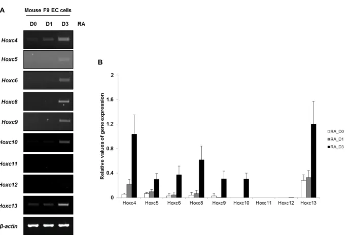
F9 배아암종 (embryonic carcinoma, EC) 세포에서 RA 에 의한 Hoxc 유전자의 순차적인 발현 변화
레티노산(RA)에 의한 Hoxc 유전자의 순차적인 발현 변화를
관찰하기 위해 생쥐 F9 EC 세포에 5×10
-7M 의 RA를 3일간
처리했다. RA를 처리 하지 않은 day0 (D0), RA를 1일 처리한
day1 (D1)과 RA를 3일간 처리한 day3 (D3), 각각 세 가지의
RNA 시료를 이용하여 RT-PCR을 수행하였다(Fig. 1A). RT-
PCR 결과, D0는 Hoxc 유전자들의 발현이 일어나지 않았고
D1의 경우 Hoxc4만 발현이 유도되었으며, D3에서는 Hoxc4부
터 -c10의 발현이 관찰되었다. Hoxc13은 D0에서부터 이미 발
현하고 있으며 D3에서는 그 발현이 증가하는 것을 볼 수 있었
다. Fig. 1B는 agarose gel에 loading한 PCR 결과물을 β-actin
A
B
Fig. 1. Expression pattern of Hoxc cluster genes following Retinoic acid (RA) treatment in F9 EC cells by RT-PCR. Mouse F9 EC cells were cultured with RA for 3 days. Total RNAs were isolated each day from Day 0 (D0), Day 1 (D1) and Day 3 (D3), and then (A) RT-PCR was performed with each Hoxc primers.
β
-actin was used as a positive control for amplifiable cDNA.(B) The RT-PCR results were normalized using the
β
-actin signal. The data are representative of the three replicate experiments.값으로 나누어 정량적으로 분석하여 그래프화한 결과로, D0 와 비교하여 D1과 D3에서 Hoxc 유전자의 발현이 앞에서부터 차례대로 증가하는 것을 확인할 수 있었다. 이 결과들을 종합 해 보면 F9 EC 세포에 RA를 처리하여 유도된 Hoxc 유전자의 발현은 시간에 따라 순차적으로 일어난다는 것을 알 수 있으 며, 이는 이전의 결과와 동일하다[12, 13].
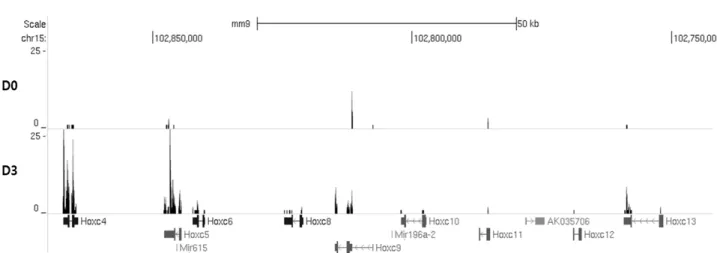
RNA-seq을 이용한 Hoxc 유전자의 발현 변화 관찰 레티노산(RA)에 의한 Hoxc 유전자 발현의 변화를 semi- quantitative RT-PCR로 관찰한 뒤, 이를 genome-wide하게 분 석하기 위해 RNA-sequencing을 수행하였다. 이전 단계의 실 험과 동일하게 생쥐 F9 EC 세포에 5x10
-7M 의 RA를 3일간 처리하였다. RA를 처리 하지 않은 D0 (대조군)와 RA를 3일간 처리한 D3 (실험군), 각각의 RNA 시료를 이용하여 RNA-se- quencing을 수행하였다. RNA-sequencing 데이터를 UCSC genome browser에 upload하여 분석한 결과, RT-PCR 결과와 동일하게 D0에서는 Hoxc 유전자의 발현이 거의 일어나지 않 았고, D3에서는 Hoxc4 부터 -c10까지 발현되어 있는 것을 관찰 할 수 있었다(Fig. 2). 또한 Hoxa, -b, -d cluster 유전자의 발현도 D0와 비교하여 D3에서 증가하는 것을 관찰하였다(data not
shown). 이와 같이 F9 EC 세포에 RA를 처리하여 유전자의 전사 정도를 genome-wide하게 분석을 한 결과, Hox 유전자는 RA에 의해 발현이 유도되며 이는 4개의 Hox 유전자 집합체 (Hoxa, -b, -c, -d cluster) 모두에게 영향을 끼치는 것을 알 수 있었다.
Hoxc 유전자의 순차적인 발현과 히스톤 변이와의 관계 Hoxc 유전자의 순차적인 발현 변화가 크로마틴 구조의 리 모델링과 관련되어 있다는 이전의 보고[12]와 더불어 Hoxc 유 전자의 발현과 히스톤 변이와는 어떤 상관관계가 있는 지 알 아보기 위해 히스톤 표지 항체로 ChIP assay를 수행하였다.
RA를 날짜 별로 처리한 D0, D1, D3 세포에서 크로마틴을 분
리하고, 전사 발현 시 히스톤에 표지 되는 마커인 H34me3
(histone H3 lysine 4 tri-methylation)과 전사 억제 시 히스톤
에 표지 되는 마커인 H3K27me3 (histone H3 lysine 27 tri-
methylation) 항체를 첨가하여 반응시켰다. 반응 후 얻어낸 각
각의 크로마틴 시료로 ChIP-PCR을 수행하였다(Fig. 3). D0의
경우 전사 억제 마커인 H3K27me3이 모든 Hoxc 좌위에서 관
찰되었고, 전사 발현 마커인 H3K4me3은 관찰되지 않았다. D1
에서는 모든 H3K27me3 변이가 사라지고 발현 중인 Hoxc4에
Fig. 2. Expression profiles of Hoxc genes following RA treatment in F9 EC cells by RNA-sequencing (RNA-seq). Total RNAs were isolated each day from D0 and D3 (D0: control cells with no RA treatment). These RNA-seq data were illustrated using UCSC genome browser. Reference genes are depicted at the lower in blue (Hoxc genes). Sequential expression pattern of Hoxc cluster genes in RA-untreated (D0) and treated (D3) samples.
Fig. 3. Histone H3 modifications at Hoxc loci in mouse F9 EC cells. ChIP assays were performed with the indicated histone antibodies on cross-linked chromatin samples from the Day 0, Day 1 and Day 3 samples. Immunoprecipitated and input DNAs were amplified by PCR using region specific primers. The data show the re- sults of one experiment of at least three independent experiments giving comparable results.
대한 H3K4me3 변이의 표지만 관찰되었다. D3에는 D1과 마찬 가지로 H3K27me3 변이가 관찰되지 않았고 모든 Hoxc 좌위에 서 H3K4me3 변이가 표지 된 것을 확인할 수 있었다. Hoxc11 과 Hoxc12의 경우 D3에서 발현하지 않음에도 불구하고 H3K4me3 변이가 관찰되고 있는데, 이는 Hoxc11과 Hoxc12 유 전자는 히스톤 변이만으로는 발현이 시작될 수 없어 다른 전 사인자단백질이 필요한 것으로 유추할 수 있다. 최근 발표된 논문에서 Hoxc11, -c12 유전자가 posterior Hoxc 유전자의 up- stream regulator의 영향을 받아 조절된다는 보고[7]가 되어 있어 단순히 히스톤 표지의 변이만으로는 설명할 수 없는 발
현 기작이 따로 존재할 것으로 보인다. Hoxc13 유전자는 D0에
서부터 이미 발현 되고 있으며, 이는 RA 처리에 의해 증가된다
(Fig. 1). 이 데이터는 1997년에 발표된 실험 결과와 유사한
것으로[3], 이 논문의 human EC 세포 또한 RA를 처리 하기
전부터 HOXC13이 이미 발현하고 있으며 RA처리 후 약간의
발현 증가를 보이는 것이 관찰되었다. 이처럼 다른 Hoxc 유전
자들의 순차적인 발현과 관계없이 Hoxc13 유전자가 따로 발현
하는 것은 EC 세포의 특징으로 보인다. Hoxc13의 경우 히스톤
변이가 유전자의 발현 양상과 동일하게 움직이지 않는 것도
관찰되었는데(Fig. 3), Hoxc11, -c12와 마찬가지로 히스톤 표지
의 변이만으로는 설명할 수 없는 후성유전학적 발현 기작[8, 24]이 있을 것으로 예상된다. 또한, Hoxc11-c13 유전자 발현과 관계없이 전체가 동일한 히스톤 마커(D0:H3K27me3, D1:no enrichment (Hoxc4 제외), D3:H3K4me3)로 표지 되어있는 것 을 볼 수 있는데, 이는 Hoxc 좌위 전체에서 히스톤 변이가 일어 나지만 posterior Hoxc 유전자(Hoxc11-c13)의 경우 anterior Hoxc 유전자(Hoxc4-c10)와 다른 기작으로 발현이 시작되며, 특 히 EC 세포에서 순차적인 발현과 관계없이 따로 독특하게 움 직이는 Hoxc13의 발현은 특이적인 경로가 있을 것으로 추측된 다. 이 과정을 밝히기 위해서는 추가적으로 심도 있는 연구가 필요할 것으로 보인다.
이상의 결과와 이전에 보고된 결과[12]를 통해 F9 EC 세포 에서 Hoxc 유전자들이 발현하지 않을 때(D0)는 크로마틴 뭉침 현상과 함께 전사 억제 마커인 H3K27me3이 표지 되어 있으 며, RA를 처리하여 Hoxc 유전자들이 발현할 때(D1, D3)는 크 로마틴 풀림 현상과 함께 H3K27me3 변이가 사라지고, H3K4me3 변이는 유전자 발현에 따라 표지 되어있는 것을 알 수 있다. 결론적으로 F9 EC 세포에서 RA에 의해 유도된 Hoxc 유전자가 시간에 따라 순차적으로 발현하게 되는 것은 Hoxc 좌위의 히스톤 메틸화가 중요한 후성유전학적 기작의 하나로 작용하여 일어나는 것으로 사료된다.
감사의 글
본 연구는 한국연구재단(2013R1A1A2008399) 및 연세대학 교 의과대학(6-2014-0147)의 연구비 지원을 받았습니다.
References
1. Boncinelli, E., Simeone, A., Acampora, D. and Mavilio, F.
1991. HOX gene activation by retinoic acid. Trends Genet.
7, 329-334.
2. Favier, B. and Dolle, P. 1997. Developmental functions of mammalian Hox genes. Mol. Hum. Reprod. 3, 115-131.
3. Flagiello, D, Gibaud A, Dutrillaux, B, Poupon, M. F. and Malfoy, B. 1997. Distinct patterns of all-trans retinoic acid dependent expression of HOXB and HOXC homeogenes in human embryonal and small-cell lung carcinoma cell lines.
FEBS Lett. 415, 263-267.
4. Gillespie, R. F. and Gudas, L. J. 2007. Retinoid regulated association of transcriptional co-regulators and the poly- comb group protein SUZ12 with the retinoic acid response elements of Hoxa1, RARbeta (2), and Cyp26A1 in F9 embry- onal carcinoma cells. J. Mol. Biol. 372, 298-316.
5. Kashyap, V., Gudas, L. J., Brenet, F., Funk, P., Viale, A. and Scandura, J. M. 2011. Epigenomic reorganization of the clus- tered Hox genes in embryonic stem cells induced by retinoic acid. J. Biol. Chem. 286, 3250-3260.
6. Kashyap, V., Laursen, K. B., Brenet, F., Viale, A. J., Scandura, J. M. and Gudas, L. J. 2013. RARgamma is essential for reti-
noic acid induced chromatin remodeling and transcriptional activation in embryonic stem cells. J. Cell Sci. 126, 999-1008.
7. Kong, K. A., Lee, J. Y., Oh, J. H., Lee, Y. and Kim, M. H.
2014. Akt1 mediates the posterior Hoxc gene expression through epigenetic modifications in mouse embryonic fibroblasts. Biochim. Biophys. Acta 1839, 793-799.
8. Kouzarides, T. 2007. Chromatin modifications and their function. Cell 128, 693-705.
9. Krumlauf, R. 1992. Evolution of the vertebrate Hox homeo- box genes. Bioessays 14, 245-252.
10. Laursen, K. B., Wong, P. M. and Gudas, L. J. 2012.
Epigenetic regulation by RARalpha maintains ligand-in- dependent transcriptional activity. Nucleic Acids Res. 40, 102-115.
11. Lee, E. R., Murdoch, F. E. and Fritsch, M. K. 2007. High histone acetylation and decreased polycomb repressive complex 2 member levels regulate gene specific transcrip- tional changes during early embryonic stem cell differ- entiation induced by retinoic acid. Stem Cells 25, 2191-2199.
12. Lee, J. Y., Min, H., Wang, X., Khan, A. A. and Kim, M.
H. 2010. Chromatin organization and transcriptional activa- tion of Hox genes. Anat. Cell. Biol. 43, 78-85.
13. Lee, Y., Lee, J. Y. and Kim, M. H. 2014. PI3K/Akt pathway regulates retinoic acid-induced Hox gene expression in F9 cells. Dev. Growth. Differ. 56, 518-525.
14. McGinnis, W. and Krumlauf, R. 1992. Homeobox genes and axial patterning. Cell 68, 283-302.
15. Min, H., Lee, J. Y. and Kim, M. H. 2012. Structural dynamics and epigenetic modifications of Hoxc loci along the ante- roposterior body axis in developing mouse embryos. Int.
J. Biol. Sci. 8, 802-810.
16. Partanen, J., Schwartz, L. and Rossant, J. 1998. Opposite phe- notypes of hypomorphic and Y766 phosphorylation site mu- tations reveal a function for Fgfr1 in anteroposterior pattern- ing of mouse embryos. Genes Dev. 12, 2332-2344.
17. Pearson, J. C., Lemons, D. and McGinnis, W. 2005.
Modulating Hox gene functions during animal body patterning. Nat. Rev. Genet. 6, 893-904.
18. Simeone, A., Acampora, D., Arcioni, L., Andrews, P. W., Boncinelli, E. and Mavilio, F. 1990. Sequential activation of HOX2 homeobox genes by retinoic acid in human embry- onal carcinoma cells. Nature 346, 763-766.
19. Soshnikova, N. and Duboule, D. 2008. Epigenetic regulation of Hox gene activation: the waltz of methyls. Bioessays 30, 199-202.
20. Soshnikova, N. and Duboule, D. 2009. Epigenetic temporal control of mouse Hox genes in vivo. Science 324, 1320-1323.
21. Soshnikova, N. and Duboule, D. 2009. Epigenetic regulation of vertebrate Hox genes: a dynamic equilibrium. Epigenetics 4, 537-540.
22. Soprano, D. R., Teets, B. W. and Soprano, K. J. 2007. Role of retinoic acid in the differentiation of embryonal carcino- ma and embryonic stem cells. Vitam. Horm. 75, 69-95.
23. Stornaiuolo, A., Acampora, D., Pannese, M., D'Esposito, M., Morelli, F., Migliaccio, E., Rambaldi, M., Faiella, A., Nigro, V., Simeone, A. and et al. 1990. Human HOX genes are dif-
초록:F9 EC 세포에서 레티노산에 의해 유도되는
Hoxc유전자의 발현에 히스톤 메틸화가 미치는 영향
민혜현․김명희*
(연세대학교 의과대학 해부학교실 발생학 실험실)
Hox 유전자는 호메오도메인을 포함한 전사인자로써, 발생 과정 중 전후축을 따라 몸의 형태 형성을 조절하는 역할을 한다. 레티노산(RA)은 발생 과정에서 필수적인 형태형성인자이며 세포의 특성을 결정하는데 중요한 조절자 이다. 특히, RA는 생쥐나 인간으로부터 만들어진 배아암종(EC)세포에서 Hox 유전자의 발현을 조절한다고 밝혀져 있다. 또한 RA에 의한 세포 분화와 유전자 조절 과정에 히스톤 변이가 중요한 역할을 하는 것으로 보고되어 있다.
히스톤 변이가 RA에 의해 유도되는 Hox 유전자의 발현에 특이적인 역할을 할 것으로 유추되기 때문에, 이 연구의 목적은 F9 생쥐배아 기형암종세포에서 RA에 의해 유도되는 Hoxc 유전자의 순차적인 발현이 히스톤 변이에 의해 일어나는 것인지를 조사하는 것이다. Hox 유전자의 발현 양상과 히스톤 변이는 semi-quantitative RT-PCR, RNA-se- quencing 과 chromatin immuno-precipitation (ChIP)-PCR 기법을 이용하여 관찰하였다. RA 처리 후(0일(D0), 1일 (D1), 3일(D3)), Hoxc4 유전자의 발현(D1)은 Hoxc5부터 –c10 유전자(D3)보다 먼저 시작되었다. Hox가 발현하지 않는 D0 샘플은 전사 억제 마커인 H3K27me3이 모든 Hoxc 좌위에 강하게 표지 되어 있었으나 D1과 D3 샘플에서는 모든 좌위의 H3K27me3 표지가 확연히 줄어들어 있었다. 전사 발현 마커인 H3K4me3가 Hoxc 유전자의 순차적인 발현과 더 연관성이 있는 것으로 보이는데 D1에서 Hoxc4 발현과 함께 H3K4me3이 표지 되어 있었고, D3에서는 Hoxc 유전자 발현과 함께 모든 좌위에서 H3K4me3 마커가 존재했기 때문이다. 모든 결과를 종합해 보았을 때 F9 세포에서 RA에 의해 유도된 Hoxc 유전자의 순차적인 발현은 Hoxc 좌위에서 H3K27me3가 사라지고, H3K4me3가 표지 되는 히스톤 메틸화의 변이에 의해 결정되는 것으로 사료된다.
ferentially activated by retinoic acid in embryonal carcino- ma cells according to their position within the four loci. Cell Differ. Dev. 31, 119-127.
24. Strahl, B. D. and Allis, C. D. 2000. The language of covalent
histone modifications. Nature 403, 41-45.
25. Young, T. and Deschamps, J. 2009. Hox, Cdx, and ante- roposterior patterning in the mouse embryo. Curr. Top. Dev.
Biol. 88, 235-255.