- 164 -
*Received: June 5, 2013 / Revised: June 17, 2013 Accepted: June 17, 2013
†Corresponding author: Na Kyung Lee. Department of Biomedical Laboratory Science, College of Medical Science, Soonchunhyang University, Asan-Si, Chungnam 336-745, Korea.
Tel: +82-41-530-3036, Fax: +82-41-530-3085 e-mail: [email protected]
ⒸThe Korean Society for Biomedical Laboratory Sciences. All rights reserved.
Brief Communication
J. Exp. Biomed. Sci. 2013, 19(2): 164~167 pISSN : 1738-3226
Gene Profiling in Osteoclast Precursors by RANKL Using Microarray
Na Kyung Lee†
Department of Biomedical Laboratory Science, College of Medical Sciences, Soonchunhyang University, Asan-Si, Chungnam 336-745, Korea
Osteoclasts are originated from hemopoietic progenitors of the monocyte/macrophage lineage and resorb mineralized tissues. Elevated osteoclast numbers and activity result in bone disease such as osteoporosis, Paget’s disease, and tumor osteolysis. In order to identify the genes that are involved in osteoclast differentiation, microarray was performed after treated with RANKL for 12 h and 24 h in osteoclast precursors. The genes that changed by RANKL treatment were grouped by biological process or molecular function. Among them, the number of genes involved in signal transduction and nucleic acid binding was 6065 and 3066, respectively. When analyzed the number of genes changed more than 1.5 fold in the cells treated with RANKL for 12 h or 24 h compared to when RANKL was not treated, 83 and 62 genes were up-regulated; 56 and 62 genes were downregulated, respectively. To verify the microarray results, real-time RT-PCR for Cxcl1 and Slfn1genes that have not been reported yet related to osteoclast differentiation, as well as Ccl2 gene associated with osteoclast differentiation were carried out. Both experiments showed a similar result of more than 1.5 fold induction of these genes by RANKL treatment. These results suggest the possibility that Cxcl1 and Slfn1 may associate with osteoclastogenesis and provide that microarray is a useful tool to analyze the profile of genes changed during osteoclast differentiation by RANKL. Moreover, this gene profile contributes to understand the regulatory mechanisms involved in osteoclast differentiation and the pathogenesis, thus developing therapeutics of bone diseases such as osteoporosis.
Key Words: Osteoclasts, Bone disease, RANKL, Microarray
뼈는 척추동물에서 내부 장기를 보호하며, 우리 몸의 형태를 지탱하는데 중요한 역할을 한다. Bone remodeling 은 뼈를 형성하는 조골세포와 뼈를 흡수하는 파골세포의 작용에 의해 조절되며, 이러한 세포들의 기능에 있어서의 균형이 골 밀도를 유지하는데 중요하다 (Teitelbaum et al., 2000). 골다공증을 포함하는 여러 골질환들은 점점 고령 화되어 가는 현대 사회에서 현대인의 삶의 질을 현저히 떨어뜨린다. 특히 파골세포의 분화에 있어서의 증가나 활 성의 증가는 골다공증의 위험을 증가시킨다.
본 연구에서는 RANKL에 의한 파골세포의 분화 과정 에서 특이적으로 발현되는 유전자의 프로파일을 확보하 고자 microarray를 수행하였다. 먼저 마우스의 대퇴골로
부터 골수세포를 분리한 후 M-CSF를 처리하여 파골전구 세포를 획득하였다 (Choi et al., 2013). 6-well plate에 세포 를 106개가 되게 분주한 후 하루 동안 배양 후 파골세포 의 분화를 유도하는 사이토카인인 RANKL을 12시간과 24시간 동안 각각 처리하였다. 각 well로부터 세포를 얻 어 RNA를 분리하여 purity를 측정하고 RNA 겔 전기영동 을 수행하여 RNA의 상태를 확인하였다. 이후 Illumina MouseRef-8 v2 Expression BeadChip (Illumina, Inc., San Diego, CA)을 이용하여 microarray를 수행하였으며 (Macrogen Inc.), 일부는 real-time RT-PCR을 수행하기 위 해 cDNA를 합성하였다 (Choi et al., 2013).
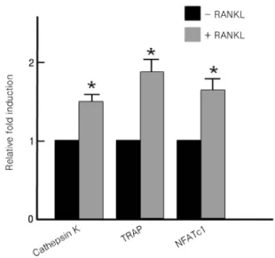
Microarray를 실시하기에 앞서 real-time RT-PCR을 수 행하여 파골세포 분화 마커로 알려진 Cathepsin K, TRAP 과 NFATc1 유전자 (Gowen et al., 1999; Takayanagi et al., 2002; Rovert et al., 2007)가 RANKL에 의해 정상적으로 발현되는지를 관찰하였다. Fig. 1에서 보는 바와 같이 RANKL을 처리한 경우 파골세포 분화마커 유전자들의 발현이 현저히 증가됨을 확인하였다.
- 165 - Fig. 1. RANKL induces the expression of osteoclast marker genes. Serum-starved preosteoclasts were treated without or with RANKL for 24 h and total RNAs were isolated from cultured samples. Reverse transcribed and synthesized cDNAs were then subjected to real-time PCR using indicated genes-specific primers. Data are presented as means standard deviations (SD) of triplicates. Statistical analyses were performed using unpaired, two-tailed Student t test. *P < 0.05 vs. cells without RANKL.
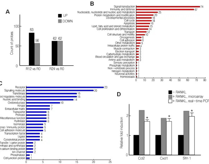
Fig. 2. Grouping of genes according to biological process or molecular function. (A and B) After microarray, genes changed by RANKL treatment were grouped according to biological process (A) or molecular function (B) and counted.
Microarray를 수행한 후 RANKL을 처리하지 않은 세포 와 비교하여 RANKL을 12시간과 24시간 각각 처리하여 발현이 변화된 유전자들을 먼저 생물학적 과정에 따라 그룹을 지어 유전자 수를 분석하였다. 그 결과 신호전달 에 관여하는 유전자 수가 6,055개로 가장 많았으며, 다음
으로 핵산 대사, 단백질 대사 및 변형에 관여하는 유전자 가 4,072개 및 3,585개로 많은 비중을 차지하였다(Fig.
2A). RANKL에 의해 발현이 변화된 유전자들을 분자적 기능에 따라 그룹을 지어 유전자 수를 분석한 경우 핵산 결합과 관련한 유전자 수가 3,066개로 가장 많았으며, 다 음으로 수용체, 전사인자 등이 2,384개, 2,185개로 많은 비중을 차지하였다 (Fig. 2B).
발현 시그널이 1.5배 이상 변화한 유전자의 수를 분석 한 결과, RANKL을 처리하지 않은 세포와 비교하여 RANKL을 12시간과 24시간 각각 처리한 경우, 증가한 유 전자 수는 각각 83개와 62개였으며, 1.5배 이상 감소한 유 전자의 수는 각각 56개와 62개였다 (Fig. 3A). 이들을 생 물학적 과정과 분자적 기능에 따라 그룹을 지어 유전자 를 분석한 결과, 생물학적 과정에서는 신호전달에 관여하 는 유전자 수가 74개로 가장 많은 비중을 차지하였으며, 분자적 기능에 따라 구분한 경우 수용체 유전자가 23개 로 가장 많은 비중을 차지하였다 (Fig. 3B and 3C).
Microarray 결과에서 발현 시그널이 1.5배 이상 증가한 유전자들 중, 파골세포 분화와 관련이 있다고 알려진 chemokine ligand 2 (Ccl2) (Miyamoto et al., 2009)와 파골 세포 분화와 관련하여 그 기능이 알려져 있지 않은 cysteine-X-cysteine family chemokine ligand 1 (Cxcl1), Schlafen1 (Slfn1)의 발현을 real-time RT-PCR을 통해 다시 한번 확인하였다. Cxcl1의 경우, 조골세포에서 형성되어 파골전구세포의 이동을 촉진하는 케모카인으로서의 역할 을 하는 것으로 보고 되었으며 (Onan et al., 2009), Slfn1
- 166 -
Fig. 3. Analysis of genes changed by RANKL treatment. (A-C) The number of genes changed more than 1.5 fold in the cells treated with RANKL for 12 h (R12) or 24 h (R24) compared to when RANKL was not treated (R0) were counted after microarray (A). The genes were analyzed by Grouping according to biological process (B) or molecular function (C).
(D) Comparison of Ccl2, Cxcl1, and Slfn1 genes induced by RANKL treatment for 24 h using microarray or real-time PCR.
*P < 0.05 vs. cells without RANKL.
은 흉선세포의 발달에 중요한 역할을 한다고 알려져 있 으나 (Schwarz et al., 1998) 두 유전자 모두 파골세포 분화 에 있어서의 역할은 보고된 바가 없다. 예상한 대로, real-time RT-PCR 결과 Ccl2, Cxcl1과 Slfn1 유전자의 발현 이 microarray 결과와 일치하게 RANKL 에 의해 현저히 증가함을 관찰할 수 있었다 (Fig. 3D). 이러한 결과는 파 골세포 분화 과정 동안 발현이 증가하는 Cxcl1과 Slfn1이 파골세포 분화 과정과 관련이 있을 가능성과 함께, 파골 세포 분화과정 동안 그들의 역할에 대한 연구의 필요성 을 제시한다.
비록 앞으로의 골질환 치료제 개발에 있어 이러한 마 우스에 존재하는 유전자들이 인간에서 높은 유사성을 가 지며 존재하는지, 아니면 기능을 하지 않는 거짓유전자 (pseudogene)는 아닌지에 대한 추가 연구 과정이 필요하
지만, 이번 연구는 파골세포 분화과정 동안 발현이 변화 되는 유전자들을 손쉽게 스크리닝함에 있어 microarray가 유용함을 보여준다. 또한 골다공증과 같은 골질환 치료제 예방 및 치료제 개발을 위한 중요한 분자적 후보 타깃을 제공하고, 앞으로 개발되는 신약 후보 물질들의 효과를 손쉽게 확인하는데 도움을 줄 수 있을 것으로 사료된다.
REFERENCES
Choi J, Choi SY, Lee SY, Lee JY, Kim HS, Lee SY, Lee NK. Caffeine enhances osteoclast differentiation and maturation through p38 MAP kinase / Mitf and DC-STAMP / CtsK and TRAP pathway. Cell Signal. 2013.
25: 1222-1227.
- 167 - Gowen M, Lazner F, Dodds R, Kapadia R, Feild J, Tavaria
M, Bertoncello I, Drake F, Zavarselk S, Tellis I, Hertzog P, Debouck C, Kola I. Cathepsin K knockout mice develop osteopetrosis due to a deficit in matrix degradation but not demineralization. J Bone Miner Res. 1999. 14: 1654-1663.
Miyamoto K, Ninomiya K, Sonoda KH, Miyauchi Y, Hoshi H, Iwasaki R, Miyamoto H, Yoshida S, Sato Y, Morioka H, Chiba K, Egashira K, Suda T, Toyama Y, Miyamoto T. MCP-1 expressed by osteoclastsstimulates osteoclasto- genesis in an autocrine/paracrine manner. Biochem Biophys Res Commun. 2009. 383:373-377.
Onan D, Allan EH, Quinn JM, Gooi JH, Pompolo S, Sims NA, Gillespie MT, Martin TJ. The chemokine Cxcl1is a novel target gene of parathyroid hormone (PTH)/ PTH-related protein in committed osteoblasts. Endocrinology. 2009. 5:
2244-2253.
Roberts HC, Knott L, Avery NC, Cox TM, Evans MJ, Hayman AR. Altered collagen in tartrate-resistant acid phosphatase (TRAP)-deficient mice: a role for TRAP in bone collagen metabolism. Calcif Tissue Int. 2007. 80: 400-410.
Schwarz DA, Katayama CD, Hedrick SM. Schlafen, a new family of growth regulatory genes that affect thymocyte development. Immunity. 1998. 5: 657-668.
Takayanagi H, Kim S, Koga T, Nishina H, Isshiki M, Yoshida H: Induction and activation of the transcription factor NFATc1 (NFAT2) integrate RANKL signaling in terminal differentiation of osteoclasts. Dev Cell. 2002. 3: 889–901.
Teitelbaum SL. Bone resorption by osteoclasts. Science. 2000.
289: 1504-1508.