INTRODUCTION
Although steady declines in the incidence and mortality rates of gastric cancer have been observed in the last century, gastric cancer is still one of the most common causes of cancer-related death in the world.
1The incidence of early gastric cancer (EGC) has increased in Korea in recent years because of the aging population, improved diagnostic pro- cedures, and regular personal check-ups.
2The safety of laparoscopy-assisted gastrectomy (LAG) in early gastric cancer has been demonstrated in many studies.
3,4However, there has been significant debate over the use of laparoscopic
resection for the management of advanced gastric cancer.
5,6Laparoscopic surgery has shown the advantages of less pain, shorter recovery time, shortened hospital stay, minimal wound and a good quality of life.
7-9For these reasons, LAG is considered an alternative treatment for gastric cancer.
Although LAG is safe, the long-term oncologic efficacy and survival of patients including early and advanced gastric cancer have not been assessed. The aim of the present study was to discuss the long-term outcomes and survival of a group of patients who underwent LAG performed by a single surgeon.
Safety of Laparoscopy Assisted Gastrectomy for Gastric Cancer, Including Advanced Cancers
Eun Mee Park, M.D., Hyung Ook Kim, M.D., Ph D., Byung Ho Son, M.D., Ph D., Jun Ho Shin, M.D., Ph D., Sung Ryol Lee, M.D., Ph D.
Department of Surgery, Sungkyunkwan University School of Medicine, Seoul, Korea
Purpose:
Laparoscopy-assisted gastrectomy (LAG) is considered an alternative treatment option for gastric cancer. LAG is safe, however the long-term oncologic efficacy and survival of patients including those with advanced gastric cancer have not been assessed. The aim of this study was to evaluate long-term outcomes and survival of patients with gastric cancer, including advanced cases, who underwent LAG performed by a single surgeon.Methods:
Between January 2006 and December 2010, 161 patients with gastric cancer underwent LAG performed by a single surgeon. Clinicopathological data were collected retrospectively along with data on survival and prognosis. Overall survival (OS) and disease-free survival (DFS) were estimated using the Kaplan-Meier method.Results:
A total of 161 patients diagnosed with gastric cancer underwent LAG. Postoperative morbidity occurred in 12 patients. The median OS was 67.0 months (range, 1.0~97.0 months), and the median DFS was 67.0 months (range, 1.0~97.0 months). T stage, N stage, TNM stage, lymphatic invasion, and venous invasion influenced overall survival and disease recurrence. The OS rates according to N stage were 96.8% for N0, 94.4% for N1, 45.5% for N2, and 42.9% for N3.Conclusion:
The current study showed that LAG for gastric cancer, including advanced gastric cancer, is technically feasible with acceptable long-term oncologic outcomes.Keywords:
Gastric cancer, Laparoscopy-assisted surgery, Overall survivalReceived July 27, 2015 Revised 1st August 30, 2015
2nd September 6, 2015 3rd September 8, 2015 Accepted September 8, 2015
Corresponding author Sung Ryol Lee Department of Surgery,
Sungkyunkwan University School of Medicine, 108 Pyung-dong, Jongno- gu, Seoul 03010, Korea
Tel: +82-2-2001-8005 Fax: +82-2-2001-2131 E-mail: [email protected]
This is an Open Access article distributed under the terms of the Creative Commons Attribution Non-Commercial License (http://
creativecommons.org/licenses/by-nc/4.0/) which permits unrestricted non-commercial use, distribution, and reproduction in any medium, provided the original work is properly cited.
Copyright © 2015 The Journal of Minimally Invasive Surgery. All rights reserved.
Journal of Minimally Invasive Surgery
J Minim Invasive Surg 2015;18(3):79-85
MATERIALS AND METHODS
Patients
Between January 2006 and December 2010, 171 patients who were diagnosed with gastric cancer underwent laparoscopic gastrectomy performed by a single surgeon in the Department of Surgery, Kangbuk Samsung Hospital, Sungkyunkwan University School of Medicine, Seoul, Korea. All cases were pathologically confirmed before surgery. In principle, all patients with gastric cancer were eligible for receiving a laparoscopic surgery. The patients pathologically diagnosed with advanced gastric cancer had to decide between laparoscopic surgery and open surgery. We explained this controversial point to the patients and have obtained their consent. Among a total of 171 patients who underwent laparoscopic gastrectomy, ten patients were converted to open gastrectomy due to abdominal adhesion (n=6) or uncontrolled bleeding in the operation field (n=4). Therefore, 161 patients were included in this study.
Patients with advanced gastric cancer received chemotherapy with intravenous 5-fluorouracil and cisplatin; patients with EGC with lymph node metastasis received oral 5-fluorouracil for adjuvant treatment. Prior to the operation, the standard workup for evaluation and staging of gastric cancer was performed and included duodenoscopy and computed tomography (CT) scans.
Surgical approach
The patients were positioned supine on the operating room table under general anesthesia. An 18-gauge nasogastric tube was inserted to decompress the stomach. Two 12-mm trocars, three 5-mm trocars and a flexible laparoscope were used during surgery. Appropriate CO
2pneumoperitoneum was achieved with 15 mmHg insufflations through a 12-mm umbilical trocar. According to the Japanese Classification of Gastric Carcinoma, extended lymph node dissection (D2) and D1+b lymph node dissection were implemented. A mini laparotomy wound was created for extracorporeal operation.
The incision was extended vertically 5 cm on the upper abdomen. The stomach was resected through the window.
The Billroth I method was performed using a circular 29 mm stapler, but the Billroth II method was performed with hand sewing. In the case of total gastrectomy, the duodenum was transected 1 or 2 cm distal to the pyloric ring using a linear stapler. After the stomach was mobilized, the esophagus was transected using a purse string clamp through mini laparotomy wound. A side-to-side jejunojejunostomy was extracorporeally performed using linear staplers. Roux-en-Y esophagojejunostomy was accomplished using a circular stapler through mini laparotomy wound. A closed suction tube
was placed in Morrison ’s pouch through the previous 5-mm trocar site.
Outcome measurement
All patients were monitored through physical examinations and laboratory tests after operation. Tumor markers, chest X-ray, abdominal CT scan and duodenoscopy were examined every three months in the first two years, every sixth months in the following year and then annually thereafter. A positron emission tomography (PET) study was not always performed for patients. When the patient required additional analysis, PET or magnetic resonance imaging (MRI) was implemented to better clarify a recurrence. Demographics and perioperative and postoperative data were retrospectively collected and analyzed from the hospital records. Follow-up data were collected from patient ’s clinical records.
Statistical analysis
Overall survival (OS) and disease-free survival (DFS) were calculated using the Kaplan-Meier method. OS was calculated from the day of surgery until death or until the end of follow- up, and DFS was calculated until relapse or end of follow-up.
The significant differences among each group of patients were determined by log rank test. p values less than or equal to 0.05 were considered to be statistically significant. SPSS ver.
18.0 (SPSS, Inc., Chicago, IL, USA) was used for the statistical calculations. Multivariate analyses for survival and recurrence were estimated using Cox ’s proportional hazard model.
RESULTS
In total, 161 patients diagnosed with gastric cancer under- went LAG. After surgery, three patients were lost to follow-up.
The patient characteristics and postoperative surgical results are summarized in Table 1. The study group consisted of 105 men and 56 women with a mean age of 62.6 years. Compared to women, men were twice as likely to develop gastric cancer.
10The average hospital stay after operation was 9.4 days. The five-year OS rate was 90.68%, and the five-year DFS rate was 93.17%.
According to the 7
thedition of the American Joint Committee
on Cancer TNM classification system, there were 96 patients
with stage IA cancer, 23 with stage IB cancer, 15 with stage
IIA cancer, 12 with stage IIB cancer, 5 with stage IIIA cancer,
8 with stage IIIB cancer, 1 with stage IIIC and one with
stage IV cancer, who died after 14 months due to disease
progression. All operations were curative resection (R0) except
one patient diagnosed with stage IV cancer. Postoperative
morbidity occurred in 12 patients. One patient died within 30 days due to duodenal stump leakage despite a second operation. The mean number of retrieved lymph nodes was 30
±6 (range, 6~68, median 31.1) per patient.
In this study, the median OS was 67.0 months (range, 1.0~97.0 months) (Fig. 1), and the median DFS was 67.0 months (range, 1.0~97.0 months) (Fig. 2). The median follow- up period for all patients was 55.0 months (range, 1.0~97.0 months). Fifteen patients died (9.31%) and 11 patients (6.83%) had a recurrence of gastric cancer during the follow-up
period. The causes of death were disease progression (n=10), pneumonia (n=1), gallbladder cancer (n=1), and aging (n=3).
Among the 11 recurred patients, 10 died. Most of the recu- rrences occurred within three years of the operation. The most common recurrent site was the peritoneum (n=4), followed by lymph node (n=3), liver (n=2), anastomosis site (n=1), and Krukenberg’s tumor (n=1). The two patients diagnosed with EGC experienced liver and lymph node recurrence, respectively.
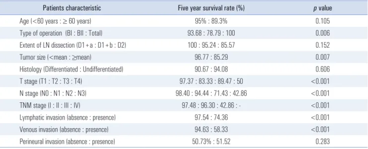
In this study, we evaluated the factors that influence OS and DFS. The different factors among the patients characteristic about five year survival rate are shown in Table 2. There were no significant differences in age, sex, or postoperative tumor differentiation among the patients groups. However, tumor size, T stage, N stage, TNM stage, lymphatic invasion, and venous invasion were significantly different. Based on univariate analysis, tumor size, T stage, N stage, lymphatic invasion, venous invasion and perineural invasion were significant prognostic factors for survival Fig. 1. Overall survival curves according to TNM stage.
0 0
100
Survivalrate(%)
1.0
0.8
0.6
0.4
0.2
20 40 60 80
Survival period (months)
Stage I (n=119) Stage II (n=27) Stage III (n=14) Stage IV (n=1) TNM stage
Table 1. Clinicopathologic characteristics and postoperative surgical results of patients who underwent laparoscopy-assisted gastrectomy
Clinicopathologic characteristics Results
Age (years, mean) 62.64±11.37
Sex (Male : Female) 105 : 56
Gross type EGC, I : IIA : IIB : IIC : III 10 : 6 : 40 : 48 : 12 Gross type AGC, 1 : 2 : 3 : 4 2 : 4 : 39 : 0
Tumor size (cm, mean) 3.33±1.95
Histology (Undifferentiated : Differentiated) 86 : 75 T stage (T1 : T2 : T3 : T4) 114 : 18 : 19 : 10 N stage (N0 : N1 : N2 : N3) 125 : 18 : 11 : 7 TNM stage (I : II : III : IV) 119 : 27 : 14 : 1 Lymphatic invasion absence : presence 122 : 39 Venous invasion absence : presence 149 : 12 Perineural invasion absence : presence 142 : 19 Proximal margin (cm, mean) 4.78±3.19
Distal margin (cm, mean) 6.35±4.47
Postoperative days (mean) 9.37±5.27 Type of operation (BI : BII : Total) 95 : 33 : 33 Extent of LN* dissection (D1+a : D1+b : D2) 10 : 84 : 67 Number of retrieved lymph nodes (mean) 30±12.87 Postoperative morbidity
Total 12
Anastomosis site stenosis 4 Anastomosis site bleeding 1
Duodenal stump leakage 2
Dysuria 1
Wound infection 2
Others 2
Postoperative mortality 1
*Lymph node.
Fig. 2. Disease free survival curves according to TNM stage.
0 0
100
Survivalrate(%)
1.0
0.8
0.6
0.4
0.2
20 40 60 80
Disease free survival period (months) Stage I (n=119) Stage II (n=27) Stage III (n=14) Stage IV (n=1) TNM stage
(Table 3). A multivariate analysis was performed to identify independent prognostic factors using the Cox's proportional hazards model with the forward method. The factors related to the academic significance or the prognosis with univariate analysis included T stage, N stage, lymphatic invasion, venous invasion, perineural invasion, and tumor histology.
Based on multivariate analysis, N2 stage was the most
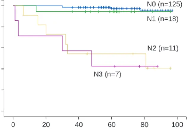
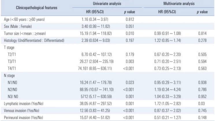
important prognostic factor (Table 3). In this study, the OS rates according to N stage were 96.8% for N0, 94.4% for N1, 45.5% for N2, and 42.9% for N3 ( p<0.001) (Fig. 3). The factors that influenced disease recurrence included most of these same factors (Table 4). However, the most powerful factor for disease recurrence was the presence of lymphatic invasion (Table 5).
Table 2. Five-year survival rate of patients with laparoscopy-assisted gastrectomy
Patients characteristic Five year survival rate (%) p value
Age (<60 years : ≥ 60 years) 95% : 89.3% 0.105
Type of operation (BI : BII : Total) 93.68 : 78.79 : 100 0.006
Extent of LN dissection (D1+a : D1+b : D2) 100 : 95.24 : 85.57 0.152
Tumor size (<mean : ≥mean) 96.77 : 85.29 0.007
Histology (Differentiated : Undifferentiated) 90.67 : 94.08 0.606
T stage (T1 : T2 : T3 : T4) 97.37 : 83.33 : 89.47 : 50 <0.001
N stage (N0 : N1 : N2 : N3) 98.40 : 94.44 : 71.43 : 42.86 <0.001
TNM stage (I : II : III : IV) 97.48 : 96.30 : 42.86 : - <0.001
Lymphatic invasion (absence : presence) 97.54 : 74.36 <0.001
Venous invasion (absence : presence) 94.63 : 58.33 <0.001
Perineural invasion (absence : presence) 50.73% : 51.52 0.283
Table 3. Univariate and multivariate analyses of risk factors for five-year survival after laparoscopy-assisted gastrectomy
Clinicopathological features Univariate analysis Multivariate analysis
HR (95%CI) p value HR (95%CI) p value
Age (<60 years : ≥60 years) 2.7 (0.8~9.7) 0.120
Sex (Male : Female) 1.6 (0.6~4.4) 0.360
Tumor size (<mean : ≥ mean) 4.29 (1.4~13.6) 0.010 0.9 (.62~1.50) 0.874
Histology (Undifferentiated : Differentiated) 1.30 (0.5~3.6) 0.610 0.3 (.049~1.43) 0.123 T stage
T2/T1 4.71 (1.051~21.069) 0.040 3.5 (.41~29.89) 0.253
T3/T1 3.25 (0.595~17.779) 0.140 1.2 (.08~16.42) 0.910
T4/T1 20.28 (5.712~72.011) <0.001 5.1 (.41~64.15) 0.206
N stage
N1/N0 2.02 (0.225~18.170) 0.530 0.83 (.06~11.37) 0.888
N2/N0 20.84 (5.854~74.160) <0.001 26.4 (2.26~308.99) 0.009
N3/N0 26.91 (6.702~108.015) <0.001 9.9 (0.58~171.32) 0.113
Lymphatic invasion (Yes/No) 10.09 (3.209~31.698) <0.001 2.13 (0.26~17.44) 0.480
Venous invasion (Yes/No) 9.08 (3.204~25.746) <0.001 1.79 (0.19~17.21) 0.613
Perineural invasion (Yes/No) 5.27 (1.87~14.85). 0.002 0.48 (0.05~4.33) 0.625
DISCUSSION
Since Kitano et al.
11first applied and described laparoscopic gastrectomy in 1994, there has been significant debate over the use of laparoscopic surgery in gastric cancer. Laparoscopic approaches in early gastric cancer have been shown to improve recovery time, lessen pain, result in faster return of gastrointestinal function, show cosmetic benefit, and result in better pulmonary function.
7-9,12Several studies have also demonstrated the efficacy of laparoscopic surgery and oncologic safety in EGC.
8,12,13However, there is still debate about LAG in advanced gastric cancer.
3,4The major concerns with the laparoscopic approach are the completeness of oncologic resection and D2 lymphadenectomy in advanced
gastric cancer. The aim of study was to evaluate long-term outcomes and survival of patients with LAG including ad- vanced gastric cancer.
The presence of lymph node metastases in EGC worsens prognosis, as reported by Kitamura et al.
14and Maehara et al.
15Also, several studies have shown that adequate lymph node dissection is the most important procedure.
16,17The majority of patients in this study were treated using laparoscopic D2 lymph node dissection except for high-risk patients with cardiopulmonary disease and old age (>75 years). Instead, D1+b lymph node dissection conducted for high-risk patients.
The mean number of retrieved LNs was 30±6 (range, 6~68, median 31.1) per patient, comparable to the 20~40 retrieved LNs reported by previous studies on the use of open conventional gastrectomy and laparoscopic gastrectomy.
17-20According to a multicenter, prospective, randomized clinical study of the Korean Laparoscopic Gastrointestinal Surgical Society that included 179 LAG patients, LAG for gastric cancer has an 11.6% early morbidity and 1% mortality rate.
21Lin et al.
18reported a morbidity rate of 13.6% and mortality rate of 0.3%. In the present study, the mortality and morbidity rates were 0.62% and 9.31%, respectively. Compared to the results of previous studies,
17-21the surgical outcome of LAG for gastric cancer including advanced gastric cancer in the present study was technically comparable.
Pak et al.
22analyzed 714 patients who underwent LAG for gastric cancer and reported five-year OS rates of 96.4%
in stage I, 83.1% in stage II, and 50.2% in stage III and five- year DFS rates of 95.8% in stage I, 83.4% in stage II, and 46.4% in stage III. Kitano et al.
23also reported on the long- Fig. 3. Overall survival curves according to N stage.
0 0
100
Survivalrate(%)
1.0
0.8
0.6
0.4
0.2
20 40 60 80
Survival period (months) N3 (n=7)
N2 (n=11) N1 (n=18) N0 (n=125)
Table 4. Disease-free survival rates of patients who underwent laparoscopy-assisted gastrectomy
Patient characteristic Disease-free survival rate (%) p value
Age (<60 years : ≥60 years) 93.75 : 92.78 0.811
Se (Male : Female) 96.19 : 94.23 0.037
Type of operation (BI : BII : Total) 92.63 : 90.90 : 96.97 0.562
Extent of LN* dissection (D1+a : D1+b : D2) 100 : 96.43 : 88.06 0.103
Tumor size (<mean : ≥ mean) 98.92 : 85.29 0.001
Histology (Undifferentiated : differentiated) 90.70 : 96.00 0.183
T stage (T1 : T2 : T3 : T4) 99.12 : 94.44 : 78.95 : 50.00 <0.001
N stage (N0 : N1 : N2 : N3) 99.20 : 88.89 : 45.45 : 71.43 <0.001
TNM stage (I : II : III : IV) 100 : 88.89 : 42.45 : - <0.001
Lymphatic invasion (absence : presence) 99.18 : 74.36 <0.001
Venous invasion (absence : presence) 95.97 : 58.33 <0.001
Perineural invasion (absence : presence) 92.00 : 63.16 0.267
*Lymph node.
term outcome of laparoscopic gastrectomy in a retrospective multicenter study on LAG for early gastric cancer, noting five- year DFS rates of 99.8, 98.7, and 85.7% for stages IA, IB, and II, respectively. This present study showed OS of stage I, II, and III of 96.63, 96.30, and 35.71%, respectively, and DFS of 100% for stage I, 88.89% for stage II and 50% for stage III.
Compared with multicenter studies,
21-24the present OS and the DFS were comparable. The oncologic outcome is similar to that of conventional open surgeries and previous laparoscopic surgeries. Therefore, these results indicate that laparoscopic gastrectomy has acceptable long-term oncologic outcomes for LAG in gastric cancer.
Based on Table 3 results, only N2 stage was a significant prognostic factor for the five-year OS. The data accumulated n the hospital show that the number of patients with greater than stage III disease was too small, and that most patients were in a relatively early stage. For this reason, it is assumed that N1 stage and N3 stage were not a significant factor for the five-year OS. Therefore, it is yet to be discussed to regard N stage as a prognostic factor for five-year OS.
Table 4 shows the factors that influenced disease recurrence.
The recurrence site varied, with peritoneum (n=4) being the most common recurrent site, and lymph node as the second most recurrent organ (n=3). These recurrent patterns are similar to those of a previous open gastrectomy study.
25Table 5 shows the prognostic factors for recurrence of gastric cancer.
Previous studies
14,15have shown that the presence of lymph node metastases in gastric cancer worsens prognosis. In the present study, lymphatic invasion was the most powerful prognostic factor of disease recurrence.
There were some limitations associated with this present study. There was no open control group against which to evaluate the safety of laparoscopic surgery compared to open gastrectomy. The data were retrospectively collected and reviewed. To overcome these limitations, a multicenter, pro- spective and randomized controlled study is necessary.
In conclusion, the present study shows that LAG for gastric cancer including advanced gastric cancer is technically feasible and has acceptable long-term oncologic outcomes.
REFERENCES
1) Roder DM. The epidemiology of gastric cancer. Gastric Cancer 2002;5 Suppl 1:5-11.
2) Lee SR, Kim HO, Son BH. Long-term outcomes and survival of patients undergoing laparoscopy-assisted distal gastrectomy for gastric cancer. J Laparoendosc Adv Surg Tech A 2012;22:432-437.
3) Mochiki E, Nakabayashi T, Kamimura H, Haga N, Asao T, Kuwano H. Gastrointestinal recovery and outcome after laparoscopy-assisted versus conventional open distal gastrectomy for early gastric cancer. World J Surg 2002;26:1145-1149.
4) Chen K, Xu XW, Mou YP, et al. Systematic review and meta-
Table 5. Univariate and multivariate analyses of risk factors for disease-free survival after laparoscopy-assisted gastrectomy
Clinicopathological features Univariate analysis Multivariate analysis
HR (95%CI) p value HR (95%CI) p value
Age (<60 years : ≥60 years) 1.16 (0.34~3.97) 0.812
Sex (Male : Female) 3.40 (0.99~11.62) 0.051
Tumor size (<mean : ≥mean) 15.19 (1.94~118.82) 0.010 0.99 (0.91~1.08) 0.814
Histology (Undifferentiated : Differentiated) 2.39 (0.634~9.03) 0.197 1.22 (0.85~1.74) 0.278 T stage
T2/T1 6.70 (0.42~107.12) 0.179 0.67 (0.20~2.20) 0.505
T3/T1 26.27 (2.934~235.19) 0.003 0.71 (0.20~2.51) 0.594
T4/T1 74.161 (8.65~636.11) <0.001 0.73 (0.25~2.13) 0.563
N stage
N1/N0 16.24 (1.47~179.78) 0.023 0.95 (0.29~3.11) 0.938
N2/N0 88.95 (10.67~741.10) <0.001 1.19 (0.34~4.24) 0.786
N3/ N0 5712 (5.17~630.59) 0.001 1.04 (0.33~3.29) 0.952
Lymphatic invasion (Yes/No) 38.05 (4.87~297.52) 0.001 1.72 (1.05~2.82) 0.03
Venous invasion (Yes/No) 12.56 (3.83~41.25) <0.001 0.87 (0.37~2.02) 0.745
Perineural invasion (Yes/No) 15.07 (4.40~51.62) <0.001 0.51 (0.21~1.27) 0.148
analysis of laparoscopic and open gastrectomy for advanced gastric cancer. World J Surg Oncol 2013;11:182.
5) Hur H, Jeon HM, Kim W. Laparoscopy-assisted distal gastrectomy with D2 lymphadenectomy for T2b advanced gastric cancers:
three years' experience. J Surg Oncol 2008;98:515-519.
6) Kawamura H, Homma S, Yokota R, et al. Inspection of safety and accuracy of D2 lymph node dissection in laparoscopy-assisted distal gastrectomy. World J Surg 2008;32:2366-2370.
7) Han HS, Kim YW, Yi NJ, Fleischer GD. Laparoscopy-assisted D2 subtotal gastrectomy in early gastric cancer. Surg Laparosc Endosc Percutan Tech 2003;13:361-365.
8) Lee JH, Han HS, Lee JH. A prospective randomized study com- paring open vs laparoscopy-assisted distal gastrectomy in early gastric cancer: early results. Surg Endosc 2005;19:168-173.
9) Asao T, Hosouchi Y, Nakabayashi T, Haga N, Mochiki E, Kuwano H. Laparoscopically assisted total or distal gastrectomy with lymph node dissection for early gastric cancer. Br J Surg 2001;88:128- 132.
10) Parkin DM, Bray FI, Devesa SS. Cancer burden in the year 2000.
The global picture. Eur J Cancer 2001;37 Suppl 8:S4-66.
11) Kitano S, Iso Y, Moriyama M, Sugimachi K. Laparoscopy-assisted Billroth I gastrectomy. Surg Laparosc Endosc 1994;4:146-148.
12) Kitano S, Shiraishi N, Fujii K, Yasuda K, Inomata M, Adachi Y.
A randomized controlled trial comparing open vs laparoscopy- assisted distal gastrectomy for the treatment of early gastric cancer:
an interim report. Surgery 2002;131:S306-311.
13) Lee JH, Yom CK, Han HS. Comparison of long-term outcomes of laparoscopy-assisted and open distal gastrectomy for early gastric cancer. Surg Endosc 2009;23:1759-1763.
14) Kitamura K, Yamaguchi T, Taniguchi H, Hagiwara A, Sawai K, Takahashi T. Analysis of lymph node metastasis in early gastric cancer: rationale of limited surgery. J Surg Oncol 1997;64:42-47.
15) Maehara Y, Orita H, Okuyama T, et al. Predictors of lymph node metastasis in early gastric cancer. Br J Surg 1992;79:245-247.
16) Zheng Z, Liu Y, Bu Z, et al. Prognostic role of lymph node metastasis in early gastric cancer. Chin J Cancer Res 2014;26:192- 199.
17) Tanimura S, Higashino M, Fukunaga Y, et al. Laparoscopic distal gastrectomy with regional lymph node dissection for gastric cancer. Surg Endosc 2005;19:1177-1181.
18) Lin JX, Huang CM, Zheng CH, et al. Surgical outcomes of 2041 consecutive laparoscopic gastrectomy procedures for gastric cancer:
a large-scale case control study. PLoS One 2015;10:e0114948.
19) Mochiki E, Kamiyama Y, Aihara R, Nakabayashi T, Asao T, Kuwano H. Laparoscopic assisted distal gastrectomy for early gastric cancer: Five years' experience. Surgery 2005;137:317-322.
20) Noshiro H, Nagai E, Shimizu S, Uchiyama A, Tanaka M.
Laparoscopically assisted distal gastrectomy with standard radical lymph node dissection for gastric cancer. Surg Endosc 2005;19:
1592-1596.
21) Kim HH, Hyung WJ, Cho GS, et al. Morbidity and mortality of laparoscopic gastrectomy versus open gastrectomy for gastric cancer: an interim report--a phase III multicenter, prospective, randomized Trial (KLASS Trial). Ann Surg 2010;251:417-420.
22) Pak KH, Hyung WJ, Son T, et al. Long-term oncologic outcomes of 714 consecutive laparoscopic gastrectomies for gastric cancer:
results from the 7-year experience of a single institute. Surg Endosc 2012;26:130-136.
23) Kitano S, Shiraishi N, Uyama I, Sugihara K, Tanigawa N. A multi- center study on oncologic outcome of laparoscopic gastrectomy for early cancer in Japan. Ann Surg 2007;245:68-72.
24) Yoo HM, Lee HH, Shim JH, et al. Long-term outcomes and survival after laparoscopy-assisted distal gastrectomy for gastric cancer: three-year survival analysis of a single-center experience in Korea. J Surg Oncol 2011;104:511-515.
25) Yoo CH, Noh SH, Shin DW, Choi SH, Min JS. Recurrence following curative resection for gastric carcinoma. Br J Surg 2000;
87:236-242.