CASE REPORT pISSN 1225-7737/eISSN 2234-8042 http://dx.doi.org/10.12701/yujm.2014.31.2.144
Yeungnam Univ J Med 2014;31(2):144-147144 YUJM VOLUME 31, NUMBER 2, DECEMBER 2014
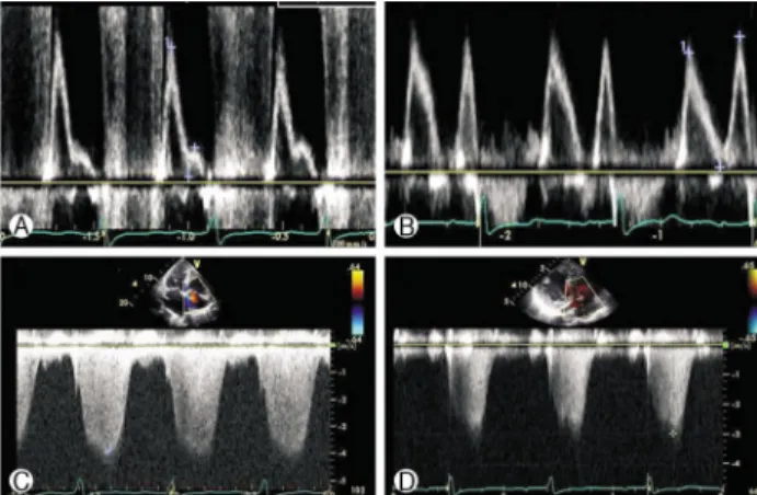
Successful emergency transcatheter aortic valve implantation
Jung-Hee Lee
1, Ah-Young Ji
1, Young Ju Kim
1, Changho Song
1, Moo-Nyun Jin
1, Sun Wook Kim
1, Myeong-Ki Hong
2, Geu-Ru Hong
11
Division of Cardiology, Yonsei University College of Medicine, Severance Cardiovascular Hospital, Yonsei University Health System;
2