Ⅰ.서 론
고립성 섬유종양은 1931년 흉막의 spindle cell neo- plasm으로 처음 보고 되었다1). 드물게 발생하는 신생물로 호발하는 부위는 흉막이며, 구강내에서 발견되는 경우는 더 욱 드물다2). 2004년 Pizzolitto 등은 1997년까지 흉막을 제외한 곳에서 발견된 Solitary fibrous tumor (SFT)는 31개라고 보고한 바 있고, 이후 2008년 Fusconi등이 문헌 고찰을 통해 구강내에서 발견된 SFT는 49 증례라고 보고 하였다3,4). 감별진단을 위해서 면역형광염색법을 사용하고 이때 사용하는 factor로는 CD34, Bcl-2, vimentin등이 있 다5-9). 종양과 연관된 부위에 따라 nasal obstruction같은 증상이 생길 수 있으며, hypoglycemia, arthralgia, osteoarthropathy, clubbing등의 전신 증상이 있을 수 있 으나 임상적, 혹은 방사선학적으로 특이 소견이 나타나지 않는 경우도 있다10).
본 교실에 협부에 발생한 종물을 주소로 내원한 60대 남
자환자를 고립성 섬유종양으로 진단하고 치험한 예가 있어 보고하고자 한다.
Ⅱ.증례보고
60대 남자환자가 좌측 협부에 발생한 종물을 주소로 하여 내원하였다. 2개월 전부터 종물이 커지기 시작했고, 초진시 왼쪽 볼이 약간 볼록하게 올라와 있는 듯이 보였으며, 종물 을 덮고 있는 상부 피부는 정상 피부조직이었다. 구외 촉진 시 약간 단단한 느낌의 연조직 덩어리가 만져졌고, 연조직 덩어리의 움직임은 없었다. 구강내에는 2cm가량의 종물이 좌측 협점막에 정상 점막 보다 약간 보랏빛을 띄는 점막으 로 덮여 있었다(Fig.1).
파노라마 방사선사진상에서는 특이 소견이 없었으며, 조 영제를 사용한 컴퓨터 단층촬영에서는 하악 좌측 구치부쪽 에 난원형의 잘 경계지워진 병소가 조영 증강된 상태로 보 였다(Fig.2).
최민혜∙윤규호∙정정권∙박관수∙신재명,∙김해린∙나혜정 인제대학교 상계백병원 구강악안면외과학교실
협부에 발생한 고립성 섬유 종양 : 증례보고
SOLITARY FIBROUS TUMOR IN BUCCAL CHEEK : CASE REPORT
Min-hye Choi, Kyu-ho Yoon, Jeong-kwon Cheong, Kwan-soo Park, Jae-myung Shin, Hae-lin Kim, Hye-jung Na Department of Oral and Maxillofacial Surgery, Inje University Sanggye -Paik Hospital
Solitary fibrous tumor (SFT) is a neoplasm that arises most commonly in pleura. Although SFT occasion- ally occurs in extrapleural locations, the incidence in the oral cavity is rare. SFT is benign in almost cases and surgical excision is the effective treatment. SFT occurred in the left cheek of a 60-year-old man pre- sented with a painless submucosal mass. The tumor was surgically removed. Immunohistochemical study showed that tumoral cells were negative for SMA, S-100, but positive for Bcl-2, CD34. SFT is easily over- diagnosed if strict criteria are not carefully applied, and strict diagnostic criteria are necessary to avoid confusion of SFT with more aggressive lesions.
Key words: Solitary fibrous tumor, CD 34, Bcl-2 Abstract
통상적인 방법으로 국소마취를 시행한 후 병소를 완전히 절제하고 봉합하였으며, 조직 검사를 시행하였다. 떼어낸 조직은 fibrous capsule로 덮여 있었으며, 잘 경계지워져 있었다(Fig.3,4). 3개월 후 경과 관찰하였으며, 특이소견은 없었다(Fig.5).
떼어낸 조직은 HE 염색과 면역 형광 염색을 하였다. HE 염색에서는 잘 경계지워진 섬유피막 (fibrous capsulation)
과 stag-horn을 관찰할 수 있었으며, 작은 fusiform cell 과 spindle cell이 pericytic pattern 및 특별한 규칙이 없이 흩어져 있는 모양이 혼재된 양상을 보였으며, 그 사이로 교 원섬유속(collagen bundle)이 발견되었다(Fig.6,7,8). 면 역형광염색법에서는 Bcl-2와 CD34에 양성반응, S-100과 SMA에 음성반응을 보였다(Fig.9,10). 수술 후 염증소견이 나 재발의 양상은 보이지 않았다.
Fig. 2.Preoperative CT scan showing a well-demarcated mass of the tumor.
Fig. 1.Preoperative photographs showing a tumor of left buccal mucosa.
Fig. 3.Post operative intrao- Fig. 4.Two pieces of specimen. Fig. 5.Post operative photograph after 3 months.
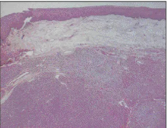
Fig. 6.Well-circumscribed nodular lesion without capsula- tion.(HE, ×10)
Fig. 7.Focally prominent staghorn vessels, Pericytic pattern, patternless pattern consisting of small fusiform and spindled cells.(HE, ×40)
Fig. 8. Hypocellular proliferation of plump spindle cells with colla- gen production and characteristic metaplastic woven bone forma- tion without osteoblastic rimming.(HE, ×100)
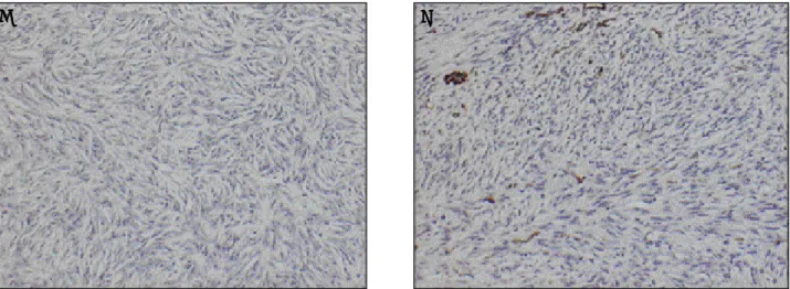
Fig. 9.IHC microphotographs of SFT. A. Positive reaction to CD34. B. Positive reaction to Bcl-2.
CD34, Bcl-2:positive reaction. S-100, SMA: negative reaction. C-kit: negative reaction. Ki-67: <1%
A
A BB
Ⅲ.고찰 및 결론
SFT는 드물게 발생하는 신생물로 1931년에 흉막 중피종 으로 처음 보고되었으며. 이전에는 localized benign mesothelioma, submesothelioma, localized fibrous tumor of pleura로 명명되기도 하였다1). SFT는 intersti- tial stem cell에서 발생하는 것으로 CD34 혹은 다른 fac- tor들을 사용함으로써 면역형광염색법으로 조직학적으로 진단할 수 있다5-9). 드물게 발생하기 때문에 감별 진단시에 배제되기 쉬우며, 감별진단할 병소로는 fibrous histiocy- toma, fibroma, schwannoma, myofibroma 같은 spin- dle cell neoplasia가 있다.
2006년 WHO 분류에서는 haemangiopericytoma 와 SFT는 구별하기 힘들다고 하였다11). SFT 중에서 두경부 영역에 발생하는 경우는 10%, 구강내 발생하는 것은 3%
정도이고, 구강내 호발부위는 협점막, 혀, 경구개이며 이중 협점막에서 가장 많이 발생한다12,13). Shimoyama등은 SFT 48증례를 분석한 결과 평균 발생 연령은 51.3세이고, 병소 의 평균적인 크기는 19.7mm라고 하였다14).
감별 진단을 하기 위해서는 HE 염색과 면역형광법을 사 용한다. HE 염색을 통해서 SFT에서 나타나는 세포배열 양 상을 보는데, 조직학적으로 비교적 일정한 형태와 크기의 방추형의 세포들이 특정한 형식 없이 배열하거나, 소용돌이 치는 성장 양식, 혈관중피종에서 관찰되는 확장된 혈관의 모습, 섬유육종 모양의 성장, 세포 밀도가 적고 호산성의 섬 유질이 풍부한 기질 등 다양한 소견들이 섞여서 관찰되는 것이 특징이다15).
면역형광 염색법으로 SFT를 진단하기 위한 요소로 Vimentin, CD34, S-100, SMA, Bcl-2, XIIIa factor등을 사용하고 있는데 명확히 규정된 방법은 없고 이들 중 몇가
지 factor들을 조합해서 사용하고 있다. 문헌에 보고된 바에 따르면 Vimentin, CD34에 양성반응을 보이는 경우, Vimentin, CD34, Bcl-2에 양성반응을 보이는 경우, CD34, Bcl-2에 양성반응을 보이는 경우 모두 SFT로 진단 하였다. Fusconi등은 spindle cell origin을 명확하게 규정지 으려면 Factor XIIIa혹은 collagen에 대한 면역형광염색법 이 필요함을 보고하였다4). SFT에는 mast cell이 풍부하고, 이것은 CD34 positive pluripotential cell에서 기원하기 때 문에 면역형광염색법에서 CD34에 강한 양성을 보인다5).
본 증례에서는 면역 형광 염색법을 한 결과 S-100과 SMA에 음성반응으로 나타나 신경 혹은 근육에서 기원한 병소를 배제하였고, CD 34와 Bcl-2에 양성 반응을 보이면 서 HE 염색시 방추모양의 세포가 밀집한 부분과 세포가 적 은 부분이 구분되어 나타나는 양상을 보이고 세포사이에 staghorn vessel이 있으면서 주변으로 collagen fiber 들이 나타나 SFT로 진단하였다.
SFT의 치료는 단순절제이고, 대부분이 양성종양으로 예 후는 좋은 것으로 알려져 있다. Jham등은 구강내에 발생한 SFT는 1증례를 제외하고 모두 양성이라고 보고하였으며, 악성으로 보고된 1증례는 혀에 발생한 것으로 2003년에 Shnayder등이 보고하였다13,16). Chan 은 SFT 의 diag- nostic criteria에서 sarcomatous area가 발견되거나 sar- coma가 있으면 악성인 것으로 간주한다고 하였다17). 본 증 례에서는 이러한 악성의 특성은 보이지 않았다.
본 증례는 왼쪽 협부에 발생한 종물을 외과적으로 절제한 후 HE염색과 CD34, Bcl-2, SMA, S-100을 이용한 면역형 광염색법을 통해 감별 진단하여 SFT로 확진하여 이를 보고 하는 바이다. 흔하지 않게 발생하는 질병으로 연구 발표가 많 지 않아 앞으로 이에 대한 연구가 더 필요할 것으로 보인다.
Fig. 10.IHC microphotographs of SFT. A. Negative reaction to S-100. B. Negative reaction to SMA.
CD34, Bcl-2:positive reaction. S-100, SMA: negative reaction. C-kit: negative reaction. Ki-67: <1%
A
A BB
References
1. Klemperer P, Rabin CB. Primary neoplasms of the pleura : A report of five cases. Arch Pathol 11: 385, 1931.
2. Sakamoto K, Sawai T, Yamaguchi A : Solitary fibrous tumor of the tongue. Oral Oncol extra. 41: 222, 2005.
3. Pizzolitto S, Falconieri G, DeMaglio G : Solitary fibrous tumor of the spinal cord : a clinicopathologic study of two cases. Ann Diagn Pathol 8: 268, 2004.
4. Fusconi M, Ciofalo A, Greco A et al : Solitary fibrous tumor of the oral cavity : case report and pathologic con- sideration. J Oral Maxillofac Surg 66: 530, 2008.
5. Veltrini VC, Etges A, Maglahaes MHCG et al : Solitary fibrous tumor of the oral mucosa morphological and immunohistochemical profile in the differential diagnosis with hemangiopericytoma. Oral Oncol 39: 420, 2003.
6. Garcia RG, Usandizaga JLGD, Nam SH et al : Solitary fibrous tumour of the oral cavity with histological features of aggressiveness. Br J Oral Maxillofac Surg 44: 543, 2006.
7. Macarenco RS, Bacchi CE, Domingues MAC : Solitary fibrous tumor with atypical histological features occurring in the palatine tonsil : an uncommon neoplasm in an uncommon site. J Oral Pathol Med 35: 602, 2006.
8. Muzio LL, Mascolo M, Capodiferro S et al : Slitary fibrous tumor of the oral cavity : the need for an extensive sam- pling for a correct diagnosis. J Oral Pathol Med 36: 538, 2007.
9. Alawi F, Stratton D, Freedman PD : Solitary fibrous tumor of the oral soft tissue. Am J Surg Pathol 25: 900, 2001.
10. Galioto S, Valentini V, Fatone FMG et al : Solitary fibrous tumours of the infratemporal fossa. Two cass reports. J Craniomaxillofac Surg 34: 494, 2006.
11. Fletcher CDM : The evolving classification of soft tissue tumours : an update based on the new WHO classifica- tion. Histopathol 48: 3, 2006.
12. Swelam W, Hiroko IY, Satoshi M et al : Solitary fibrous tumor of the lower lip involving minor salivary gland com- ponent : Report of a case and review of the literature of salivary gland cases. Oral Oncol extra 40: 107, 2004.
13. Jham BC, Salles JMP, Soares JMA et al : Solitary fibrous tumour of the buccal mucosa : case report and review of the literature. Br J Oral Maxillofac Surg 45: 232, 2007.
14. Shimoyama T, Horie N, Ide F : Solitary fibrous tumor of the palate : A case report and review of the literature. J Oral Maxillofac Surg 62: 895, 2004.
15. Woo SM, Lee KM : Solitary fibrous tumor clinico-patholog- ical analysis of 11cases. J Korean Cancer Assoc 31: 847, 1999.
16. Shnayder Y, Greenfield BJ. Oweity T et al : Malignant solitary fibrous tumor of the tongue. Am J Otolaryngol 24:
246, 2003.
17. Chan JK : Solitary fibrous tumor-everywhere, and a diag- nosis in vogue. Histopathol 31: 568, 1997.
저자 연락처
우편번호 139-707
서울특별시 노원구 상계7동 761-1
인제대학교 상계백병원 구강악안면외과학교실 박 관 수
원고 접수일 2009년 04월 08일 게재 확정일 2009년 05월 20일
Reprint Requests Kwan-Soo Park
Dept. of OMFS, Sanggye Paik Hospital, Inje University 761-1 Sanggye 7-dong Nowon-gu, Seoul, 139-707, Korea Tel: 82-2-950-1167
E-mail: [email protected] Paper received 04월 08일 Paper accepted 05월 20일