Ⅰ. 서 론
양성 섬유성 조직구종은 섬유모세포와 조직구세포의 분화 를 보이는 간엽 세포에서 유래된 종양으로,
1)1967년 Stout 와 Lattes가 연조직의 양성 섬유성 조직구종에 대하여 처음 기술하였으며,
2)1972년 Kempson과 Kyriakos가 섬유아 세포와 조직구로 구성된 종양을 섬유성조직구종이라 명칭 하고 이들을 양성과 악성으로 구분하였다
3).
양성 섬유성 조직구종은 주로 태양에 노출된 사지의 피부 에서 발생하며,
2)발생 빈도는 하지 50%, 상지 20%, 후복 막 20%, 안와 부위 순이다. 골에 발생하는 경우는 드물며, 주로 발생하는 부위는 골반, 대퇴골, 경골이다
4-6). 양성 섬유 성 조직구종이 악골에서 발생하는 경우는 극히 드물며,
7-11)구강에서 발생하는 경우의 대부분이 연조직에 발생한다.
본 교실에서는 하악에 발생한 양성 섬유성 조직구종을 치 험하였기에 임상적, 방사선학적, 조직학적인 소견을 문헌 고찰과 함께 보고한다.
Ⅱ. 증례 보고
32세 여자 환자가 본원 내원 2주전 하악 우측부위 우리한 통증을 주소로 타병원에서 방사선 사진을 촬영한 후 우측 하악골 부위 광범위한 방사선 투과성 병소로 조직검사를 시 행하였다. 채취한 조직이 검사에 적절하지 못해 조직검사 결과가 나오지 않아 재조직 검사 및 치료를 위해 본원으로 의뢰되었다.
환자 병력상 특별한 기왕력은 없었다. 일반 혈액 검사, 간 기능 검사, 신기능 검사, 혈당, 전해질, 뇨검사, 심전도 검 최소영∙김진욱∙권대근∙신홍인*∙변기정**∙김진수
경북대학교 치과대학 구강악안면외과학교실, *경조직-바이오치아 재생 연구소, 구강병리학교실
**울산대학교병원 치과
하악골에 발생한 양성 섬유성 조직구종의 치험례
BENIGN FIBROUS HISTIOCYTOMA OF MANDIBLE - A CASE REPORT-
So-Young Choi, Jin-Wook Kim, Tae-Geon Kwon, Hong-In Shin
*
, Ki-Jeong Byeon**
, Chin-Soo KimDepartment of Oral & Maxillofacial Surgery,
*IHBR, Dept. of Oral Pathology, School of Dentistry, Kyungpook National University
**Department of Dentistry, Ulsan University Hospital
Benign fibrous histiocytoma(BFH) is a mesenchymal cell-originated tumor composed of cells with fibrob- lastic and histiocytic differentiation. BFH occurs predominantly on sun-exposed skin of extremities. Oral BFH lesions are uncommon. The majority of oral lesions includes the soft tissue but not the jaw bones. The lesion appears as well-defined multilocular radiolucencies associated with bony swelling when it occurs on the jaw. The lesion induces the thinning and expansion of the cortex and shows many thin, indistinct septa in the lesion. Surgical excision is the choice of treatment. The recurrence rate is low and metastasis has not been reported. We report the clinical, radiographic and microscopic findings of a BFH case occurred in the mandible with literature reviews.
Key words: Benign fibrous histiocytoma(BFH)
Abstract사, 흉부 방사선 검사 등은 정상범주였다.
방사선 사진상에 부분적으로 경계가 구분이 되나 전반적 으로 불명확한 경계를 보이는 다방성의 방사선투과성 병소 가 하악지, 근돌기, 하악과두 경부에서 관찰되며 하악지 전 연으로의 비박과 팽윤을 동반하고 병소에 이환된 하악관의 피질골성 벽이 소실되어 보인다. 컴퓨터 단층 촬영상에는 두께가 불균일한 협설측 피질골의 비박과 팽윤이 관찰되며 부분적인 천공이 관찰된다(Fig. 1). 술 전 조직 검사를 시행 하였으며 조직 검사 결과 양성 섬유성 조직구종으로 진단되 어 전신 마취하에 하악 우측 제1대구치를 발치하면서 제 1
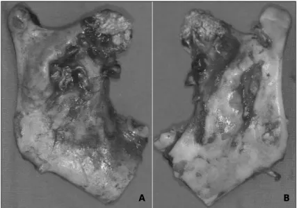
대구치 후방부에서 편측 하악골 절제술을 시행하고 선부자 를 통해 악관고정을 시행하였다. 종물은 피질골의 비박과 팽윤이 관찰되었으며 하악지 부위와 오훼돌기 부위에 피직 골 천공이 부분적으로 관찰되었다(Fig. 2).
조직학적 소견은 소포성 핵을 가진 방추형의 잘 분화된 섬 유모세포가 나선형으로 배열을 이루고 있다. 면역조직화학 적 검사상 산재한 조직구 형태의 세포들이 CD68에 양성반 응을 보이고 있다(Fig. 3).
술 후 9개월간 계속적인 추적 관찰 중이며 현재까지 재발 은 없는 상태이다. 저작에 대한 특별한 불편감을 호소하지
Fig. 1.A. Panoramic radiograph showing ill-defined multilocular radiolucent lesion on right ramal, coronoid process areas. B. Computed tomogram showing bucco-lingual expainsion with thinning and partial perforation of the cortical bone.
A B
Fig. 2.Specimen showing bucco-lingual expainsion with thinning of the cortical bone. Notice the focal perforation of ramal and coronoid process areas.
A. Outer surface, B. Inner surface
A B
는 않고 있지만 우측 안면부가 함몰되어 보여 외관상의 이 유로 하악골 절제부위에 대한 재건술을 계획하고 있다.
Ⅲ. 고 찰
섬유성 조직구종은 지질을 함유하는 조직구와 방추형의 섬유아세포가 다양한 비율로 구성되는 연조직 종양으로 사 지부의 피부에 호발한다. 두경부에서는 비교적 드물게 발생 하며 대부분 양성이다. 심부조직에 발생시 예후가 불량하며 국소적인 침윤과 재발을 나타낸다
12). 골조직에 발생하는 섬 유성 조직구종은 드물며, 주로 장골에서 발생된다. 악골에 발생하여 보고된 증례는 상악에 1증례, 하악에 5증례로 두 개골과 안면골에 발생하는 경우는 매우 드물다(Table 1).
골조직에 발생한 경우 방사선 소견상 경계가 뚜렷한 방사 선 투과성의 병소로 피질골의 비박 및 팽윤은 있으나 골막 반응이나 종양의 석회화 소견이 없는 것이 특징이다. 본 증 례와 같이 경계가 불명확한 방사선 투과성 병소를 가지며, 피질골의 천공과 벌레먹은 양상(moth-eaten)이 관찰되거 나, 인접 정상 골조직으로의 침윤을 보일 경우 악성 병소로 오인될 수 있으며, 예후가 불량할 것으로 예측할 수 있다
13).
섬유성 조직구종은 다양한 조직학적 소견 및 명칭이 있는 데 1967년 Stout와 Lattes가 연조직의 양성 섬유성 조직 구종에 대하여 처음 기술하였으며,
2)1972년 Kempson과 Kyriakos가 섬유아세포와 조직구로 구성된 종양을 섬유성 조직구종이라 명칭하고 이들을 양성과 약성으로 구분하였 다
3). 1967년 Stout와 Lattes로 인해 연조직 기원의 섬유성 조직구종에 대한 분류를 더 잘 이해할 수 있었지만 골에서 발생할 경우 정확한 분류는 여전히 논란이 되고 있다. 1989 년 Cale이 골에 발생한 섬유성 조직구종을 분류하기 위해
새로운 분류 체계를 제안하였다(Table 2)
7). Cale의 분류에 따르면 본 증례는 양성분류에서 소분류인 양성 섬유성 조직 구종에 속한다.
섬유성 조직구종은 일반적으로 성숙 교원질을 합성하는 길고 방추형의 섬유모세포와 조직구를 나타내는 난원형 핵 과 풍부한 세포질을 갖는 큰 세포로 구성된다. 변연부는 종 종 침윤성이지만 부분적으로 피막에 싸여있거나 경계가 잘 되어 있다. 조직내에 좀 더 표면쪽에 위치한 병소는 섬유모 세포성 세포들이 바람개비(pinwheel) 모양의 배열을 보이 는데 이를 회오리형(storiform pattern)이라 하며, 섬유조 직구 병소의 특징이다. 감수분열은 흔지 않으며 조직구 세 포가 다형핵을 보일 수 있다. 세포질내 지방의 존재로 인해 거품 세포질을 가진 황색종 세포(xanthoma cell)를 관찰 할 수 있다.
정확한 진단을 위해서는 면역조직화학적 검사가 도움이 된다. 면역조직 화학적 검사중 조직구에 특이적 반응을 보 이는 CD68이 섬유성 조직구종 진단에 도움이 된다. 본 증 례의 경우 조직구양 세포들이 CD68 면역 염색에는 양성반 응을 보였고, trichrome 염색에서 섬유모세포들이 푸른색 으로 염색을 보여 섬유성 조직구종으로 진단되었다.
섬유성 조직구종의 치료는 광범위한 외과적 절제가 선택 적 치료이다. 완전한 절제가 이루어진 경우, 재발율은 낮으 며 두경부 영역에서의 전이는 아직 보고되어 있지 않다
14). Bielamowicz 등
15)의 보고에 의하면 국소적 절제술을 시행 한 양성 섬유성 조직구종의 18명의 환자에서 2명(11%)의 재발을 보였으며, Clarke 등
4)의 보고에 의하면 8명의 환자 중 3명이 재발을 보였다. 이들 3명의 환자들은 소파술 및 골 이식을 한 경우였다. Peter B
19)등의 보고에 의하면 18명 의 환자에서 외과적 절제술을 시행한 경우 재발이 없었다고
Fig. 3.A. Note the characteristic storiform arrangement of highly proliferated spindle-shaped fibroblastic cells with vesicular nuclei (H-E, original magnification ×100). B. The scattered histiocyte-like cells reveal positive immunoactivity for CD68 (original magnification ×200).A B
보고하고 있다. 이러한 점을 고려해 볼 때 종양의 완전한 절 제가 재발을 방지하기 위한 최선의 치료 방법으로 생각 된다.
Ⅳ. 결 론
저자 등은 우측 하악골에 생긴 양섬 섬유성 조직구종을 외 과적 절제술로 치험하여 양호한 결과를 얻었기에 이를 보고 하는 바이며 완전한 절제가 이루어진 경우 재발율이 낮다고 보고되고 있으나 추후 계속적인 추적 조사가 필요하다고 사 료된다. 양성 섬유성 조직구종이 악골에 발생하는 경우는 매우 드물기 때문에 이러한 병소의 성격을 더욱 잘 이해하 기 위해서는 앞으로 더 많은 증례들이 수집되어져야 할 것 으로 보인다.
REFERENCES
1. Calonje E, Mentzel T, Fletcher CD : Cellular benign fibrous histiocytoma. Am J Surg Pathol 18 : 668, 1994.
2. Stout AP, Lattes R : Tumors of the soft tissues. Atlas of tumor pathology, second series. Washington DC, United States Armed Forces Institute of Pathology, 1967, p. 38.
3. Kempson RL, Kyriakos M : Fibroxanthosarcoma of the soft tissues : A type of malignant fibrous histiocytoma.
Cancer 29 : 961, 1972.
4. Clarke BE, Xipell JM, Thomas DP : Benign fibrous histio- cytoma of bone. Am J Surg Pathol 9 : 806, 1985.
5. Bertoni F, Calderoni P, Bacchini P et al : Benign fibrus histiocytoma of bone. J Bone Joint Surg Am 68 : 1225, 1986.
6. Hamada T, Ito H, Araki Y et al : Benign fibrous histiocy- toma of the femur: review of three cases. Skeletal Radiol 23 : 23, 1996.
7. Cale AE, Freedman PD, Kerpel SM et al : Benign fibrous histiocytoma of the Maxilla. Oral Surg Oral Med Oral Table 1. Benign fbrous histiocytoma in the jaw
Case Year Author Patient Anatomic location Treatment Prognosis
(age/sex)
1 1986 White 29/F Lt. mandible curettage 2 year f/u,
RD8 (body and ramus) no recurrence
2 1989 Cale 13/M Lt. maxilla Excision ?
AE7 (from the deciduous canine to the deciduous second molar)
3 2003 Ertas18 32/M Mandible curettage ?
(from Rt. 1st incisor to Lt. 1st premolar)
4 2004 Heo MS9 42/M Lt. mandible Hemimandibulectomy 1 year f/u
(body and ramus) and a fibular flap
5 2005 Kishino 49/F Lt. mandible Excision 2 years and
M10 (from the molar 11months f/u,
to the condyle) no recurrence
6 2007 Wataru 48/M Rt. mandible Excision after 1 year f/u,
K11 (condylar process) removal of the no recurrence
cortical bone
Table 2. New category for the classification of fibrohistiocytic lesions of bone (1989, Cale7)
Benign 1. Metaphyseal fibrous defect a. Fibrous cortical defect b. Nonossifying fibroma 2. Benign fibrous histiocytoma
Malignant 1. Malignant fibrous histiocytoma
Pathol 68 : 444, 1989.
8. White RD, Markar J : Xanthofibroma of the mandible. J Oral Maxillofac Surg 44 : 1010, 1986.
9. Heo MS, Cho HJ, Kwon KJ et al : Benign fibrous histiocy- toma in the mandible. Oral Surg Oral Med Oral Pathol Oral Radiol Endod 97 : 276, 2004.
10. Kishino M, Murakami S, Toyosawa S et al : Benign fibrous histiocytoma of the mandible. J Oral Pathol Med 34 : 109, 2005.
11. Wataru K, Mitsuhiro N, Mitsunobu K : Benign fibrous histiocytoma in the condylar process of the mandible: Case report. British J Oral Maxillofac Surg 46 : e1-e2, 2008.
12. Jordan MB, Soamest JV : Fibrous histiocytoma of the lar- ynx. J of Laryngology and Otology 103 : 216, 1989.
13. Thompson SH, Shear M : Fibrous histiocytomas of the oral and maxillofacial regions. J of Oral Pathology 13 : 282, 1984.
14. Hong KH, Kim YK, Park JK : Benign fibrous histiocytoma
of the floor of the mouh. Otolaryngol Head Neck Sur 121 : 330, 1999.
15. Bielamowicz S, Dauer MS, Chang B et al : Noncutaneous benign fibrous histiocytoma of the head and neck.
Otolaryngol Head Neck Surg 113 : 140, 1995.
16. Remagen W, Nidecker A, Prein J : Case report 359:
Gigantic benign fibrous histiocytoma (nonossifying fibro- ma) Skeletal Radiol 15 : 251, 1986.
17. Harsanyi BB, Larsson A : Xanthomatous lesion of the mandible. osseous expression of non-X histiocytosis and benign fibrous histiocytoma. Oral Surg Oral Med Oral Pathol 65 : 551, 1988.
18. Ertas U, Buyukkurt MC : Benign fibrous histiocytoma ; report of case. J Contemp Dent Pract 4 : 74, 2003.
19. Peter B, Arthur S, Michael J : Benign fibrous histiocytoma of the oral/perioral regions ; report of a case and review of 17 additional cases. J Oral Maxillofac Surg 50 : 1239, 1992.
저자 연락처
우편번호 700-422 대구광역시 중구 삼덕동 2가
경북대학교 치과대학 구강악안면외과학교실 김 진 수
원고 접수일 2008년 6월 10일 게재 확정일 2008년 7월 8일
Reprint Requests Chin-Soo Kim
Dept. of OMFS, School of Dentistry, Kyungpook National Univ.
101 Dong In-Dong 2 Ga, Jung-Gu, Daegu, 700-422, Korea Tel: 82-53-420-5911, 5914 Fax: 82-53-426-5365 E-mail: [email protected]
Paper received 10 June 2008 Paper accepted 8 July 2008